
This chapter follows the KMU learning outcomes and explains soft-tissue and muscle tumours in a logical sequence. First understand how each tumour behaves and appears morphologically, then revise the high-yield distinguishing features.
Topic 11 — Soft-Tissue and Muscle Tumours
MSK Module — Classification, clinical behaviour and morphology of adipocytic, fibrous, fibroblastic and muscle tumours
Topic Introduction
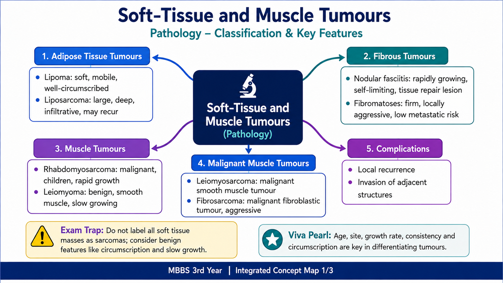
Soft-tissue tumours arise from the tissues that support, connect and move the body, including adipose tissue, fibrous tissue and skeletal or smooth muscle. They range from common, harmless lesions such as lipoma to aggressive malignant tumours such as liposarcoma and rhabdomyosarcoma. Their names usually reflect the type of tissue or cell that the tumour resembles. Understanding these lesions requires three linked ideas: the tissue of origin, whether the tumour is benign or malignant, and its characteristic gross and microscopic appearance. This chapter explains the classification and salient clinical features of soft-tissue tumours, followed by the important morphology and behaviour of lipoma, liposarcoma, nodular fasciitis, fibromatoses, rhabdomyosarcoma, leiomyoma, leiomyosarcoma and fibrosarcoma.
A. Classification and General Behaviour of Soft-Tissue Tumours
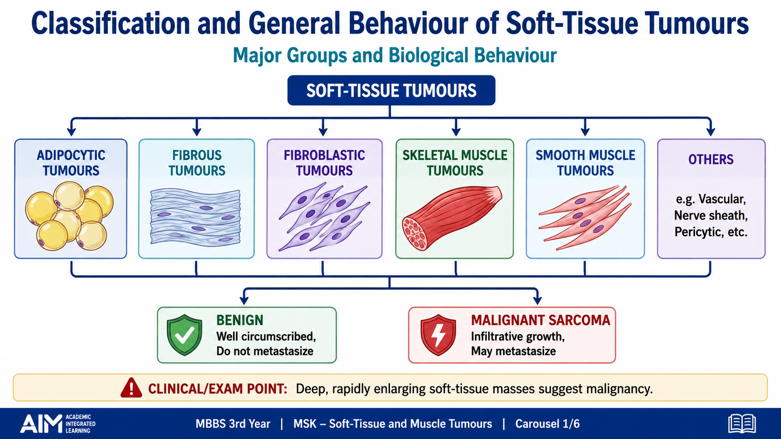
Soft-tissue tumours are classified mainly according to the normal adult tissue or cell type that they resemble. This classification does not always prove the exact cell from which a tumour originated. Instead, it describes the direction of differentiation shown by the tumour cells. Each major group may contain benign, locally aggressive or malignant lesions. Benign tumours usually remain localized and resemble normal mature tissue. Malignant soft-tissue tumours are called sarcomas. They may infiltrate surrounding structures, recur after removal and spread through the bloodstream, particularly to the lungs.
Basic classification
- Adipocytic tumours: lipoma and liposarcoma.
- Fibrous and fibroblastic tumours: nodular fasciitis, fibromatoses and fibrosarcoma.
- Skeletal muscle tumours: rhabdomyoma and rhabdomyosarcoma.
- Smooth muscle tumours: leiomyoma and leiomyosarcoma.
Salient clinical features
Most soft-tissue tumours present as a mass or swelling. A benign lesion is often superficial, mobile, slowly growing and painless. A malignant lesion may be deep, progressively enlarging, firm or fixed. Pain may occur when the tumour compresses nerves, invades surrounding structures or undergoes necrosis.
- Superficial, small and mobile masses are more often benign.
- Deep-seated or rapidly enlarging masses raise concern for malignancy.
- Local recurrence is especially important in infiltrative lesions such as fibromatosis.
- Blood-borne metastasis is a major feature of many sarcomas.
- The final diagnosis depends on morphology and, where necessary, immunohistochemical or molecular confirmation.
B. Tumours of Adipose Tissue: Lipoma and Liposarcoma
Adipocytic tumours show differentiation towards fat cells. A lipoma is a benign tumour composed mainly of mature adipocytes, whereas a liposarcoma is a malignant tumour showing adipocytic differentiation. Despite their similar names, liposarcoma usually develops independently and is not the usual result of malignant transformation within a lipoma.
Lipoma
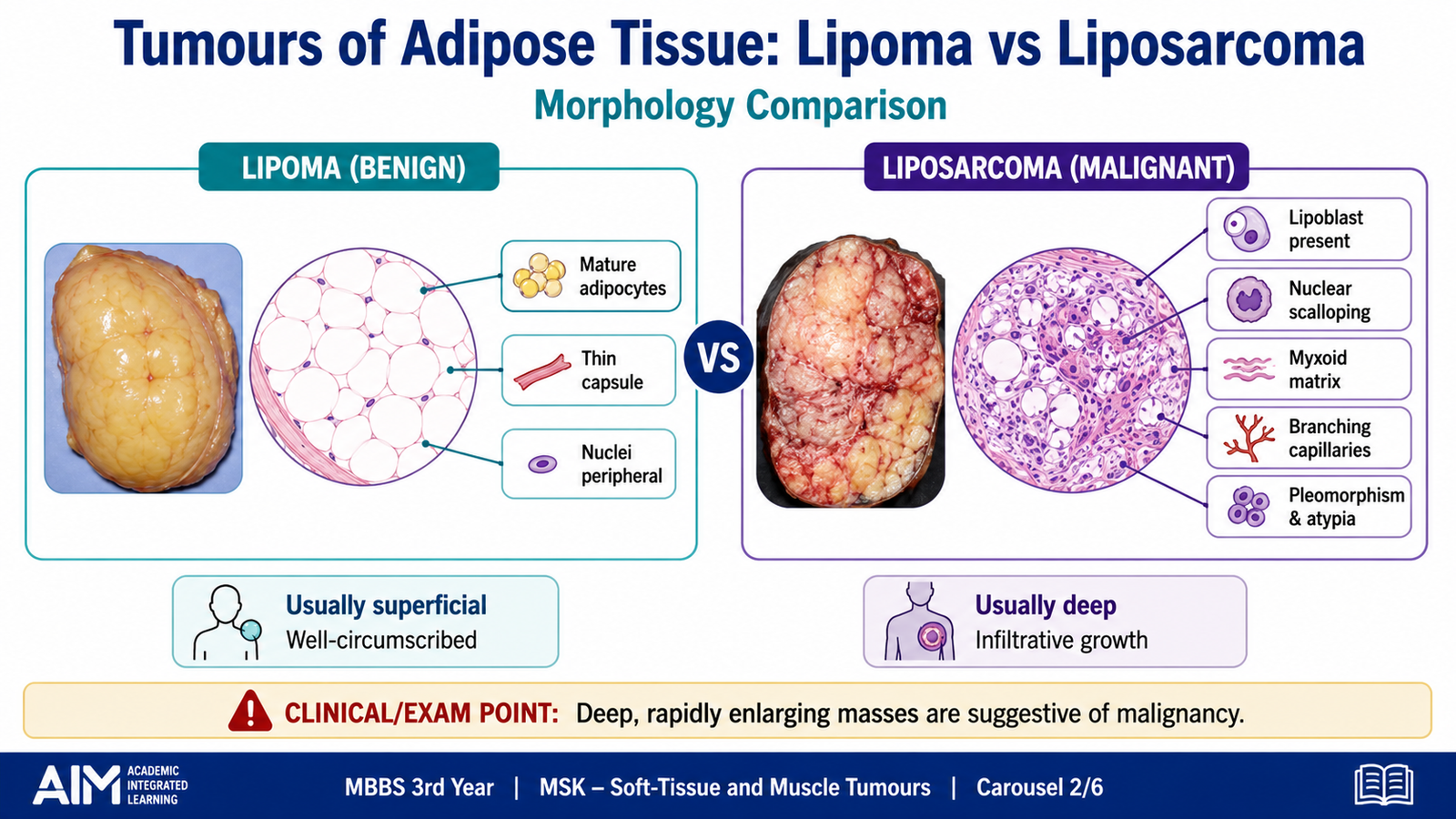
Lipoma is one of the most common benign soft-tissue tumours. It usually arises in the subcutaneous tissue of adults, particularly over the trunk, neck and proximal limbs. Because it grows slowly and remains well circumscribed, it commonly presents as a soft, freely mobile and painless swelling.
Gross morphology
- Usually a well-circumscribed or thinly encapsulated mass.
- Soft, yellow and lobulated.
- The cut surface resembles normal adipose tissue.
- Most lesions are located in superficial subcutaneous tissue.
Microscopic morphology
- Composed of mature adipocytes arranged in lobules.
- The cells closely resemble normal fat cells.
- Nuclei are small and peripheral without significant atypia.
- Fibrous septa may separate the lobules.
- Mitotic activity and tumour necrosis are absent.
The close resemblance to normal fat explains the soft consistency and yellow colour of the lesion. Complete removal is usually curative, although recurrence can occur if the lesion is incompletely removed.
Liposarcoma
Liposarcoma is a malignant adipocytic tumour that usually occurs in adults. Unlike lipoma, it commonly develops in deep soft tissues, especially the thigh or retroperitoneum. Retroperitoneal tumours may become very large before detection because there is considerable space for expansion. Liposarcoma includes several morphological patterns. Their clinical behaviour varies from relatively indolent, locally recurrent tumours to highly aggressive sarcomas with metastatic potential.
Important morphological forms
| Type | Important morphology | General behaviour |
|---|---|---|
| Well-differentiated liposarcoma | Mature adipocytes with variation in cell size, atypical stromal cells and occasional lipoblasts | Mainly locally aggressive; recurrence is important |
| Myxoid liposarcoma | Abundant myxoid matrix, delicate branching capillaries and primitive cells with lipoblasts | Intermediate malignant potential |
| Pleomorphic liposarcoma | Marked cellular pleomorphism, bizarre tumour cells and pleomorphic lipoblasts | High-grade aggressive sarcoma |
Lipoblast
A lipoblast is a tumour cell showing adipocytic differentiation. It contains one or more cytoplasmic lipid vacuoles that indent or scallop the nucleus. Lipoblasts support the diagnosis of liposarcoma, but the diagnosis depends on the overall architecture and cytological features rather than on a single cell alone.
Clinical features and complications
- Presents as a deep, enlarging and often painless mass.
- Retroperitoneal lesions may produce abdominal fullness or pressure symptoms.
- Local recurrence may occur because deep tumours can be difficult to remove completely.
- High-grade forms can metastasize through the bloodstream.
- Large tumours may compress adjacent organs, vessels or nerves.
C. Fibroblastic Proliferations: Nodular Fasciitis and Fibromatoses
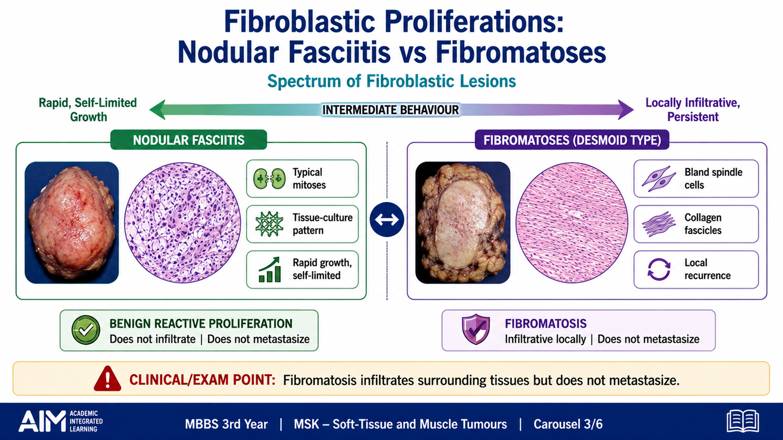
Nodular fasciitis and fibromatoses are fibroblastic or myofibroblastic proliferations. They are important because both can grow rapidly or infiltrate surrounding tissue and may therefore be mistaken for sarcoma. However, nodular fasciitis is a self-limited benign proliferation, while fibromatoses are locally aggressive lesions that do not metastasize.
Nodular fasciitis
Nodular fasciitis is a benign, rapidly growing proliferation of fibroblasts and myofibroblasts. It often affects young adults and commonly occurs in the superficial fascia of the upper limbs, trunk, head or neck. Its rapid appearance can create concern for malignancy, but the lesion is usually small and well circumscribed.
Clinicopathological features
- Rapidly growing, solitary mass developing over a short period.
- Usually superficial and relatively small.
- May be tender or painful.
- A history of preceding trauma may occasionally be present, but it is not required.
- Does not metastasize and may regress or be cured by local excision.
Microscopic morphology
- Plump spindle-shaped fibroblasts and myofibroblasts.
- Cells arranged in short, irregular bundles producing a tissue-culture-like pattern.
- Myxoid background with extravasated red blood cells.
- Numerous mitotic figures may be present.
- Mitotic figures are usually typical rather than atypical.
- Marked pleomorphism and destructive necrosis are absent.
The high mitotic activity reflects rapid growth, not necessarily malignancy. The combination of rapid growth and numerous mitoses is the main reason nodular fasciitis can be confused with a sarcoma.
Fibromatoses
Fibromatoses are clonal fibroblastic proliferations that lie between benign fibrous lesions and malignant fibrosarcoma. They do not metastasize, but they can infiltrate surrounding skeletal muscle, fascia and other structures. This infiltrative growth makes complete surgical removal difficult and explains their tendency to recur.
Classification
- Superficial fibromatoses: arise in superficial fascia and are usually smaller and less aggressive.
- Deep fibromatosis: also called a desmoid tumour; arises in deep musculoaponeurotic tissues and is more infiltrative.
Superficial fibromatoses
These lesions form firm nodules or bands in superficial fascia. Examples include palmar and plantar fibromatosis. Their contraction may produce deformity or limitation of movement because the abnormal fibrous tissue shortens over time.
Deep fibromatosis
Deep fibromatosis commonly presents as a firm, poorly defined mass in the abdominal wall, shoulder girdle, trunk or limbs. It consists of bland fibroblasts embedded in abundant collagen. Although the cells appear cytologically benign, the tumour sends long extensions into adjacent muscle and soft tissue.
Morphology
- Firm, grey-white and poorly circumscribed mass.
- Long sweeping fascicles of uniform spindle cells.
- Abundant collagen between tumour cells.
- Minimal cytological atypia.
- Low or moderate mitotic activity without atypical mitoses.
- Infiltration of surrounding skeletal muscle and soft tissue.
Complications
- Repeated local recurrence after incomplete removal.
- Compression or entrapment of nerves and vessels.
- Functional impairment due to invasion of muscle or fascia.
- Serious local effects when the lesion involves vital anatomical structures.
- No distant metastasis.
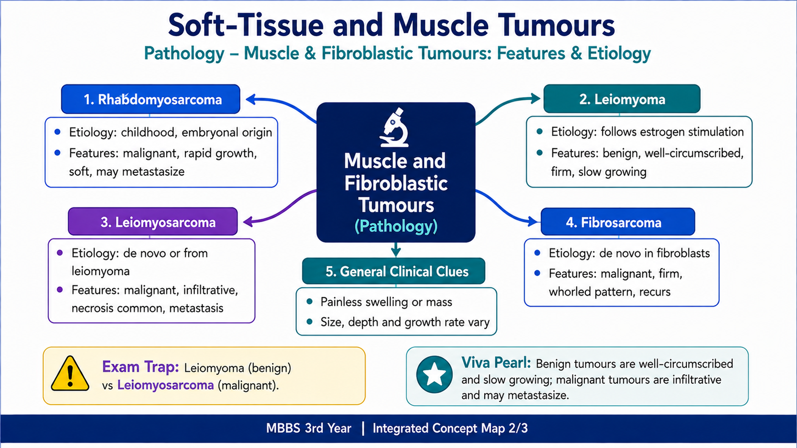
D. Skeletal Muscle Tumours and Rhabdomyosarcoma
Skeletal muscle tumours show differentiation towards striated muscle. They are broadly classified into benign rhabdomyoma and malignant rhabdomyosarcoma. Rhabdomyoma is uncommon, while rhabdomyosarcoma is an important malignant tumour of childhood. Rhabdomyosarcoma may arise in locations where mature skeletal muscle is sparse because the tumour develops from primitive mesenchymal cells capable of skeletal muscle differentiation.
Etiology and pathogenesis
Rhabdomyosarcoma develops when primitive mesenchymal cells acquire genetic alterations that disturb normal growth and muscle differentiation. The abnormal cells continue to proliferate while showing partial development towards skeletal muscle. The exact molecular abnormality varies among its morphological types.
Clinical features
Rhabdomyosarcoma occurs mainly in children and adolescents. The presenting feature depends strongly on the anatomical site. Common locations include the head and neck region, genitourinary tract and extremities.
- Head and neck: may produce a rapidly enlarging facial, orbital or nasopharyngeal mass.
- Genitourinary tract: may present with urinary obstruction, bleeding or a polypoid mass.
- Extremities: usually presents as a deep enlarging soft-tissue mass.
- Local invasion may cause pain, obstruction or loss of function.
- Metastasis may occur through blood or lymphatic channels.
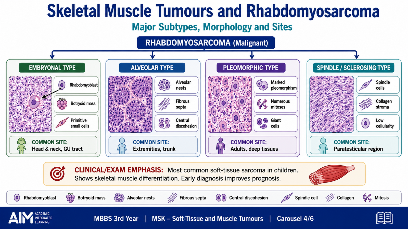
Major morphological types
Embryonal rhabdomyosarcoma
Embryonal rhabdomyosarcoma is the most common pattern in younger children. Its cells resemble developing embryonic skeletal muscle. The tumour may contain small primitive cells together with larger cells showing eosinophilic cytoplasm and greater skeletal-muscle differentiation.
- Commonly affects the head and neck or genitourinary region.
- Shows variable cellularity in a myxoid stroma.
- Contains primitive cells and rhabdomyoblasts.
- Rhabdomyoblasts may have deeply eosinophilic cytoplasm.
- Cross-striations may occasionally be visible but are not always present.
Alveolar rhabdomyosarcoma
Alveolar rhabdomyosarcoma occurs more often in older children and adolescents, particularly in the deep soft tissues of the extremities. Tumour cells form nests separated by fibrous septa. Cells in the centre of the nests may lose attachment, producing spaces that resemble pulmonary alveoli.
- Nests of relatively uniform tumour cells.
- Fibrous septa divide the tumour into compartments.
- Central loss of cohesion creates an alveolar pattern.
- Usually behaves more aggressively than typical embryonal tumours.
Pleomorphic rhabdomyosarcoma
Pleomorphic rhabdomyosarcoma mainly occurs in adults. It contains highly atypical and bizarre tumour cells with evidence of skeletal-muscle differentiation. It is a high-grade malignant tumour.
Complications
- Destruction of adjacent muscles and soft tissues.
- Obstruction of hollow organs when arising in the genitourinary tract.
- Neurological or visual problems in head and neck lesions.
- Local recurrence.
- Lymphatic and blood-borne metastasis.
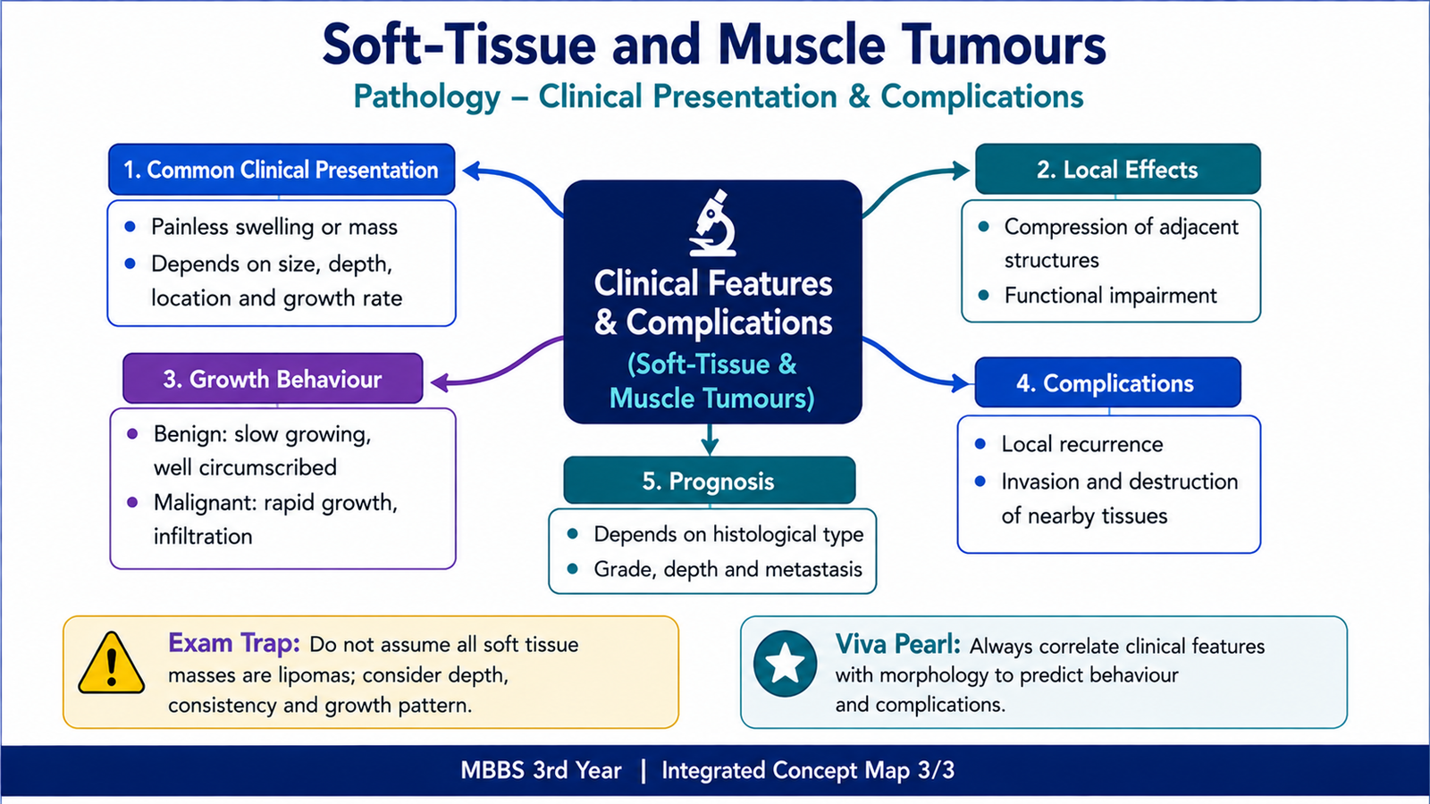
E. Smooth Muscle Tumours: Leiomyoma and Leiomyosarcoma
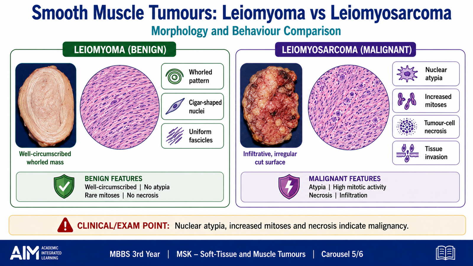
Smooth muscle tumours are classified as benign leiomyoma and malignant leiomyosarcoma. They may arise wherever smooth muscle is present, including the uterus, gastrointestinal tract, walls of blood vessels and skin. Their distinction depends mainly on cellular atypia, mitotic activity, tumour-cell necrosis and infiltrative behaviour.
Leiomyoma
Leiomyoma is a benign tumour composed of well-differentiated smooth muscle cells. The uterus is the most common site. Uterine leiomyomas may be single or multiple and often form sharply circumscribed masses within or around the uterine wall.
Gross morphology
- Well-circumscribed, firm and rubbery mass.
- Usually grey-white.
- Characteristic whorled appearance on the cut surface.
- May show degenerative change when the lesion outgrows its blood supply.
Microscopic morphology
- Intersecting fascicles of uniform spindle-shaped smooth muscle cells.
- Cells have eosinophilic cytoplasm.
- Nuclei are elongated with blunt ends, often described as cigar-shaped.
- Minimal or no cytological atypia.
- Low mitotic activity.
- No coagulative tumour-cell necrosis.
Clinical features and complications
Clinical effects depend on tumour size, number and location. Uterine leiomyomas may cause abnormal uterine bleeding, pelvic pressure, pain or reproductive difficulty. These manifestations occur because the mass distorts the uterine cavity, increases the bleeding surface or compresses nearby structures.
Leiomyosarcoma
Leiomyosarcoma is a malignant tumour showing smooth-muscle differentiation. It may arise in the uterus, deep soft tissues, gastrointestinal tract or large blood vessels. Uterine leiomyosarcoma usually develops as a separate malignant tumour rather than through transformation of an ordinary leiomyoma.
Etiology and pathogenesis
Genetic abnormalities lead to uncontrolled proliferation of smooth-muscle cells. As the tumour becomes more aggressive, the cells lose their normal uniform appearance, divide rapidly and invade surrounding tissue. Rapid growth may exceed the available blood supply, producing tumour-cell necrosis.
Gross morphology
- Large, soft or fleshy mass.
- May be poorly circumscribed and infiltrative.
- Cut surface may show haemorrhage and necrosis.
- The typical whorled pattern of leiomyoma may be absent or less distinct.
Microscopic morphology
- Spindle cells showing smooth-muscle differentiation.
- Moderate to marked nuclear atypia.
- Increased mitotic activity, sometimes with atypical mitoses.
- Coagulative tumour-cell necrosis may be present.
- Infiltration of surrounding tissue.
Clinical features and complications
- Progressively enlarging mass.
- May produce pain, bleeding or pressure symptoms depending on site.
- Can recur locally after treatment.
- May spread through the bloodstream, particularly to the lungs and other distant organs.
- Necrosis and haemorrhage may contribute to rapid enlargement.
F. Fibrosarcoma
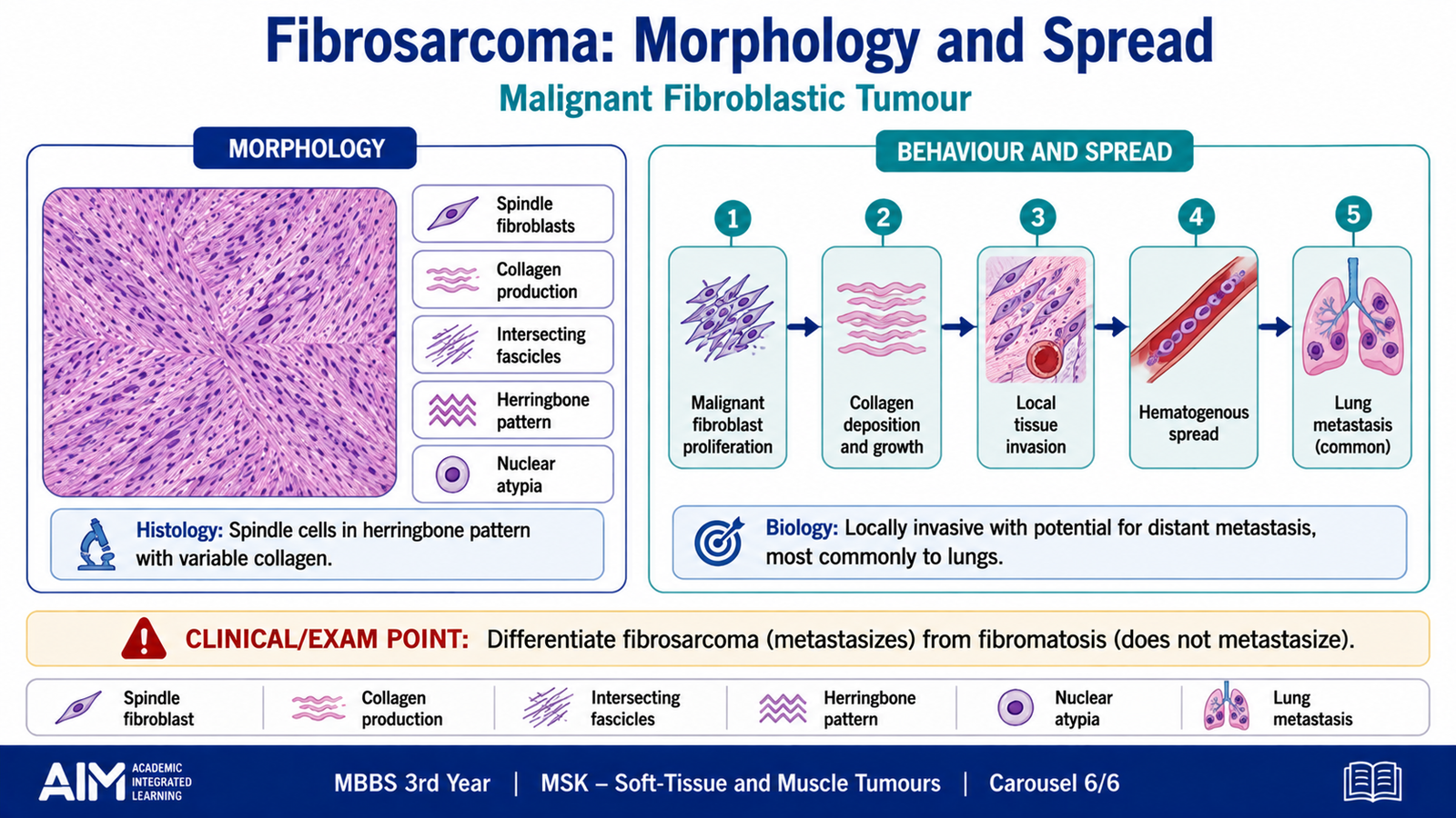
Fibrosarcoma is a malignant tumour composed of fibroblasts that produce variable amounts of collagen. It must be distinguished from benign fibroblastic proliferations and fibromatosis. Unlike fibromatosis, fibrosarcoma shows definite malignant cytological features and has the ability to metastasize.
Etiology and pathogenesis
Fibrosarcoma develops when fibroblastic cells acquire genetic alterations that promote uncontrolled growth and malignant behaviour. The tumour expands and infiltrates surrounding soft tissue. More aggressive lesions show increasing cellularity, atypia, mitotic activity and necrosis.
Clinical features
- Usually presents as a deep, enlarging soft-tissue mass.
- May occur in the extremities or trunk.
- The swelling may initially be painless.
- Pain or functional impairment may develop due to local invasion.
- Large tumours may compress nerves, vessels or nearby structures.
Gross morphology
- Unencapsulated and infiltrative mass.
- Grey-white, firm or fleshy cut surface.
- May show haemorrhage and necrosis in high-grade lesions.
Microscopic morphology
- Malignant spindle-shaped fibroblasts.
- Cells arranged in intersecting fascicles.
- Classic fascicles may form a herringbone pattern.
- Variable collagen deposition between tumour cells.
- Nuclear atypia and increased mitotic activity.
- High-grade lesions may contain atypical mitoses and necrosis.
The herringbone pattern is produced when intersecting fascicles meet at sharp angles, resembling the arrangement of fish bones. It is a useful classical clue but should be interpreted together with evidence of malignant spindle-cell morphology.
Complications
- Infiltration and destruction of nearby tissues.
- Local recurrence after incomplete excision.
- Haematogenous metastasis, especially to the lungs.
- Loss of function when the tumour involves muscles, nerves or major vessels.
Integrated Mechanism Flow
Important Comparisons
Lipoma versus Liposarcoma
| Feature | Lipoma | Liposarcoma |
|---|---|---|
| Behaviour | Benign | Malignant |
| Usual location | Superficial subcutaneous tissue | Deep soft tissue or retroperitoneum |
| Clinical growth | Slow, soft and mobile | Progressively enlarging deep mass |
| Microscopy | Uniform mature adipocytes | Atypical adipocytic tumour with subtype-specific features |
| Necrosis and atypia | Absent | May be prominent in high-grade types |
| Outcome | Usually cured by removal | May recur or metastasize |
Nodular Fasciitis, Fibromatosis and Fibrosarcoma
| Feature | Nodular fasciitis | Fibromatosis | Fibrosarcoma |
|---|---|---|---|
| Nature | Benign, self-limited proliferation | Locally aggressive proliferation | Malignant fibroblastic tumour |
| Growth | Rapid but usually circumscribed | Infiltrative | Infiltrative and destructive |
| Cell appearance | Plump spindle cells without marked atypia | Uniform bland spindle cells | Malignant spindle cells with atypia |
| Mitoses | Frequent but typical | Usually low to moderate | Increased; may be atypical |
| Metastasis | Absent | Absent | Possible, especially to lungs |
| Major concern | Mistaken for sarcoma | Local recurrence | Recurrence and metastasis |
⭐ AIM High-Yield Review
- Soft-tissue tumours are classified according to the adult tissue or cell type they resemble.
- Malignant soft-tissue tumours are called sarcomas and commonly spread through the bloodstream.
- ⭐ Lipoma is a soft, mobile and usually superficial benign tumour composed of mature adipocytes.
- Liposarcoma commonly arises in deep soft tissue or the retroperitoneum and does not usually develop from a lipoma.
- A lipoblast contains lipid vacuoles that indent or scallop its nucleus.
- ⭐ Nodular fasciitis may grow rapidly and contain many mitoses, but the mitoses are typically normal and the lesion does not metastasize.
- Fibromatosis is cytologically bland but locally infiltrative, frequently recurrent and non-metastatic.
- Rhabdomyosarcoma is an important childhood sarcoma showing skeletal-muscle differentiation.
- Sarcoma botryoides is a grape-like mucosal form of embryonal rhabdomyosarcoma.
- ⭐ Alveolar rhabdomyosarcoma shows tumour-cell nests separated by fibrous septa with central loss of cohesion.
- Leiomyoma is well circumscribed and whorled, with uniform cigar-shaped nuclei and low mitotic activity.
- Marked atypia, increased mitoses and coagulative tumour-cell necrosis favour leiomyosarcoma.
- Ordinary uterine leiomyosarcoma usually arises independently rather than from a pre-existing leiomyoma.
- ⭐ Fibrosarcoma is a malignant fibroblastic tumour that may show a herringbone fascicular pattern.
- Fibrosarcoma can metastasize, whereas fibromatosis remains locally aggressive without distant spread.
Soft-Tissue and Muscle Tumours
Review the classification, clinical behaviour and pathological features of important soft-tissue sarcomas.