
📖 AIM Learning Material
Osteomyelitis Made Simple
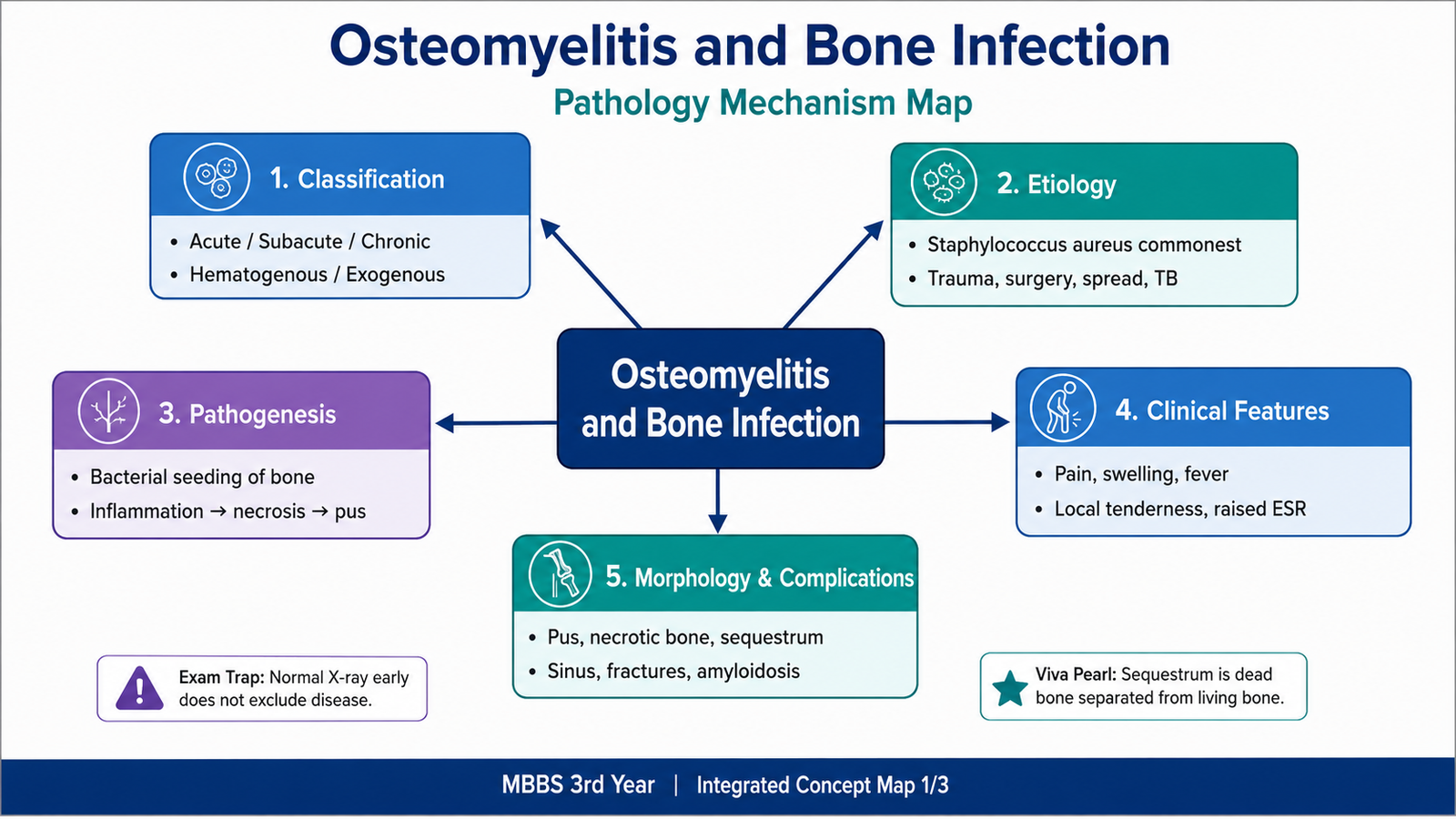
Osteomyelitis is infection-associated inflammation of bone and bone marrow. It may begin when microorganisms reach bone through the bloodstream, enter directly after trauma or surgery, or spread from nearby infected tissue. Once established, the infection produces pus and raises pressure inside the rigid bone. This damages blood vessels and may cause ischemic death of bone. The dead bone can separate as a sequestrum, while surrounding viable bone attempts repair by forming an involucrum. In this chapter, you will learn how osteomyelitis is classified, which organisms and routes are important, how acute infection becomes chronic, what the gross and microscopic changes are, how patients present, and why serious complications may develop.
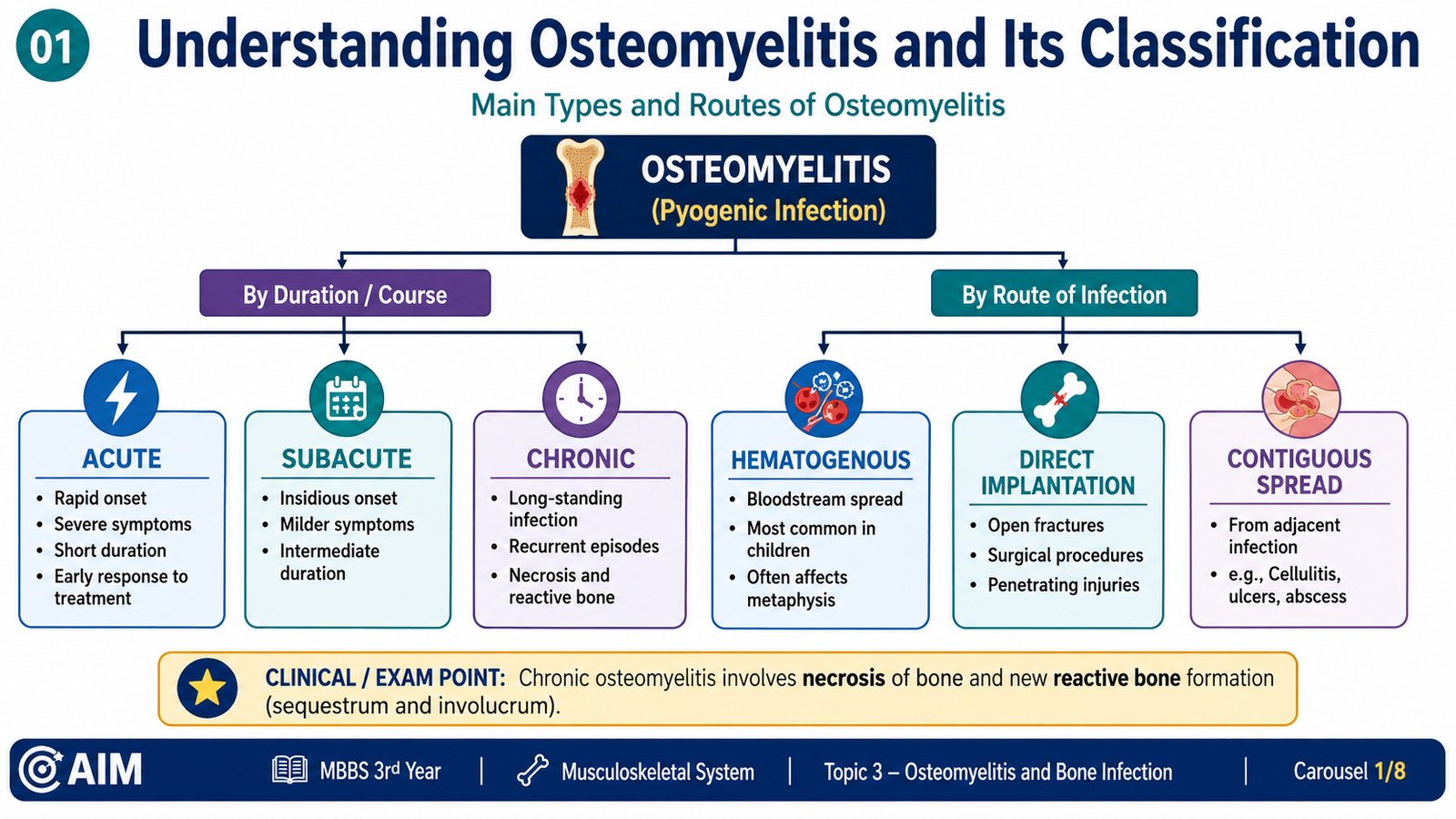
Understanding Osteomyelitis and Its Classification
Definition and core concept
Osteomyelitis is inflammation of bone and bone marrow caused most commonly by infection. The process may involve the marrow cavity, cancellous bone, cortical bone and periosteum. Although bacteria are the usual agents, mycobacteria and fungi can also produce osteomyelitis in appropriate clinical settings.
The central pathological problem is not merely the presence of organisms. Infection produces suppuration, vascular thrombosis, ischemic bone death and reactive repair. These changes explain why inadequately treated infection may persist even after systemic symptoms improve.
Classification
| Basis | Categories | Key interpretation |
|---|---|---|
| Clinical duration | Acute, subacute and chronic | Reflects the tempo of inflammation, necrosis and repair. |
| Route of infection | Hematogenous, contiguous spread and direct implantation | Identifies how organisms reached the bone. |
| Causative agent | Pyogenic, tuberculous, fungal and other specific infections | Correlates with inflammatory pattern and clinical course. |
| Anatomical pattern | Localized or diffuse involvement | Describes the extent of medullary and cortical disease. |
Acute osteomyelitis
Acute osteomyelitis is dominated by rapid bacterial multiplication and neutrophil-rich suppurative inflammation. Severe localized pain, fever, tenderness and elevated inflammatory markers are typical. Bone necrosis can begin early if vascular compromise develops.
Subacute osteomyelitis
Subacute osteomyelitis follows a less aggressive course, often because of reduced organism virulence, partial host resistance or incomplete antimicrobial exposure. A localized intraosseous abscess, classically termed a Brodie abscess, may develop.
Chronic osteomyelitis
Chronic osteomyelitis is characterized by persistent infection, dead bone, fibrosis, reactive new-bone formation and recurrent drainage. Its persistence is promoted by avascular sequestra and poorly penetrated infected spaces.
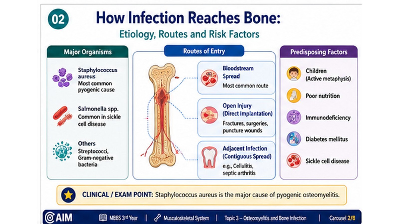
How Infection Reaches Bone: Etiology, Routes and Risk Factors
Major causative organisms
The causative organism varies with age, route of infection, immune status and underlying disease. Staphylococcus aureus is the most important cause of pyogenic osteomyelitis because it can adhere to bone matrix, evade host defense and produce destructive suppurative inflammation.
- Staphylococcus aureus: the most common cause across most age groups.
- Group B streptococci and enteric gram-negative bacilli: important in neonates.
- Haemophilus influenzae: historically important in incompletely immunized young children.
- Salmonella species: classically associated with sickle-cell disease.
- Gram-negative bacilli, including Pseudomonas: may follow penetrating wounds, healthcare exposure or particular environmental contamination.
- Mycobacteria and fungi: usually produce more indolent or granulomatous disease, particularly in immunocompromised patients.
Routes by which infection reaches bone
Age-related anatomical distribution
| Group | Typical site or pattern | Reason |
|---|---|---|
| Infants | Metaphysis with possible extension across the growth plate | Transphyseal vessels can permit spread into epiphysis and joint. |
| Children | Metaphysis of long bones, especially near the knee | Slow flow in metaphyseal vascular loops favors bacterial seeding. |
| Adults | Vertebrae commonly affected by hematogenous spread | Organisms reach vertebral bodies through the bloodstream. |
| Patients with ulcers or trauma | Underlying or adjacent bone | Contiguous extension or direct inoculation. |
Predisposing factors
- Recent bacteremia or systemic infection
- Open fracture, penetrating trauma or orthopedic surgery
- Prosthetic material or implanted orthopedic devices
- Diabetes mellitus with chronic foot ulceration
- Peripheral vascular insufficiency and poor tissue perfusion
- Sickle-cell disease
- Immunosuppression
- Chronic skin or soft-tissue infection adjacent to bone
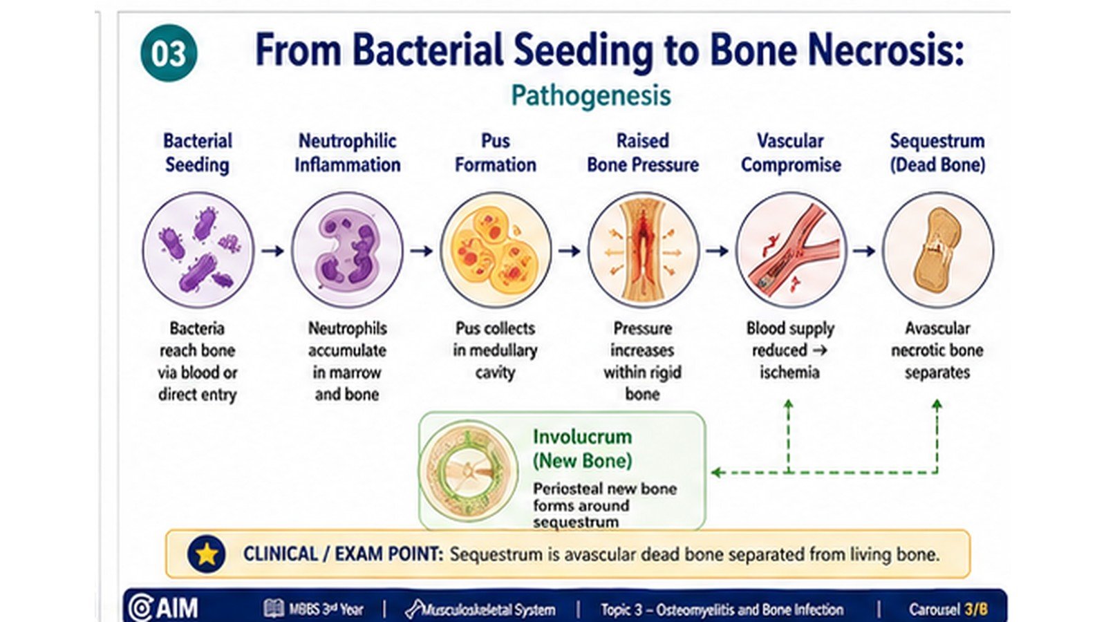
From Bacterial Seeding to Bone Necrosis: Pathogenesis
Initial microbial localization
In hematogenous osteomyelitis, circulating bacteria lodge in small vessels within bone. In children, metaphyseal vascular loops provide a favorable site because blood flow is slow and phagocytic activity is relatively limited. Organisms adhere, multiply and initiate acute inflammation.
Development of suppurative inflammation
Bacterial products and tissue injury activate innate immune responses. Neutrophils migrate into the marrow spaces, and accumulation of inflammatory exudate produces pus. Neutrophil enzymes, bacterial factors and pressure within the rigid bony compartment damage marrow tissue and trabeculae.
Spread through bone
The purulent exudate spreads through medullary spaces and along vascular channels. It may pass through the cortex and collect beneath the periosteum. In children, the relatively loose periosteal attachment allows rapid formation of a subperiosteal abscess.
Accumulating pus separates the periosteum from cortical bone. This disrupts the periosteal blood supply, while inflammation and thrombosis interfere with medullary vessels. The combined loss of blood flow produces segments of necrotic bone.
Formation of a sequestrum
A fragment of devitalized bone may separate from the living bone and become a sequestrum. Because it is avascular, immune cells and antimicrobial agents reach it poorly. It can therefore act as a protected surface for persistent microorganisms.
Reactive new-bone formation
Surviving periosteal osteoblasts attempt repair by depositing new bone around the infected and necrotic focus. This shell of reactive bone is called an involucrum. Openings in the involucrum may allow pus to escape toward the skin, creating a draining sinus.
Why chronic infection develops
Chronic osteomyelitis develops when organisms persist within necrotic bone, fibrotic tissue, poorly vascularized cavities or foreign material. Recurrent cycles of inflammation and drainage are accompanied by fibrosis, bone destruction and attempted repair.
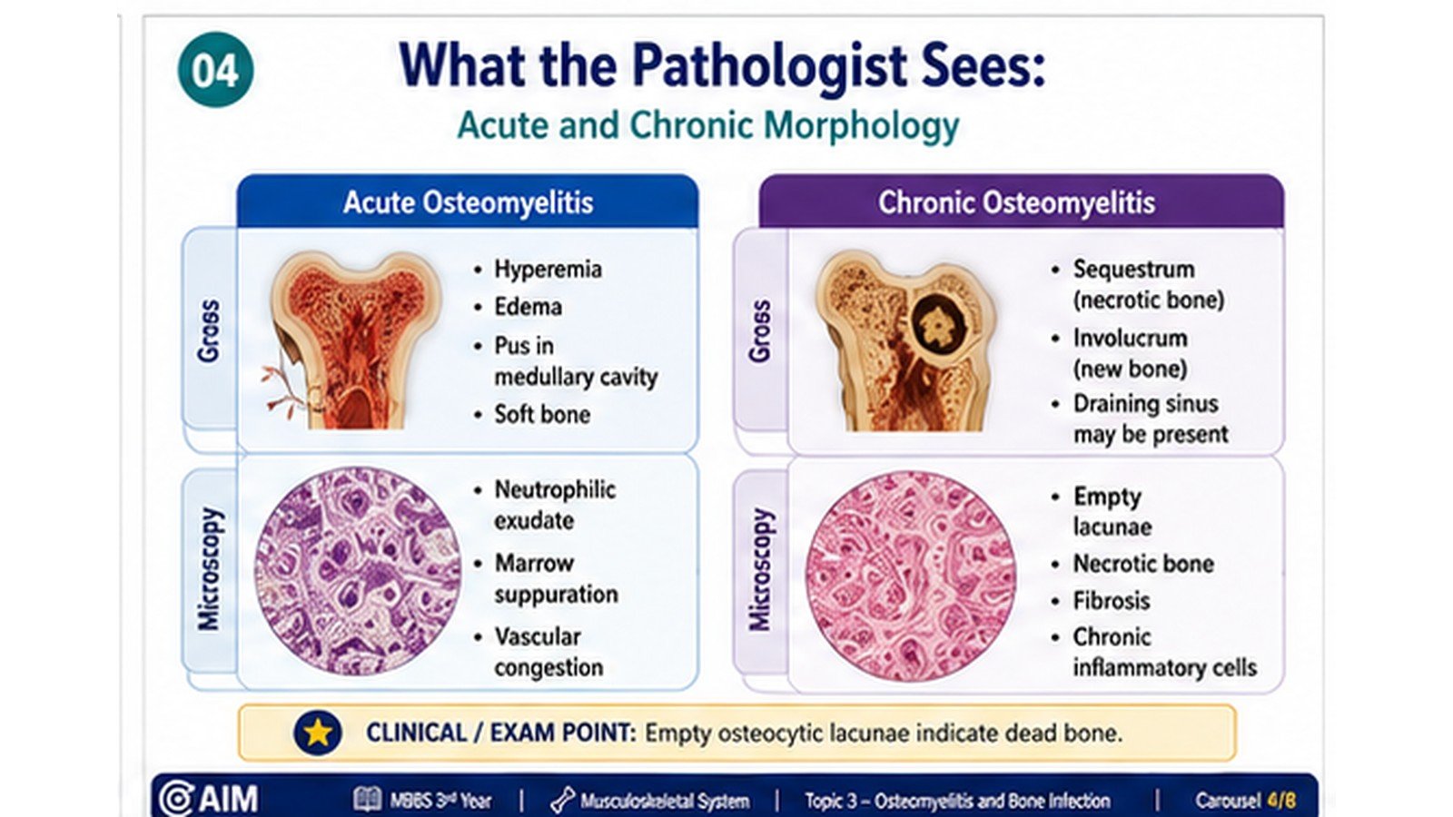
What the Pathologist Sees: Acute and Chronic Morphology
Acute pyogenic osteomyelitis
- Congested, edematous marrow
- Purulent material within the medullary cavity
- Foci of yellow-white suppuration
- Cortical destruction in progressive disease
- Subperiosteal collection of pus
- Early areas of devitalized bone
- Numerous neutrophils in marrow spaces
- Necrosis of marrow cells and bone trabeculae
- Bacterial colonies may be present
- Vascular congestion and thrombosis
- Osteocyte loss from lacunae in devitalized bone
- Reactive changes at the viable margin
Chronic osteomyelitis
- Irregular cavities containing necrotic debris
- Dense fragment of dead bone forming a sequestrum
- Reactive shell of new bone forming an involucrum
- Fibrosis and cortical thickening
- Sinus tracts extending through soft tissue to skin
- Distortion and weakening of affected bone
- Necrotic bone with empty lacunae
- Mixed inflammatory infiltrate
- Lymphocytes, plasma cells and macrophages
- Fibrosis and granulation tissue
- Reactive woven-bone formation
- Neutrophils during active exacerbations
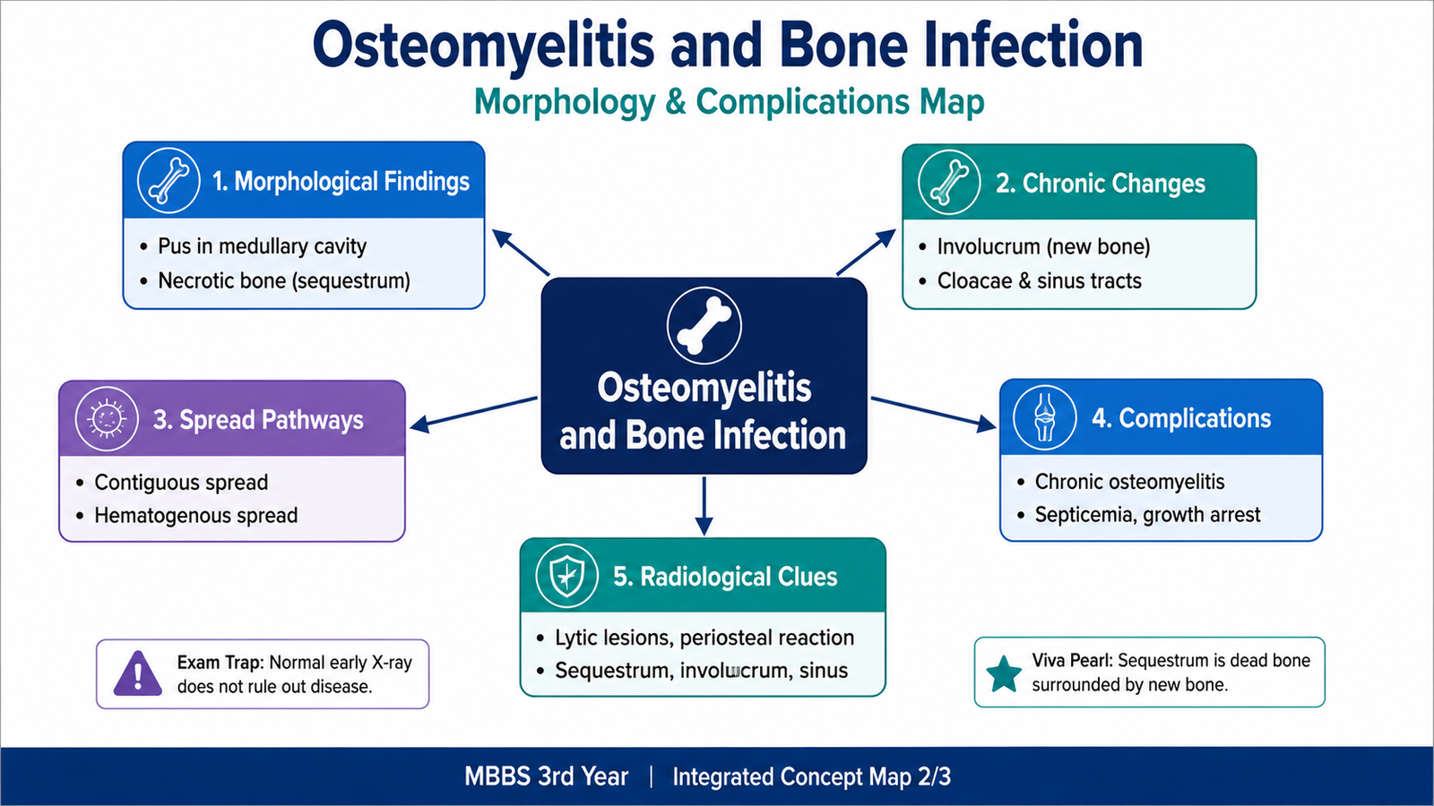
Sequestrum, involucrum and cloaca
| Term | Pathological meaning | Clinical or diagnostic relevance |
|---|---|---|
| Sequestrum | Separated fragment of necrotic bone | Acts as a persistent avascular focus of infection. |
| Involucrum | Reactive new bone surrounding a sequestrum | Represents attempted repair and may appear as cortical thickening. |
| Cloaca | Opening in the involucrum through which inflammatory material escapes | May communicate with a draining sinus tract. |
Subacute localized abscess
A Brodie abscess is a localized intraosseous abscess associated with subacute osteomyelitis. It usually consists of a central suppurative or necrotic focus surrounded by granulation tissue and reactive sclerotic bone. Its relatively indolent presentation can mimic a bone tumor.
How Pathology Produces the Clinical Presentation
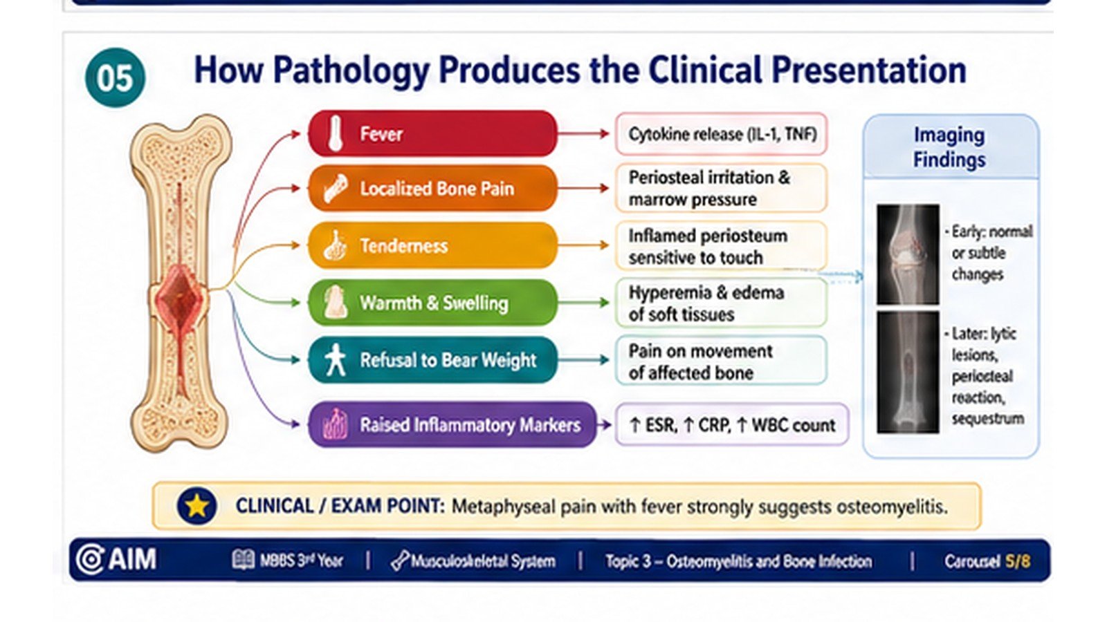
Clinical features of acute osteomyelitis
- Fever, malaise and systemic illness
- Severe localized bone pain
- Point tenderness over the affected site
- Warmth, swelling and erythema
- Restricted movement of the adjacent limb or joint
- Refusal to use the limb or inability to bear weight in children
- Elevated inflammatory markers and leukocytosis may be present
Why these manifestations occur
Clinical features of chronic osteomyelitis
- Persistent or recurrent localized pain
- Intermittent swelling
- Recurrent fever during exacerbations
- Non-healing wound or chronic discharging sinus
- Localized deformity or thickened bone
- Reduced limb function
- History of previous trauma, surgery, ulcer or inadequately treated infection
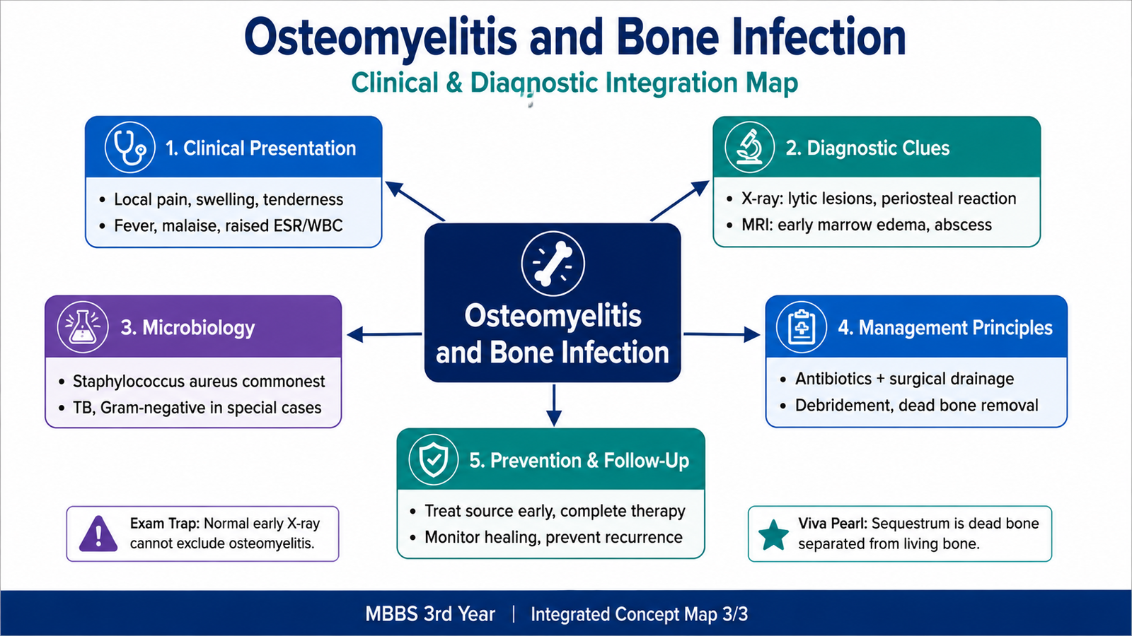
Basic diagnostic approach
Diagnosis requires integration of history, examination, inflammatory markers, microbiological sampling and imaging. No single feature should be interpreted in isolation.
- Blood cultures: may identify the organism, especially in hematogenous acute disease.
- Inflammatory markers: support active inflammation and may help monitor response, but they are not specific.
- Imaging: defines the site, extent, cortical destruction, periosteal reaction and chronic structural changes.
- Bone or deep-tissue sampling: provides the strongest microbiological correlation when superficial contamination is a concern.
- Histopathology: demonstrates acute suppuration, necrotic bone, chronic inflammation, fibrosis or reactive bone formation.
Radiological correlation
Early plain radiographs may not immediately show major structural abnormalities. As disease progresses, bone destruction, cortical irregularity, periosteal reaction and reactive sclerosis may become visible. Chronic disease may show sequestrum, involucrum, cortical thickening and sinus-related changes.
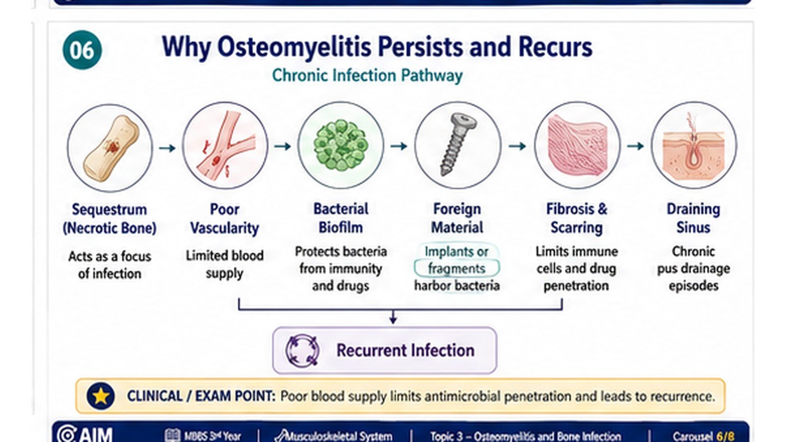
Why Osteomyelitis Persists and Recurs
Avascular necrotic bone
The sequestrum lacks an effective blood supply. Host leukocytes and systemically administered antimicrobial agents cannot reach organisms within it efficiently. Persistence of this protected nidus is a major reason chronic osteomyelitis may not resolve without removal of devitalized tissue.
Biofilm and foreign material
Bacteria may adhere to necrotic bone or implanted devices and form organized microbial communities within an extracellular matrix. This biofilm reduces bacterial susceptibility to host defenses and antimicrobial therapy, contributing to persistence and recurrence.
Poor vascular supply
Diabetes mellitus, vascular insufficiency, extensive scarring and previous tissue damage reduce perfusion. Poor blood flow limits oxygen delivery, immune-cell access, antimicrobial penetration and tissue repair.
Sinus formation and recurrent drainage
Persistent pus may track through cortical defects, soft tissue and skin. Drainage can temporarily reduce pressure and symptoms without eliminating the deep infected focus. Closure of the sinus may therefore be followed by renewed accumulation and another clinical exacerbation.
Cycles of injury and repair
Chronic inflammation causes continuing bone resorption, fibrosis and architectural distortion. At the same time, periosteal and endosteal osteoblasts produce reactive bone. The result is an irregular combination of destruction, sclerosis, sequestration and attempted reconstruction.
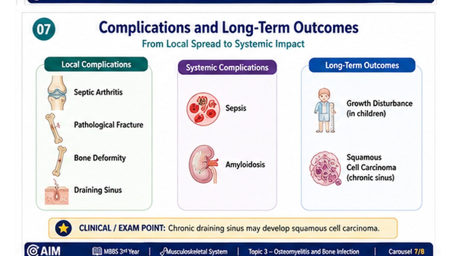
Complications and Long-Term Outcomes
Local extension of infection
- Subperiosteal abscess: pus dissects beneath the periosteum and further compromises cortical blood supply.
- Soft-tissue abscess: infection breaks through the cortex and spreads into surrounding tissue.
- Septic arthritis: infection reaches an adjacent joint, particularly where the metaphysis lies within the joint capsule or where transphyseal spread occurs in infants.
- Cellulitis and sinus formation: infection tracks toward the skin and creates chronic drainage.
Systemic spread
Organisms may enter the bloodstream from the infected bone, causing bacteremia or sepsis. Metastatic infection can occur at distant sites when bacteremia is sustained.
Structural complications
- Pathological fracture: destruction and remodeling weaken the bone until normal stress produces fracture.
- Growth disturbance: damage to the growth plate may cause limb shortening or angular deformity in children.
- Bone deformity: repeated destruction and irregular repair alter normal architecture.
- Chronic pain and disability: fibrosis, deformity and recurrent inflammation impair limb function.
Complications of chronic inflammation
- Secondary amyloidosis: prolonged inflammatory activity may promote systemic deposition of serum amyloid-associated protein derivatives.
- Squamous-cell carcinoma: a long-standing draining sinus may undergo malignant transformation.
Mechanistic summary of major complications
| Complication | Pathological basis | Clinical consequence |
|---|---|---|
| Septic arthritis | Extension into adjacent joint | Painful restricted joint with risk of cartilage damage |
| Pathological fracture | Loss of cortical and trabecular strength | Fracture after minor stress |
| Growth deformity | Physeal injury | Limb shortening or angular deformity |
| Amyloidosis | Persistent systemic inflammatory stimulus | Progressive organ dysfunction, particularly renal involvement |
| Squamous carcinoma | Long-standing epithelial irritation in a sinus tract | Malignant transformation of chronic sinus epithelium |
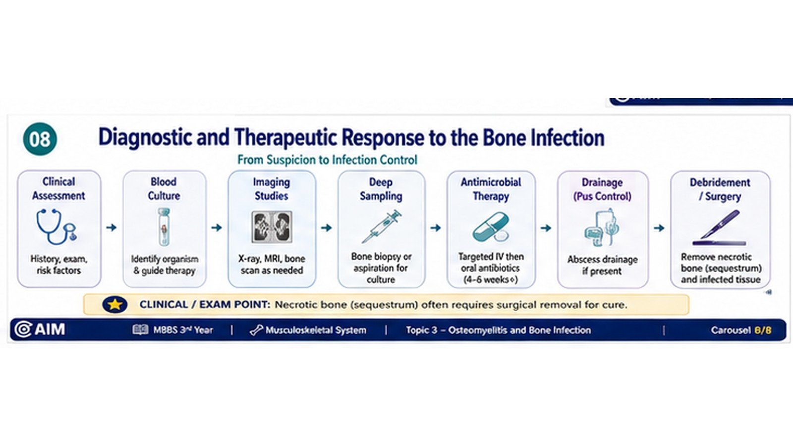
Clinical and Diagnostic Correlation Visual
This preserved visual reinforces how clinical findings, microbiological sampling and imaging relate to the underlying bone lesion.
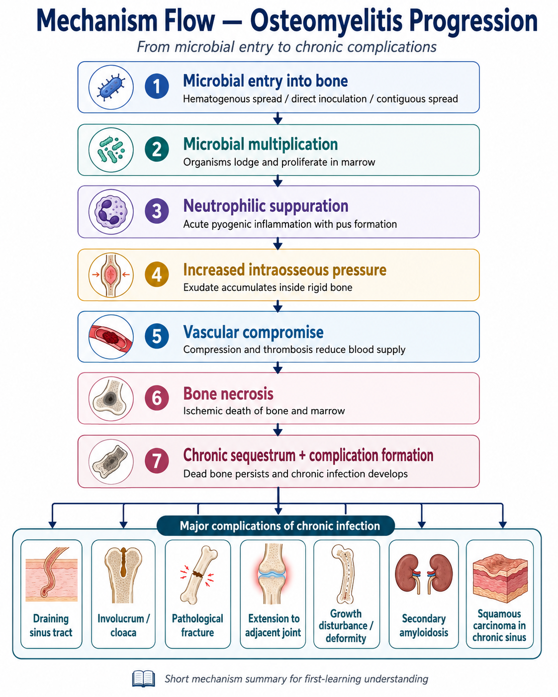
From Microbial Entry to Chronic Bone Damage
Essential Points for Revision
- Osteomyelitis is infection-associated inflammation involving bone and marrow.
- It is classified by clinical course, route of infection, causative organism and anatomical extent.
- Staphylococcus aureus is the most important cause of pyogenic osteomyelitis.
- Hematogenous infection commonly affects the metaphysis of long bones in children and vertebral bodies in adults.
- Salmonella is a classic association in patients with sickle-cell disease.
- Neutrophilic suppuration and raised intraosseous pressure damage vessels and cause ischemic bone necrosis.
- A sequestrum is separated dead bone; an involucrum is reactive new bone around it; a cloaca is an opening through which pus may escape.
- Acute osteomyelitis shows pus, neutrophils, marrow necrosis and vascular thrombosis.
- Chronic osteomyelitis shows necrotic bone, chronic inflammation, granulation tissue, fibrosis, reactive bone and sinus formation.
- Empty osteocytic lacunae are an important microscopic clue to dead bone.
- Typical acute features are fever, severe localized bone pain, tenderness, warmth, swelling and reduced limb use.
- Chronic disease may present with recurrent pain, deformity and a persistent or intermittent draining sinus.
- Major complications include septic spread, septic arthritis, pathological fracture, growth disturbance, deformity and chronic disability.
- Long-standing disease may rarely lead to secondary amyloidosis or squamous-cell carcinoma arising in a chronic sinus.
Recommended Video — Osteomyelitis and Bone Infection
Use this video after reading the chapter to strengthen your understanding of classification, etiology, pathogenesis, morphology, clinical features and complications of osteomyelitis.
- First understand how infection reaches bone and why the metaphysis is commonly affected in children.
- Focus on the sequence from microbial entry to suppuration, vascular compromise and bone necrosis.
- Revise the key pathological terms: sequestrum, involucrum and cloaca.
- Correlate acute and chronic morphology with the usual clinical features and complications.
- Use the video as reinforcement, not as a substitute for the written AIM learning material.
Admin note: Replace VIDEO_ID in the iframe link with the YouTube ID of your selected osteomyelitis teaching video before pasting into LMS.
Concept Not Clear? Ask AIM AI
Use the AIM Learning Assistant to clarify difficult concepts from this topic before proceeding to assessment.
You may ask AIM AI to:
- Explain a difficult concept in simpler words.
- Describe a mechanism step by step.
- Compare two confusing diseases, drugs or findings.
- Provide a clinically relevant example.
- Highlight a high-yield KMU examination point.
- Explain a common MCQ trap related to the topic.
Best use: Ask one focused question at a time. AIM AI supports your understanding but does not replace the complete learning material.