
This chapter follows the supplied KMU learning outcomes. First understand how the different skin lesions are recognized and distinguished, then use the final high-yield review to revise the most important examination points.
Premalignant Skin Lesions, Keratinocyte Carcinomas, Nevi and Malignant Melanoma
MSK Module — recognition, morphology and basic diagnostic assessment of important premalignant and malignant skin lesions and melanocytic nevi.
Topic Introduction
Skin lesions range from harmless nevi to premalignant changes and invasive cancers. In this topic, the main epithelial malignancies are squamous cell carcinoma (SCC) and basal cell carcinoma (BCC), while the major melanocytic malignancy is malignant melanoma. Their appearances may overlap, so students need to connect the clinical appearance of a lesion with its underlying morphology. You will also learn the main premalignant epithelial lesions, important types of nevocellular nevi, the special significance of dysplastic nevi, and the basic investigations used when a suspicious skin lesion is assessed.
A. Premalignant Epithelial Lesions of the Skin
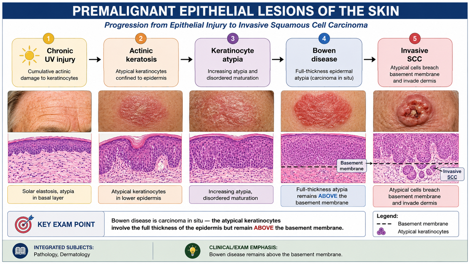
A premalignant skin lesion is a lesion in which epithelial cells show abnormal proliferation or atypia and have an increased possibility of progressing to invasive malignancy. The most important relationship in this topic is between chronic ultraviolet-induced epidermal damage and the subsequent development of squamous cell carcinoma. Not every premalignant lesion becomes cancer, but recognition is important because persistent or changing lesions may require biopsy.
Important premalignant conditions
- Actinic keratosis: a common sun-induced premalignant keratinocytic lesion occurring on chronically exposed skin.
- Bowen disease: squamous cell carcinoma confined to the epidermis, also called SCC in situ; it can progress to invasive SCC.
- Chronic scars and long-standing ulcers: persistent epithelial injury and regeneration can predispose to SCC.
- Certain chronic radiation- or chemical-associated keratoses: prolonged carcinogenic injury may produce epithelial atypia and later carcinoma.
Actinic keratosis
Actinic keratosis develops mainly because of cumulative ultraviolet radiation. Ultraviolet injury produces genetic damage in epidermal keratinocytes. Abnormal keratinocytes then proliferate within the epidermis, producing a rough, scaly lesion. The lesion therefore represents dysplastic keratinocytic growth rather than simply dry skin. Typical clinical appearance:
- Occurs mainly on chronically sun-exposed sites such as the face, scalp, ears, neck, forearms and backs of the hands.
- Usually appears as a small rough or gritty macule, papule or plaque.
- May be erythematous, tan or brown.
- Scale and surface keratosis are common.
- A persistent thickening, ulcer or enlarging nodule raises concern for progression to SCC.
Bowen disease
In Bowen disease, atypical squamous cells involve the full thickness of the epidermis but have not crossed the basement membrane. It is therefore an intraepidermal carcinoma. Clinically, it commonly appears as a persistent, well-defined, erythematous and scaly plaque. Once malignant cells penetrate through the basement membrane into the dermis, the lesion is considered invasive SCC.
Clinical differential diagnosis
Premalignant lesions may resemble inflammatory or benign keratotic conditions. Important clues are persistence, progressive change, surface roughness, ulceration, bleeding or failure of an apparently inflammatory lesion to resolve.
- Actinic keratosis: rough, sun-exposed lesion.
- Bowen disease: persistent sharply defined scaly erythematous plaque.
- Seborrheic keratosis: usually has a waxy or “stuck-on” appearance and is benign.
- Chronic dermatitis: usually has an inflammatory pattern and may be multiple or associated with itching.
- Invasive SCC: increasing induration, nodularity, ulceration or tissue invasion suggests progression beyond an intraepidermal lesion.
Relevant investigations
The lesion is first assessed clinically. Dermoscopy may help evaluate surface and vascular features, but when malignancy cannot be excluded, histopathological examination of an appropriate biopsy is the definitive investigation. Histology determines whether atypia remains limited to the epidermis or has become invasive.

B. Squamous Cell Carcinoma of the Skin
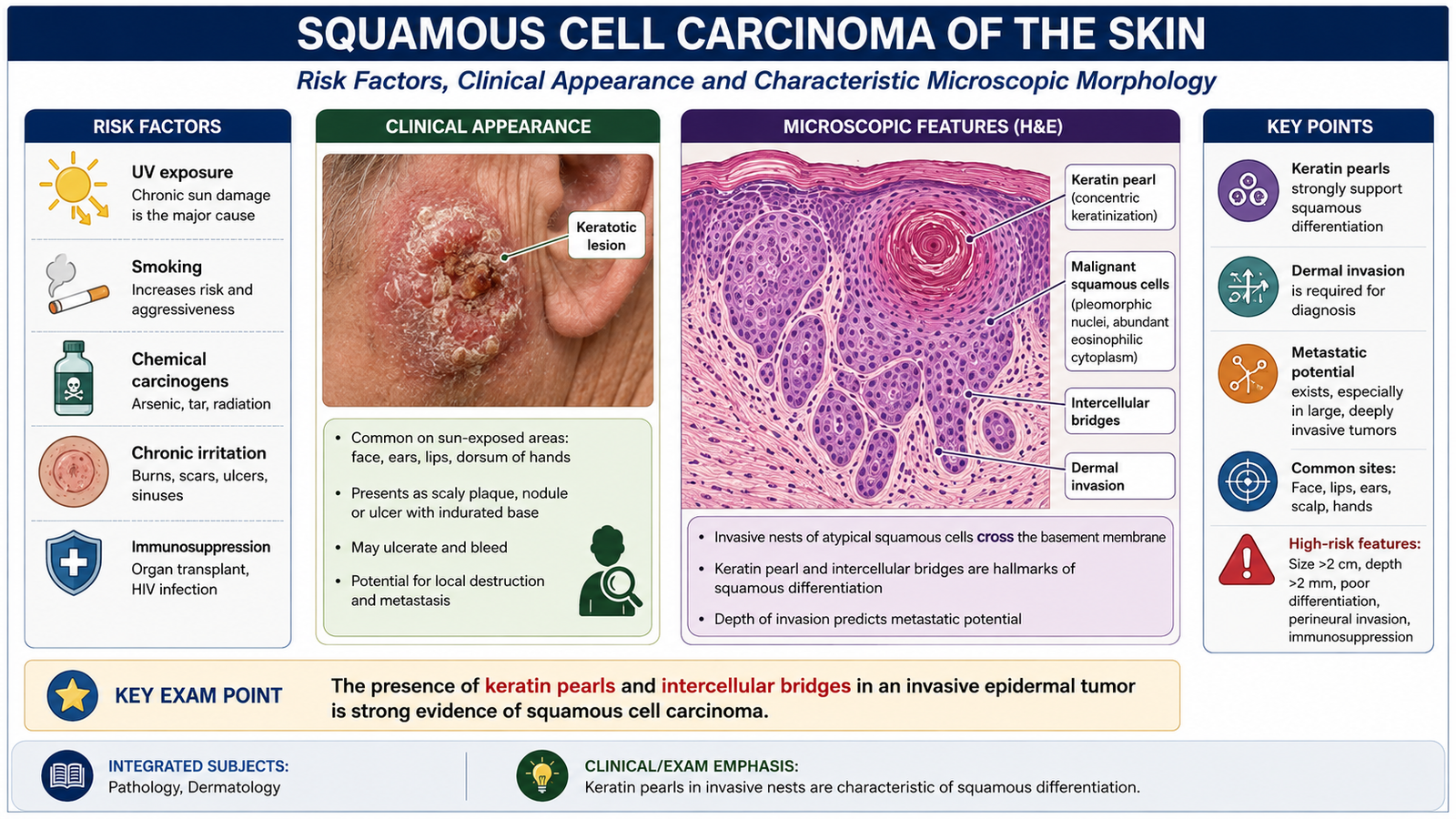
Squamous cell carcinoma is a malignant tumor of epidermal keratinocytes showing squamous differentiation. It commonly arises on sun-damaged skin and may develop from a precursor lesion such as actinic keratosis or SCC in situ. Unlike basal cell carcinoma, SCC has a definite capacity for metastasis, although the risk varies according to the site and characteristics of the tumor.
Predisposing factors
The important risk factors act mainly by producing repeated DNA damage, chronic epithelial injury or reduced immune control over abnormal keratinocytes.
- Chronic ultraviolet radiation exposure, particularly cumulative sun exposure.
- Fair skin and reduced protective melanin.
- Increasing age, reflecting accumulated environmental injury.
- Immunosuppression, which reduces immune surveillance against abnormal cells.
- Actinic keratosis and Bowen disease.
- Chronic ulcers, draining sinuses and old burn scars.
- Ionizing radiation and certain chronic chemical carcinogenic exposures.
- Some HPV-associated lesions, especially at particular anatomical sites.
Pathogenesis
Repeated carcinogenic injury causes genetic damage in keratinocytes. A clone of abnormal cells develops and expands within the epidermis. With increasing cellular atypia, the lesion may become SCC in situ. Once the malignant keratinocytes breach the basement membrane and enter the dermis, invasive SCC has developed.
Clinical features
- Usually develops on sun-exposed skin such as the face, ears, scalp, lower lip and dorsum of the hands.
- May appear as a firm erythematous or hyperkeratotic papule, plaque or nodule.
- The surface may be scaly, crusted or ulcerated.
- More advanced lesions may become indurated and infiltrative.
- A lesion arising in a chronic scar or ulcer deserves particular attention.
Gross morphology
SCC may form an irregular, firm, keratotic mass. Some tumors are exophytic, while others become ulcerated with an indurated base. The gross appearance reflects the abnormal production of keratin together with invasion of the dermis.
Microscopic morphology
The tumor is composed of malignant squamous cells extending from the epidermis into the dermis. The cells show cytological atypia and abnormal maturation. Well-differentiated tumors retain the ability to produce keratin.
- Nests and cords of atypical squamous cells invade the dermis.
- Keratin pearls may be present in well-differentiated SCC.
- Intercellular bridges support squamous differentiation.
- Nuclear pleomorphism and abnormal mitotic activity may be seen.
Investigations
Clinical inspection and palpation identify suspicious morphology and induration. Dermoscopy can support assessment, but the diagnosis is established by biopsy with histopathology. Histology confirms invasive squamous differentiation and distinguishes SCC from other keratinocytic lesions.
C. Basal Cell Carcinoma
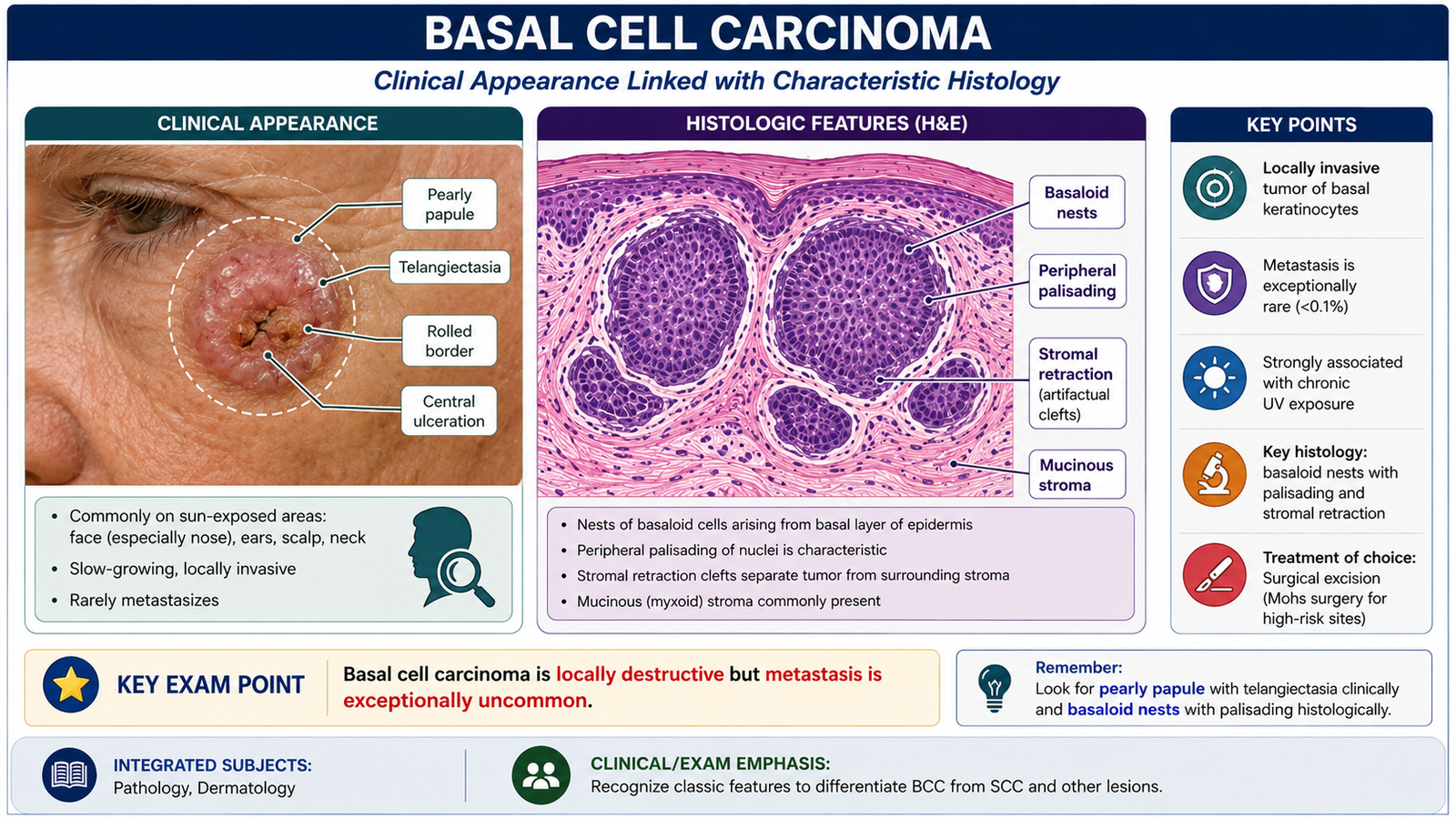
Basal cell carcinoma is a malignant keratinocytic skin tumor characterized by nests of basaloid cells. It is strongly associated with ultraviolet radiation and commonly develops on sun-exposed skin. BCC is locally invasive and can destroy surrounding tissue if neglected, but metastatic spread is exceptionally uncommon. This difference in biological behavior is important when comparing it with SCC.
Clinical features
- Usually occurs on chronically sun-exposed parts of the head and neck.
- Classically begins as a pearly or translucent papule.
- Fine surface telangiectatic vessels may be visible.
- The edge may become raised or rolled.
- Central ulceration may develop as the lesion enlarges.
- The tumor usually grows slowly but may cause significant local tissue destruction.
Gross morphology
The classic lesion is a pearly nodule with a smooth surface and small dilated vessels. With time, central ulceration may produce an ulcer surrounded by a raised border. Other lesions may appear more superficial or pigmented, so clinical appearance is not always identical.
Microscopic morphology
Histologically, BCC is composed of nests or cords of basaloid tumor cells extending into the dermis. A characteristic feature is the organized arrangement of nuclei at the outer edge of tumor nests.
- Basaloid tumor cells form nests in the dermis.
- Peripheral palisading of nuclei is characteristic.
- Cleft-like retraction spaces may appear between tumor nests and surrounding stroma.
Differential diagnosis and investigations
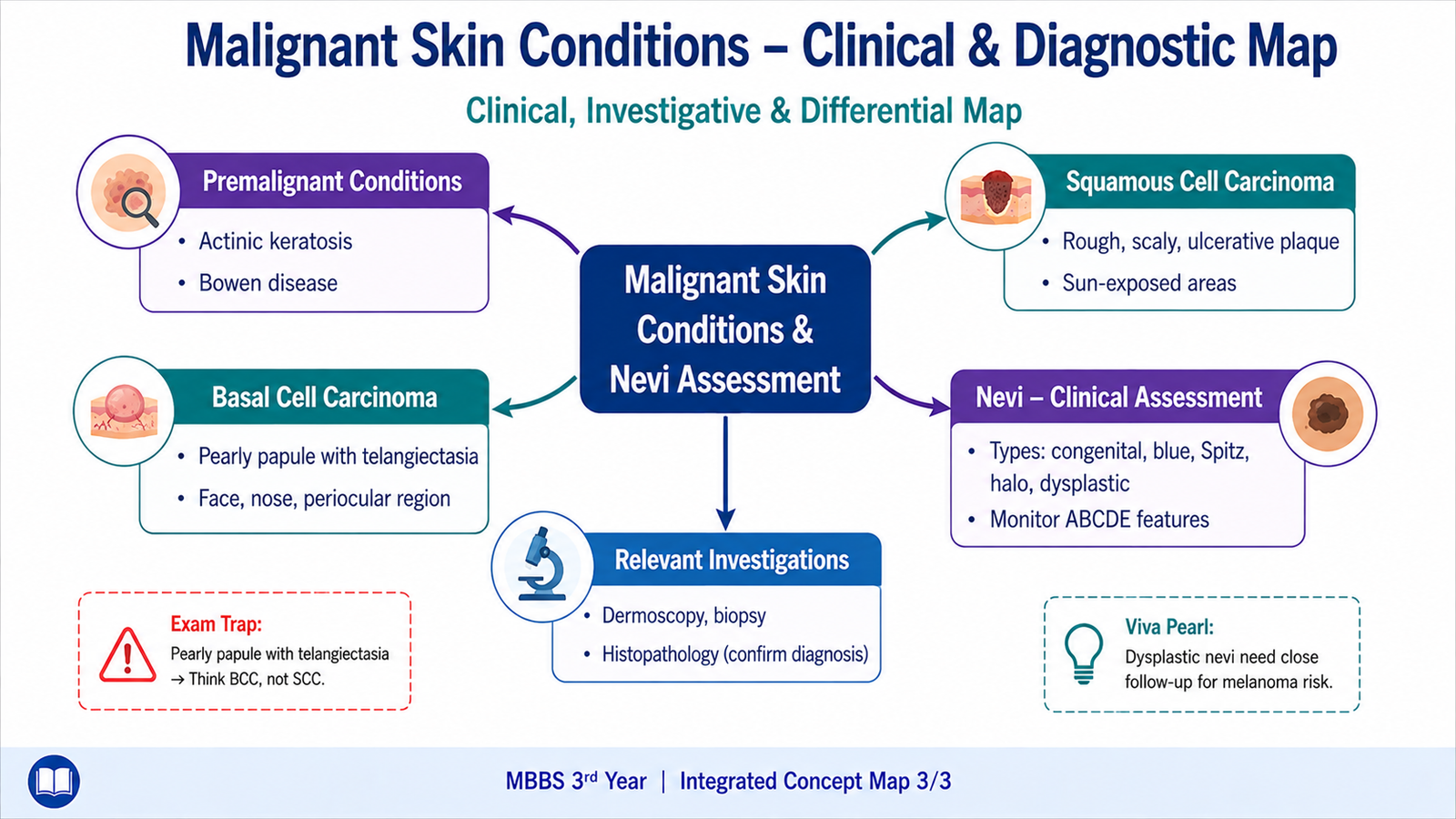
A pearly papule with telangiectasia and a rolled border strongly suggests BCC, whereas a rough keratotic or indurated lesion favors SCC. However, ulcerated or pigmented lesions may overlap clinically. Suspicious lesions are therefore assessed by clinical examination and, where appropriate, dermoscopy followed by biopsy for histopathological confirmation.
D. Nevocellular Nevi and Their Clinical Significance
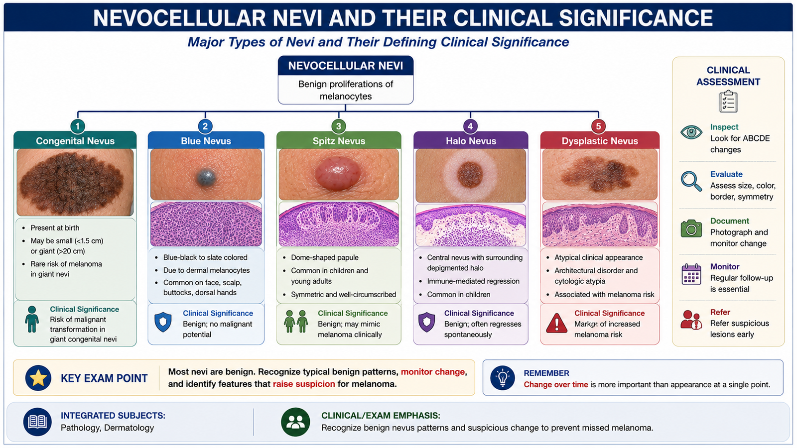
A nevocellular nevus is a benign proliferation of melanocytic nevus cells. Nevi vary greatly in color, elevation, age of appearance and microscopic location. Most remain benign. Their importance lies in recognizing characteristic benign patterns while identifying lesions whose clinical appearance overlaps with melanoma or which indicate increased melanoma risk.
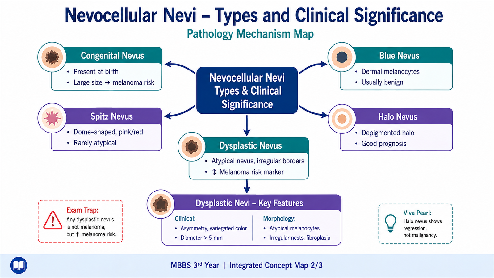
Congenital nevus
A congenital melanocytic nevus is present at birth or becomes apparent early in life. It may range from a small pigmented lesion to a large lesion involving a substantial area of skin. Larger congenital nevi are clinically important because melanoma risk is greater than in ordinary small acquired nevi.
Blue nevus
A blue nevus is usually a small, well-circumscribed blue to blue-black papule. Its blue appearance results from melanin being located relatively deep within the dermis, which alters the way light is scattered back through the skin. Most are benign.
Spitz nevus
Spitz nevus is usually a benign lesion of children or young people and commonly appears as a pink, red-brown or pigmented papule. Microscopically, its cells can look strikingly atypical and may resemble melanoma. Correct clinicopathological interpretation is therefore important.
Halo nevus
A halo nevus is surrounded by a sharply defined area of depigmentation. The pale halo results from an immune response against melanocytes and nevus cells. The central nevus may gradually regress. Its symmetrical halo around an otherwise typical nevus is characteristic.
Dysplastic nevus
A dysplastic nevus is an acquired melanocytic nevus with atypical clinical and histological features. It is particularly important because patients with multiple dysplastic nevi may have an increased risk of melanoma. Dysplastic nevi themselves should not be regarded as lesions that inevitably progress to melanoma.
Clinical assessment of a nevus
Most benign nevi are relatively symmetrical, have regular borders and show fairly uniform pigmentation. A lesion becomes more concerning when it is new, changing or clinically atypical.
- Assess symmetry and overall shape.
- Look for border regularity.
- Assess whether color is uniform or variegated.
- Compare the lesion with the patient’s other nevi.
- Ask whether it is enlarging or changing in appearance.
- Note ulceration, bleeding or other unexplained change.
Differential diagnosis and investigations
A pigmented nevus may need to be differentiated from melanoma, seborrheic keratosis, lentigo and other pigmented lesions. Clinical inspection is the starting point. Dermoscopy improves visualization of pigment and structural patterns. If a lesion has suspicious clinical or dermoscopic features, histopathological examination of an appropriate biopsy provides definitive morphological assessment.
E. Dysplastic Nevi — Clinical and Morphological Features
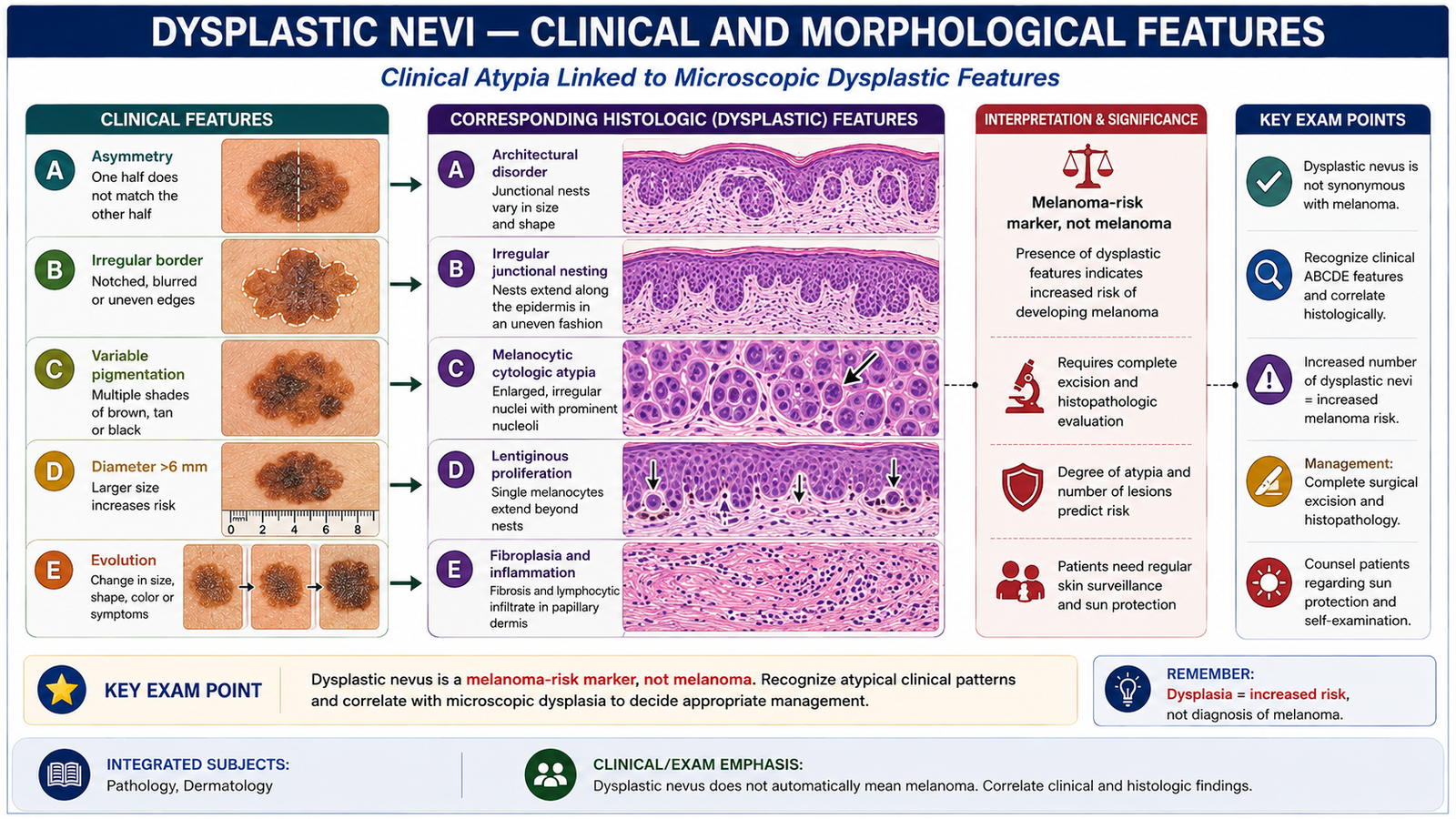
Dysplastic nevi deserve separate attention because they may clinically resemble melanoma. They are usually larger and less regular than ordinary acquired nevi and may show both flat and slightly raised components. The key idea is atypia without automatically implying malignancy: a dysplastic nevus is still a nevus, but its appearance and association with melanoma risk require careful assessment.
Clinical morphology
- Often larger than ordinary common nevi.
- May be asymmetrical.
- Borders may be irregular or indistinct.
- Pigmentation may be variable, with different shades of brown or tan.
- A flat pigmented area may contain a slightly raised central component.
- Multiple lesions may be present.
Microscopic morphology
Histologically, dysplastic nevi show both architectural disorder and cytological atypia. Melanocytes form irregular nests along the dermoepidermal junction, and the lesion may show abnormal growth across adjacent rete ridges. Stromal changes and lymphocytic inflammation may also accompany the atypical melanocytic proliferation.
- Irregularly distributed melanocytic nests.
- Architectural disorder at the dermoepidermal junction.
- Variable cytological atypia of melanocytes.
- Associated fibroplasia and inflammatory changes may occur in the superficial dermis.
Clinical significance
The importance of a dysplastic nevus is twofold. First, an individual lesion may be difficult to distinguish clinically from melanoma. Second, the presence of numerous dysplastic nevi identifies a person who may have an increased overall risk of melanoma. Any lesion showing significant change or suspicious morphology therefore requires careful assessment.
F. Malignant Melanoma
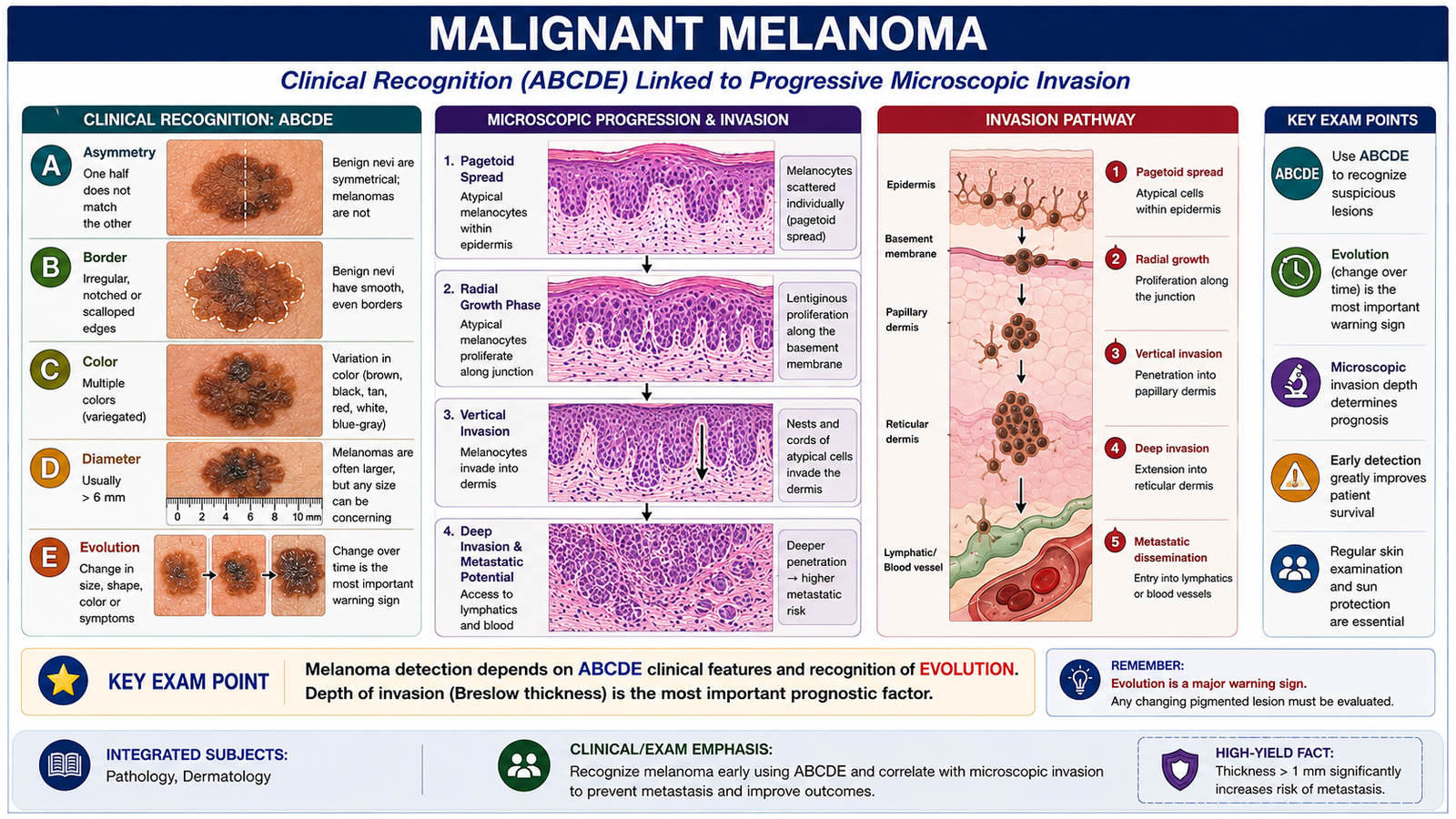
Malignant melanoma is a malignant tumor of melanocytes. It is clinically important because it can invade and metastasize, and early recognition is therefore essential. Melanoma often develops in the skin, either in association with a pre-existing melanocytic lesion or as a new lesion. Recognition depends on change over time, asymmetry, irregular borders, uneven pigmentation and other suspicious morphological features.
Frequent sites of origin
Melanoma can arise on many skin surfaces. Its distribution varies with patterns of sun exposure and melanoma subtype.
- The trunk, particularly the back, is an important site in men.
- The lower limbs are an important site in women.
- The head and neck may be involved, especially in chronically sun-damaged skin.
- Palms, soles and subungual sites are important sites for acral melanoma.
- Melanoma may occasionally arise at non-cutaneous melanocyte-containing sites, although cutaneous melanoma is the main focus here.
Clinical features
A melanoma may present as a new pigmented lesion or as a change within a pre-existing lesion. The most useful general warning sign is evolution: a lesion that changes in size, shape, color or overall character deserves assessment.
- A — Asymmetry: one half does not resemble the other.
- B — Border irregularity: edges are uneven, notched or poorly defined.
- C — Color variation: more than one shade or uneven pigment distribution.
- D — Diameter: larger lesions may be more suspicious, but size alone does not diagnose melanoma.
- E — Evolution: enlargement or another significant change over time is particularly important.
Additional concerning features include persistent change in surface, unexplained ulceration or bleeding, and a lesion that looks noticeably different from the patient’s other nevi.
Gross morphology
Melanomas commonly show irregular shape, uneven borders and variegated pigmentation. Colors may include different shades of brown, black and tan, with occasional red, blue or pale areas. Some melanomas are only lightly pigmented, so absence of dark pigmentation does not exclude the diagnosis.
Microscopic morphology
Melanoma consists of atypical melanocytes showing disordered proliferation within the epidermis and, in invasive lesions, extension into the dermis. Early lesions may show a prominent horizontal or radial growth pattern. Acquisition of deeper invasive growth is called the vertical growth phase and reflects a greater capacity for invasion and spread.
- Atypical melanocytes are present singly and in irregular nests.
- Cells may show enlarged, irregular nuclei and prominent nucleoli.
- Abnormal melanocytes may extend upward through the epidermis, producing pagetoid spread.
- Invasive tumor cells extend into the dermis during vertical growth.
- Pigment production is variable; some lesions contain abundant melanin while others contain little.
Investigations
Clinical examination is supported by dermoscopy when available. A lesion suspicious for melanoma requires tissue diagnosis. Histopathological examination assesses the melanocytic architecture, cytological atypia and presence and depth of invasion. These morphological findings distinguish melanoma from benign or dysplastic melanocytic nevi.
Important Comparison — Squamous Cell Carcinoma vs Basal Cell Carcinoma
SCC and BCC are both keratinocyte carcinomas and commonly occur on sun-exposed skin, but their clinical appearance, histology and biological behavior differ. These differences are frequently useful in examinations and clinical recognition.
| Feature | Squamous Cell Carcinoma | Basal Cell Carcinoma |
|---|---|---|
| Main tumor cells | Malignant squamous keratinocytes | Basaloid tumor cells |
| Typical clinical appearance | Scaly, keratotic, firm or ulcerated plaque/nodule | Pearly papule/nodule with telangiectasia and rolled border |
| Characteristic histology | Keratin pearls and intercellular bridges | Peripheral palisading and stromal retraction |
| Local invasion | Present | Present and may be locally destructive |
| Metastatic potential | Definite potential | Exceptionally uncommon |
| Important precursor association | Actinic keratosis / SCC in situ | No equivalent common precursor lesion emphasized |
⭐ AIM High-Yield Review
- Actinic keratosis is an important UV-related premalignant keratinocytic lesion associated with SCC.
- Bowen disease is SCC in situ: atypical squamous cells involve the epidermis but have not invaded through the basement membrane.
- Major SCC risk factors include chronic UV exposure, fair skin, immunosuppression, precursor lesions and chronic scars or ulcers.
- ⭐ Keratin pearls and intercellular bridges are classic morphological clues to SCC.
- ⭐ A pearly papule with telangiectasia and a rolled border strongly suggests BCC.
- Peripheral palisading of basaloid cells and stromal retraction are characteristic microscopic features of BCC.
- BCC is locally destructive but metastasis is exceptionally uncommon; SCC has greater metastatic potential.
- Important nevus types in this topic are congenital, blue, Spitz, halo and dysplastic nevi.
- A blue nevus appears blue because its melanocytes and melanin are located relatively deep in the dermis.
- A halo nevus has surrounding depigmentation related to an immune response against melanocytes.
- Dysplastic nevi may show asymmetry, irregular borders and variable pigmentation and are important markers of increased melanoma risk.
- ⭐ For melanoma, remember ABCDE: Asymmetry, Border irregularity, Color variation, Diameter and especially Evolution.
- Melanoma microscopy may show atypical melanocytes, irregular nests, pagetoid spread and dermal invasion.
- Histopathology is the definitive method for distinguishing suspicious premalignant, keratinocytic and melanocytic lesions.
🎥 AIM Video Learning
Premalignant Skin Lesions, Keratinocyte Carcinomas, Nevi and Malignant Melanoma