
📖 AIM Learning Material
Topic 9 — Infectious Arthritis, Rheumatic Fever and Bone–Joint Infections
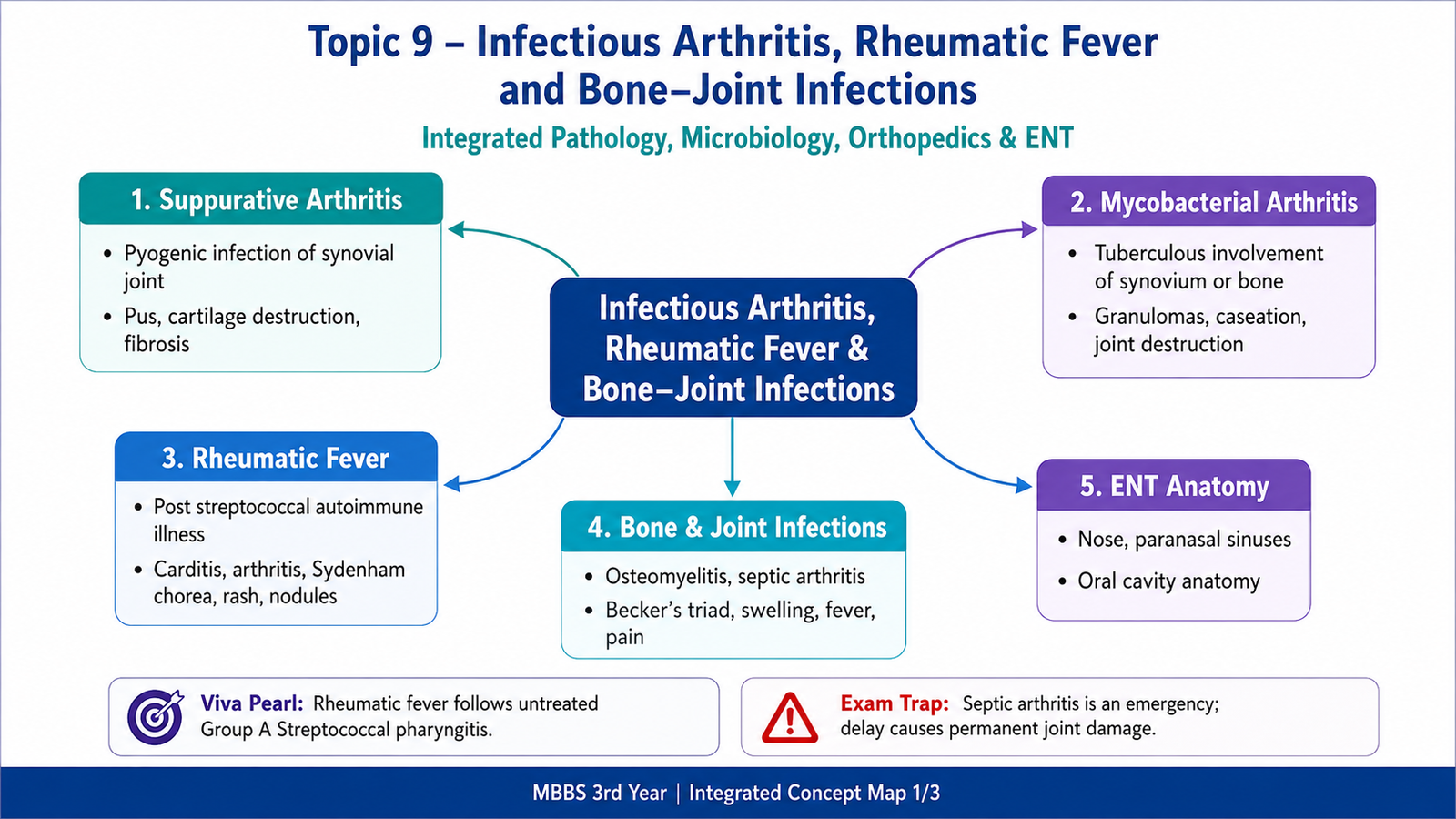
Bone and joint disease may result from direct microbial invasion or from an immune response that continues after an infection has ended. Suppurative arthritis is an acute pyogenic infection that can rapidly destroy articular cartilage, while mycobacterial arthritis usually causes a slower granulomatous process. Osteomyelitis affects bone and marrow and may progress from acute inflammation to necrotic bone and chronic sinus formation. Group A Streptococcus pyogenes is also important because a preceding throat infection can trigger rheumatic fever through cross-reactive immunity. This chapter connects etiology, pathogenesis, morphology, clinical features, investigations and complications so that these conditions can be understood and distinguished clearly.
A. Routes and Basic Pathogenesis of Bone–Joint Infection
The central infectious problem
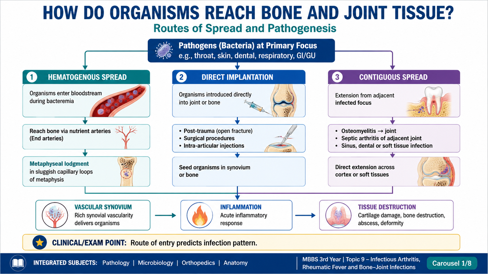
Bone and joint infections develop when microorganisms enter susceptible tissue, overcome local defense mechanisms and initiate inflammation. Infection may remain localized, spread between adjacent structures or enter the bloodstream. The resulting lesion depends on the organism, route of entry, host age, immunity, vascular anatomy and duration of infection.
Major routes of infection
- Hematogenous spread: organisms circulate in blood and lodge in synovium or bone, especially in children and patients with bacteremia.
- Direct implantation: penetrating trauma, open fracture, surgery, injection or prosthetic implantation introduces organisms directly.
- Contiguous spread: infection extends from nearby skin, soft tissue, ulcer, sinus tract or another infected anatomical compartment.
Host and local risk factors
- Extremes of age, impaired immunity, malnutrition or chronic systemic disease
- Damaged or previously diseased joints
- Prosthetic joints or recent orthopedic procedures
- Open wounds, fractures, pressure sores or chronic ulcers
- Persistent bacteremia or infection elsewhere in the body
A vascular synovium can rapidly receive circulating organisms. Once purulent exudate accumulates within a closed joint, raised pressure compromises perfusion while neutrophil enzymes and bacterial products damage cartilage. In bone, vascular obstruction, inflammation and necrosis can separate dead bone from viable tissue and promote chronic infection.
B. Suppurative Arthritis
Definition and core concept
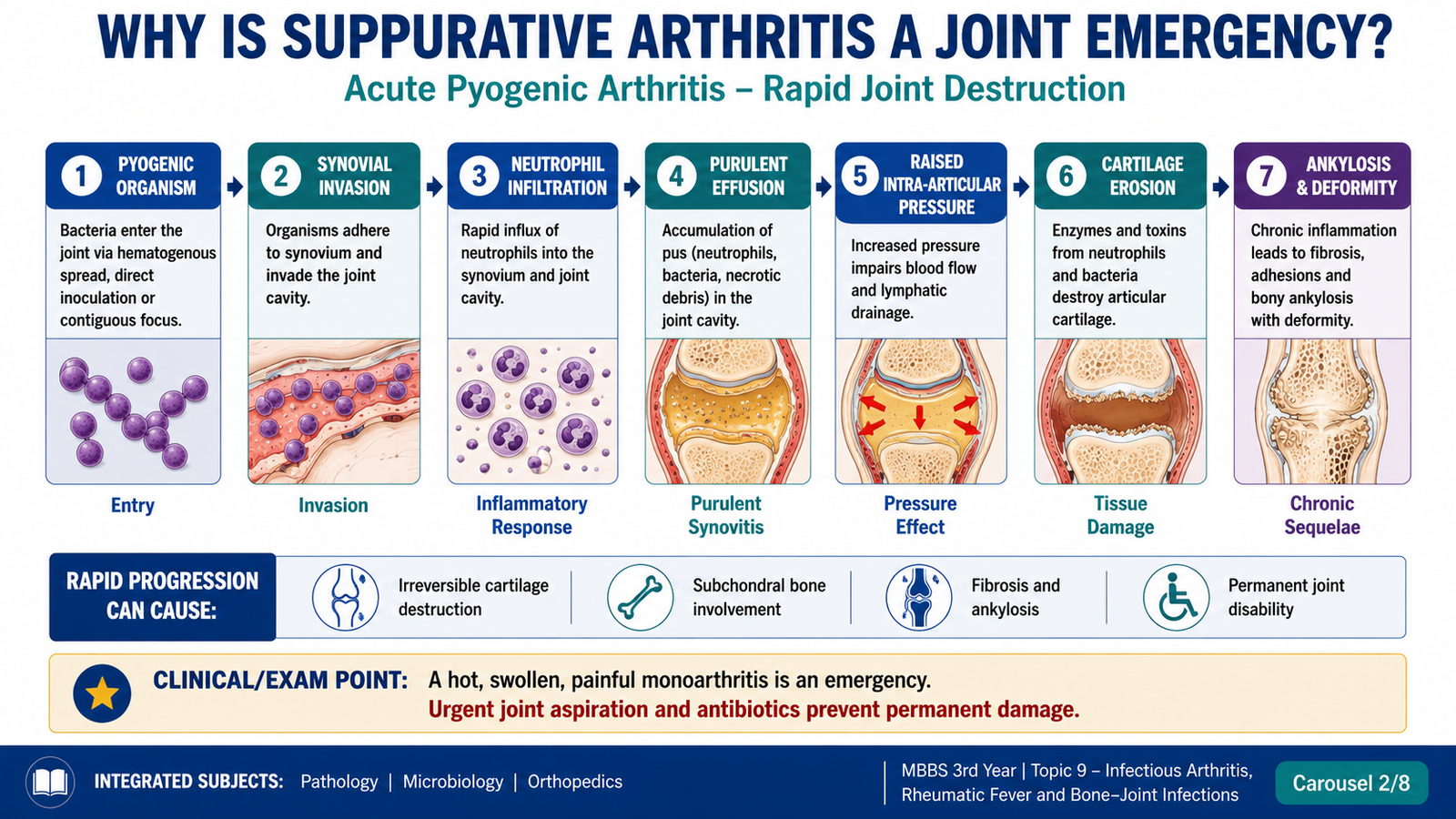
Suppurative, pyogenic or septic arthritis is an acute infection of a joint caused by pyogenic organisms and characterized by intense neutrophilic inflammation with purulent exudate. It is a medical and orthopedic emergency because articular cartilage may be damaged rapidly.
Etiology
The responsible organism varies with age, exposure, immune status and route of entry. Important etiological groups include:
- Pyogenic cocci: especially Staphylococcus aureus; streptococci are also important.
- Age- or exposure-related organisms: organisms associated with infancy, sexual transmission, penetrating injury or immunocompromise may be encountered in appropriate settings.
- Directly introduced organisms: following trauma, injection, surgery or prosthetic implantation.
Pathogenesis
The synovial membrane becomes congested, edematous and infiltrated by neutrophils. Pus enters the joint space and may extend into adjacent cartilage, subchondral bone, ligaments and periarticular soft tissue. The inflammatory exudate also raises intra-articular pressure, which can impair blood supply and accelerate tissue injury.
Morphology
- Swollen joint capsule
- Congested, dull or granular synovium
- Cloudy or frankly purulent joint fluid
- Erosion or ulceration of articular cartilage
- Possible extension into adjacent bone and soft tissue
- Dense neutrophilic infiltrate
- Fibrin and necrotic debris
- Synovial edema and vascular congestion
- Surface ulceration and tissue necrosis
- Repair by granulation tissue and fibrosis in later stages
Clinical features
- Acute severe pain, usually involving one major joint
- Swelling, warmth, tenderness and marked restriction of movement
- Fever, malaise and other evidence of systemic infection
- Joint held in a position that minimizes capsular tension
- Infants or debilitated patients may show less obvious local signs
Complications
- Rapid articular cartilage destruction and permanent loss of joint function
- Extension into adjacent bone, producing osteomyelitis
- Periarticular abscess or soft-tissue spread
- Fibrous ankylosis followed by possible bony ankylosis
- Growth disturbance in children when the epiphysis or growth plate is damaged
- Septicemia and systemic deterioration
C. Mycobacterial Arthritis
Definition and etiology
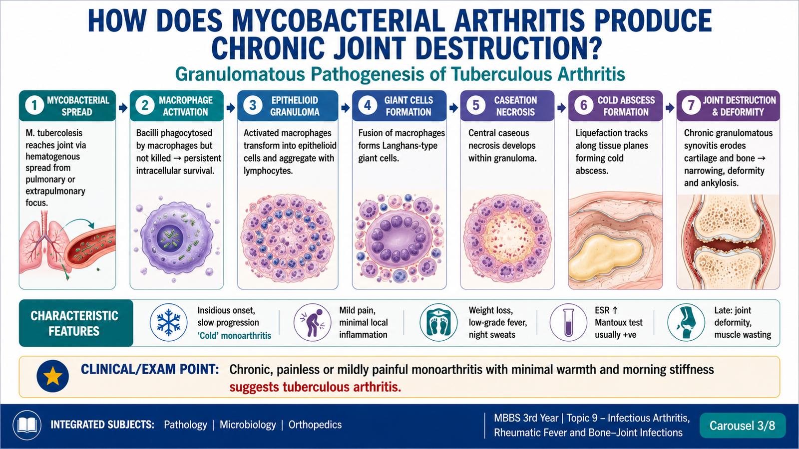
Mycobacterial arthritis is a chronic granulomatous infection of a joint, most commonly associated with Mycobacterium tuberculosis. It usually develops through hematogenous dissemination from a primary or latent focus, or by spread from adjacent infected bone.
Pathogenesis
Unlike acute pyogenic infection, the inflammatory response evolves slowly. Activated macrophages become epithelioid cells and may fuse into multinucleated giant cells. Caseous necrosis may develop within granulomas. The thickened synovium spreads over articular cartilage and gradually erodes it. Granulation tissue and caseous material may extend through the capsule and form a cold abscess or draining sinus.
Morphology
- Thickened, pale or congested synovium
- Granular or nodular synovial surface
- Caseous material in advanced disease
- Cartilage erosion and marginal bone destruction
- Cold abscess or sinus formation
- Epithelioid cell granulomas
- Langhans-type giant cells
- Central caseous necrosis when present
- Lymphocytes and chronic inflammatory cells
- Fibrosis in healing or longstanding lesions
Clinical features
- Slowly progressive monoarthritis, commonly affecting a large weight-bearing joint
- Pain, stiffness, swelling and progressive limitation of movement
- Muscle wasting around the affected joint
- Relatively little warmth or erythema compared with acute pyogenic arthritis
- Constitutional symptoms may be absent, mild or nonspecific
- Cold abscess or discharging sinus in advanced disease
Complications
- Progressive cartilage and subchondral bone destruction
- Joint deformity, instability or ankylosis
- Cold abscess and sinus tract formation
- Secondary bacterial infection of a sinus or damaged joint
- Pathological fracture when adjacent bone is severely weakened
- Chronic disability and growth disturbance in children
D. Osteomyelitis and Other Bone Infections
Definition and etiological pathways
Osteomyelitis is infection and inflammation of bone and marrow. It may be acute or chronic and may follow hematogenous spread, direct implantation or extension from adjacent infected tissue.
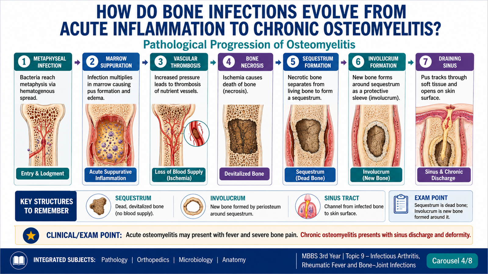
Pathological sequence
The rigid bony cortex limits expansion. Accumulating exudate compresses vessels and promotes thrombosis, resulting in ischemic bone necrosis. A separated fragment of dead bone is called a sequestrum. The periosteum and surviving bone may produce a shell of reactive new bone around the lesion, termed an involucrum. An opening in the involucrum through which pus escapes is a cloaca.
Distribution and age-related anatomy
- In children, hematogenous infection commonly lodges in the metaphysis of long bones because of slow, looping blood flow.
- In infants, vascular connections may permit extension into the epiphysis and nearby joint.
- In adults, vertebral involvement and infection related to surgery, trauma, ulcers or prosthetic material become more relevant.
- Contiguous infection is important near chronic ulcers, wounds and exposed bone.
Clinical presentation
| Pattern | Typical presentation | Pathological basis |
|---|---|---|
| Acute osteomyelitis | Fever, localized deep pain, tenderness, swelling and reduced limb use | Acute suppuration, edema and rising intraosseous pressure |
| Chronic osteomyelitis | Persistent or recurrent pain, drainage, sinus tract and intermittent fever | Sequestrum, fibrosis, involucrum and persistent microbial focus |
| Vertebral infection | Back pain, spinal tenderness and possible neurological symptoms | Vertebral body destruction, collapse or epidural extension |
Complications
- Chronic osteomyelitis with sequestrum and sinus formation
- Pathological fracture and structural deformity
- Spread to an adjacent joint or soft tissue
- Growth disturbance in children
- Sepsis in severe acute infection
- Secondary amyloid deposition in longstanding suppurative disease
- Malignant transformation in the epithelium of a longstanding draining sinus, although uncommon
E. Streptococcus pyogenes: Structure, Virulence and Diagnosis
Key structural and microbiological features
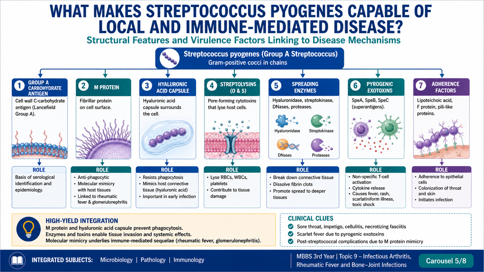
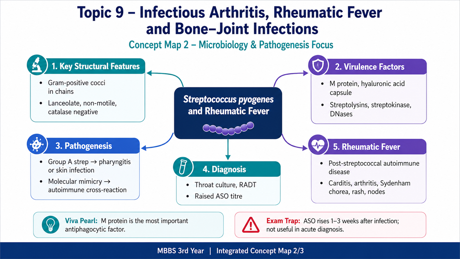
- Gram-positive spherical cocci arranged characteristically in chains
- Beta-hemolytic growth pattern on blood agar
- Group A carbohydrate antigen in the cell wall
- Non-motile and non-spore-forming organism
- Catalase-negative, helping distinguish streptococci from staphylococci
- Some strains possess a hyaluronic acid capsule that resembles host connective-tissue material
Virulence factors and their effects
| Virulence factor | Main action | Clinical or pathological importance |
|---|---|---|
| M protein | Interferes with phagocytosis and contributes to adhesion | Major virulence determinant; certain epitopes participate in cross-reactive immunity |
| Hyaluronic acid capsule | Reduces recognition and phagocytosis | Facilitates survival in tissue |
| Streptolysins | Damage host cell membranes | Contribute to hemolysis and tissue injury; antibodies aid evidence of recent infection |
| Streptokinase | Promotes fibrin breakdown | May facilitate spread through tissue |
| DNases | Degrade extracellular DNA | Reduce viscosity of pus and aid spread; antibodies may support recent-infection assessment |
| Pyrogenic exotoxins | May act as superantigens | Associated with fever, rash and severe toxin-mediated illness |
Modes of pathogenesis
- Direct local infection: adherence, multiplication and inflammation produce pharyngitis or skin and soft-tissue disease.
- Invasive spread: enzymes and immune evasion facilitate deeper tissue involvement.
- Toxin-mediated disease: exotoxins produce systemic effects without requiring direct invasion of every affected organ.
- Post-infectious immune disease: immune responses generated against streptococcal antigens cross-react with host tissues.
Laboratory diagnosis of streptococcal pharyngitis and recent infection
- Specimen: a properly collected throat swab is used when active pharyngitis is suspected.
- Direct antigen detection: may identify group A streptococcal antigen in appropriate clinical settings.
- Culture: demonstrates beta-hemolytic colonies and permits organism identification using established laboratory principles.
- Evidence of preceding infection: rising or elevated antibodies to streptococcal products support a recent infection when the organism is no longer present in the throat.
F. Rheumatic Fever
Etiology and defining principle
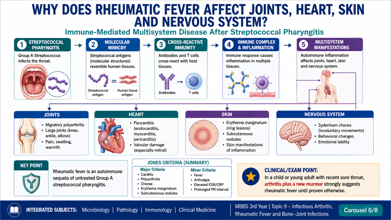
Acute rheumatic fever is a delayed, non-suppurative, immune-mediated inflammatory disease that follows pharyngeal infection by rheumatogenic strains of group A Streptococcus pyogenes. The affected tissues are not being directly invaded by the organism.
Pathogenesis: molecular mimicry
Antibodies and T cells generated against streptococcal components recognize structurally similar host antigens. In the heart, this produces inflammatory injury involving endocardium, myocardium and pericardium. Recurrent episodes amplify valvular scarring and create chronic rheumatic heart disease.
Clinical features
| Manifestation | Clinical clue | Underlying process |
|---|---|---|
| Migratory polyarthritis | Large joints become painful sequentially, with one improving as another becomes involved | Transient immune-mediated synovial inflammation without purulent destruction |
| Carditis | Tachycardia, murmur, cardiac enlargement, failure or pericardial features | Inflammation may involve all cardiac layers |
| Sydenham chorea | Involuntary movements, emotional lability and impaired motor control | Cross-reactive immune effects on neural tissue |
| Erythema marginatum | Evanescent, non-pruritic, ring-like rash | Cutaneous inflammatory manifestation |
| Subcutaneous nodules | Small, firm nodules over extensor surfaces or bony prominences | Focal inflammatory lesion associated with significant carditis |
Cardiac morphology
Acute rheumatic carditis: The characteristic myocardial lesion is the Aschoff body, a focus of fibrinoid change surrounded by lymphocytes, plasma cells and activated macrophages. Some macrophages have distinctive central chromatin and are called Anitschkow cells. Endocardial inflammation commonly affects valves, where small sterile vegetations may develop along lines of closure.
Chronic rheumatic heart disease: Repeated inflammation heals by fibrosis, producing leaflet thickening, commissural fusion and shortening or fusion of chordae tendineae. The mitral valve is most commonly and most severely affected.
Diagnosis
Diagnosis is clinical and requires a compatible pattern of manifestations together with evidence of preceding group A streptococcal infection. The diagnostic reasoning commonly combines:
- Major manifestations such as carditis, migratory polyarthritis, chorea, characteristic rash or subcutaneous nodules
- Supporting manifestations such as fever, arthralgia, inflammatory-marker elevation or conduction disturbance
- Evidence of recent streptococcal infection from microbiological or serological testing
- Exclusion of important competing diagnoses
Complications
- Acute cardiac failure in severe carditis
- Recurrent rheumatic fever following subsequent streptococcal infections
- Chronic valvular stenosis or regurgitation
- Atrial enlargement, arrhythmia, mural thrombosis and embolic complications secondary to chronic valve disease
- Infective endocarditis developing on previously damaged valves
- Progressive pulmonary vascular and right-heart consequences of severe left-sided valve disease
G. Investigations and Diagnostic Differentiation
Immediate diagnostic priorities
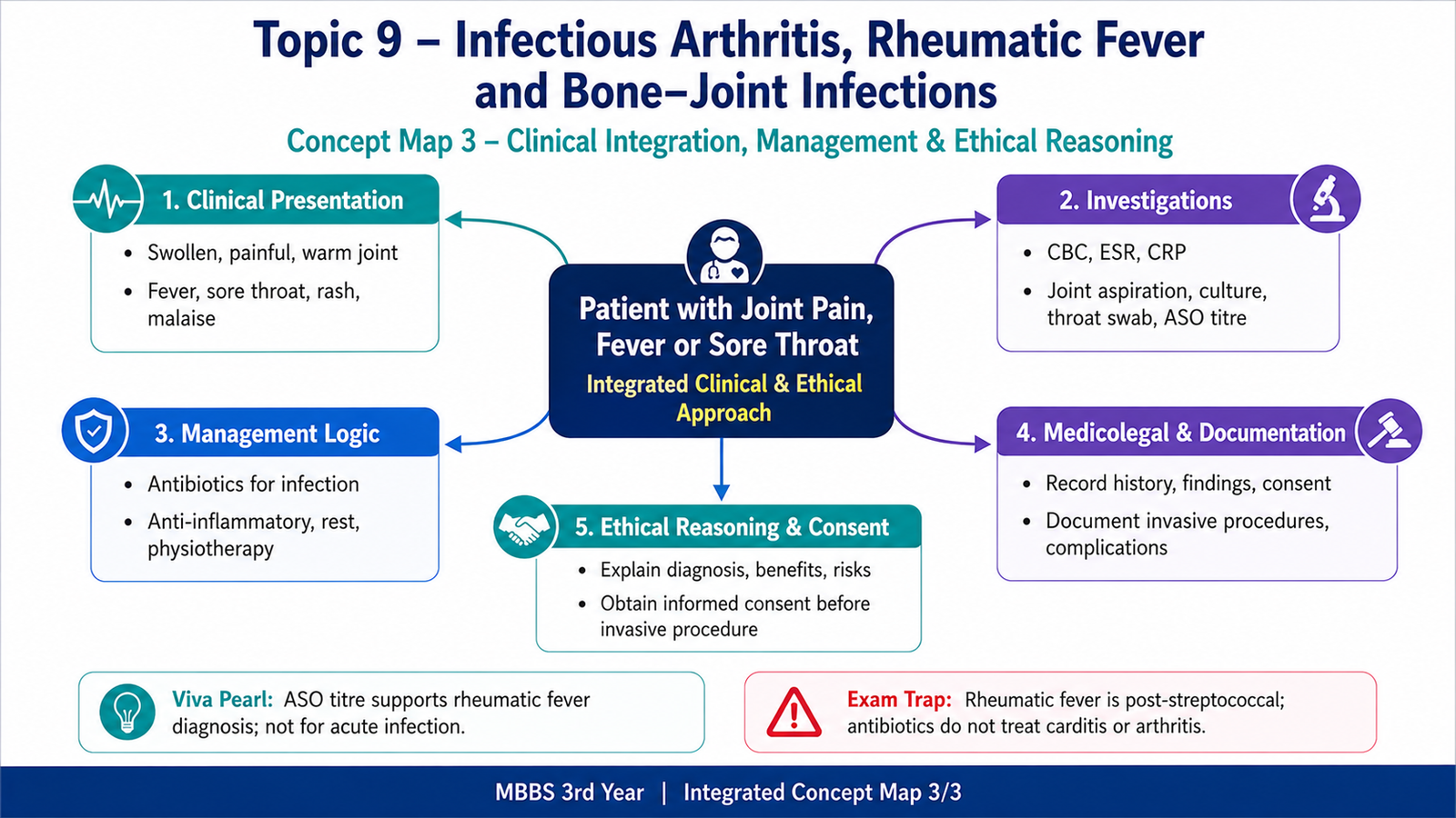
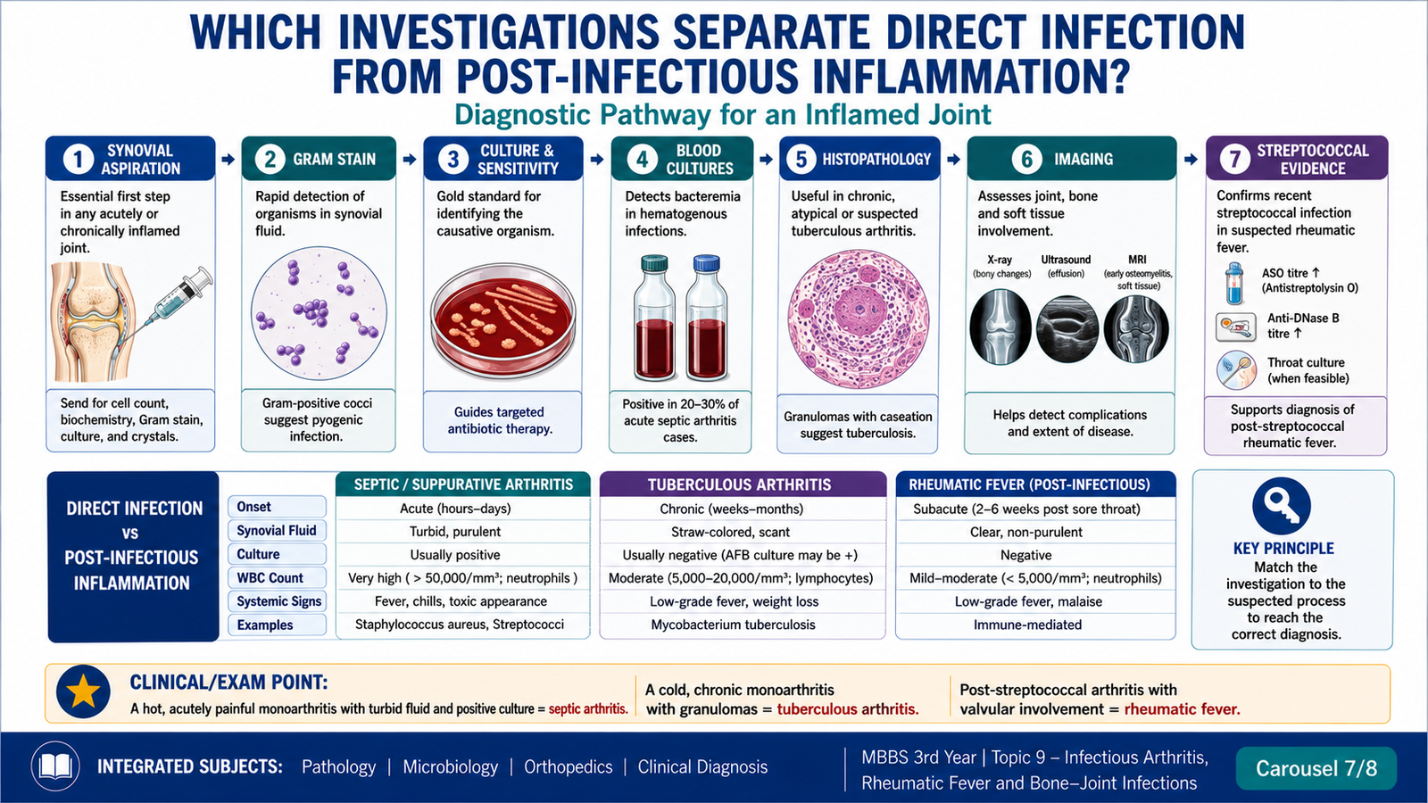
Investigation should answer four questions: Is infection present? Which tissue is involved? Which organism is responsible? Has structural damage or systemic spread occurred? Specimens should be obtained promptly and handled according to local laboratory protocols, without delaying urgent care in an unstable patient.
Joint-focused investigations
- Synovial fluid examination: gross appearance, cell pattern, microscopy, Gram stain and culture are central when septic arthritis is suspected.
- Crystal assessment: helps distinguish crystal-induced arthritis, but identification of crystals does not automatically exclude coexisting infection.
- Blood cultures: useful when fever, bacteremia or systemic illness is present.
- Inflammatory markers and blood count: support inflammation and help monitor progression but are not organism-specific.
- Imaging: ultrasound can detect an effusion and guide aspiration; plain radiography provides a baseline and may show later destruction; advanced imaging better demonstrates deep soft-tissue or early osseous involvement when clinically required.
Bone-infection investigations
- Plain radiography for baseline structural assessment, although early changes may be absent
- Imaging capable of identifying marrow, cortical, soft-tissue or vertebral involvement when indicated
- Blood cultures in suspected hematogenous disease
- Aspirated material, tissue or bone specimen for microbiological culture and histopathological examination
- Assessment for a primary source, implant, ulcer, wound or distant focus
Mycobacterial disease investigations
- Synovial tissue or appropriate aspirated material for histopathology and mycobacterial studies
- Microscopy for acid-fast organisms, recognizing that sensitivity may be limited
- Culture and validated molecular identification methods where available
- Imaging to assess gradual cartilage loss, bone erosion, abscess and adjacent involvement
- Evaluation for tuberculosis elsewhere in the body
Rheumatic-fever investigations
- Evidence of preceding group A streptococcal infection through throat testing or antistreptococcal antibodies
- Inflammatory markers as supportive evidence of systemic inflammation
- Electrocardiographic assessment for conduction abnormalities
- Echocardiographic assessment for valvular involvement or carditis
- Clinical evaluation for joint, skin and neurological manifestations
High-yield comparison
| Feature | Suppurative arthritis | Mycobacterial arthritis | Rheumatic arthritis |
|---|---|---|---|
| Onset | Acute | Chronic, insidious | Acute after pharyngitis |
| Joint pattern | Usually severe monoarthritis | Usually chronic monoarthritis | Migratory large-joint polyarthritis |
| Inflammatory pattern | Neutrophilic and purulent | Granulomatous, often caseating | Sterile immune-mediated inflammation |
| Key specimen | Synovial fluid | Synovial tissue or appropriate aspirate | Evidence of recent streptococcal infection plus clinical assessment |
| Cartilage destruction | Rapid if untreated | Slow and progressive | Arthritis usually resolves without destructive suppuration |
| Major extra-articular clue | Bacteremia or systemic sepsis | Cold abscess or tuberculosis elsewhere | Carditis, chorea, rash or nodules |
H. Regional Anatomy of the Nose, Paranasal Sinuses and Oral Cavity
Nasal cavity
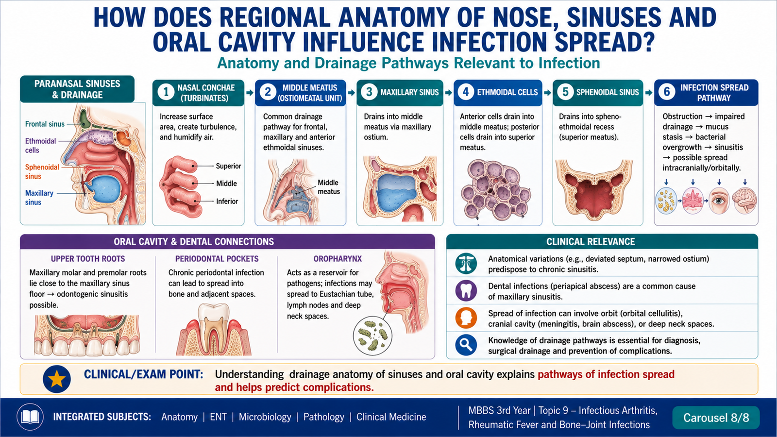
The nasal cavity extends from the nostrils to the choanae and is divided by the nasal septum. The lateral wall contains the superior, middle and inferior conchae, which increase mucosal surface area and create corresponding meatuses. Most paranasal sinuses drain through openings in these regions, making local anatomy important in sinus ventilation and infection.
- The respiratory mucosa warms, humidifies and filters inspired air.
- Mucociliary clearance moves secretions toward the pharynx.
- Mucosal edema can obstruct narrow drainage pathways and promote retained secretions.
- The olfactory area occupies the superior part of the nasal cavity.
Paranasal sinuses
| Sinus | Important relation | Drainage relevance |
|---|---|---|
| Frontal | Above the orbit | Drains toward the middle meatus through the frontal recess region |
| Maxillary | Closely related to upper teeth and orbit | Its opening lies relatively high on the medial wall, so drainage depends strongly on mucociliary action |
| Ethmoidal air cells | Between nasal cavity and orbit | Drain into superior or middle meatal regions according to the cell group |
| Sphenoidal | Near the pituitary region, optic structures and major vessels | Drains into the sphenoethmoidal recess |
Oral cavity
The oral cavity includes the vestibule and oral cavity proper. Its boundaries include the lips and cheeks, hard and soft palate, tongue and floor of mouth. Teeth are anchored in the alveolar processes, and their roots have clinically important relationships with adjacent spaces and, particularly for upper posterior teeth, the maxillary sinus.
- Salivary ducts enter the oral cavity and contribute to lubrication, buffering and antimicrobial defense.
- Dental, gingival or periodontal infection may spread into adjacent facial or deep neck spaces.
- Upper dental infection may involve the maxillary sinus because of close root relationships.
- Oropharyngeal and tonsillar infection is clinically relevant to group A streptococcal pharyngitis.
Anatomical links to systemic and regional infection
Mucosal infection in the pharyngeal region provides the antigenic trigger for rheumatic fever. Local sinonasal or dental infections more commonly cause contiguous regional complications, whereas bone and joint infection usually requires bacteremia, direct implantation or local extension from nearby tissue. Anatomy therefore explains the source and route but does not mean every upper-airway infection spreads directly to a joint.
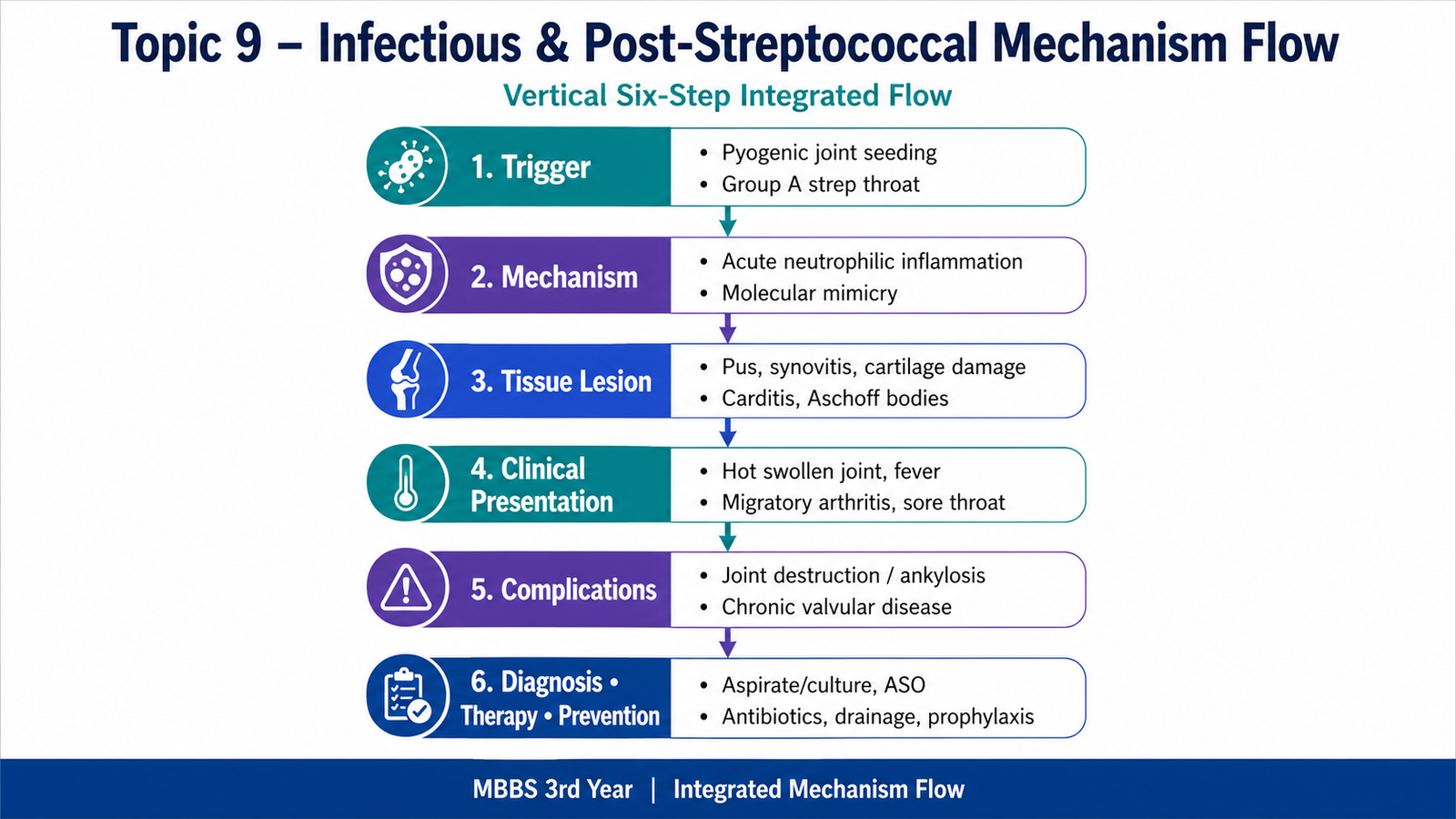
- Three main routes: bone and joint infection reaches tissue through hematogenous spread, direct implantation or contiguous extension.
- ⭐ Suppurative arthritis: acute hot monoarthritis with purulent, neutrophil-rich synovial fluid; rapid cartilage destruction makes it an emergency.
- Mycobacterial arthritis: usually a chronic, relatively cold monoarthritis with caseating granulomatous synovitis, progressive erosion and possible sinus formation.
- Osteomyelitis: suppuration and rising intraosseous pressure impair blood supply and produce bone necrosis.
- ⭐ Key terms: sequestrum is separated dead bone, involucrum is reactive new bone around it, and a cloaca is an opening through which pus drains.
- Streptococcus pyogenes is a group A, beta-hemolytic, catalase-negative Gram-positive coccus that typically forms chains.
- M protein is a major antiphagocytic virulence factor and also contributes to molecular mimicry in rheumatic fever.
- ⭐ Rheumatic fever: follows group A streptococcal pharyngitis and is immune-mediated; the organism does not directly multiply in the joints or heart.
- Rheumatic pathology: Aschoff bodies and Anitschkow cells characterize acute carditis, while repeated injury produces fibrotic valvular deformity, especially of the mitral valve.
- Diagnostic specimen: urgent synovial fluid analysis and microbiological testing are central in suspected septic arthritis.
- Imaging point: early radiographs may be normal or nonspecific in osteomyelitis; imaging must be interpreted with the clinical and laboratory findings.
- Anatomical link: mucosal edema can obstruct sinus drainage, upper dental infection may extend to the maxillary sinus, and pharyngeal infection provides the antigenic trigger for rheumatic fever.