
📖 AIM Learning Material
Topic 5 — Mechanical Wounds, Medicolegal Assessment and Battered Baby Syndrome
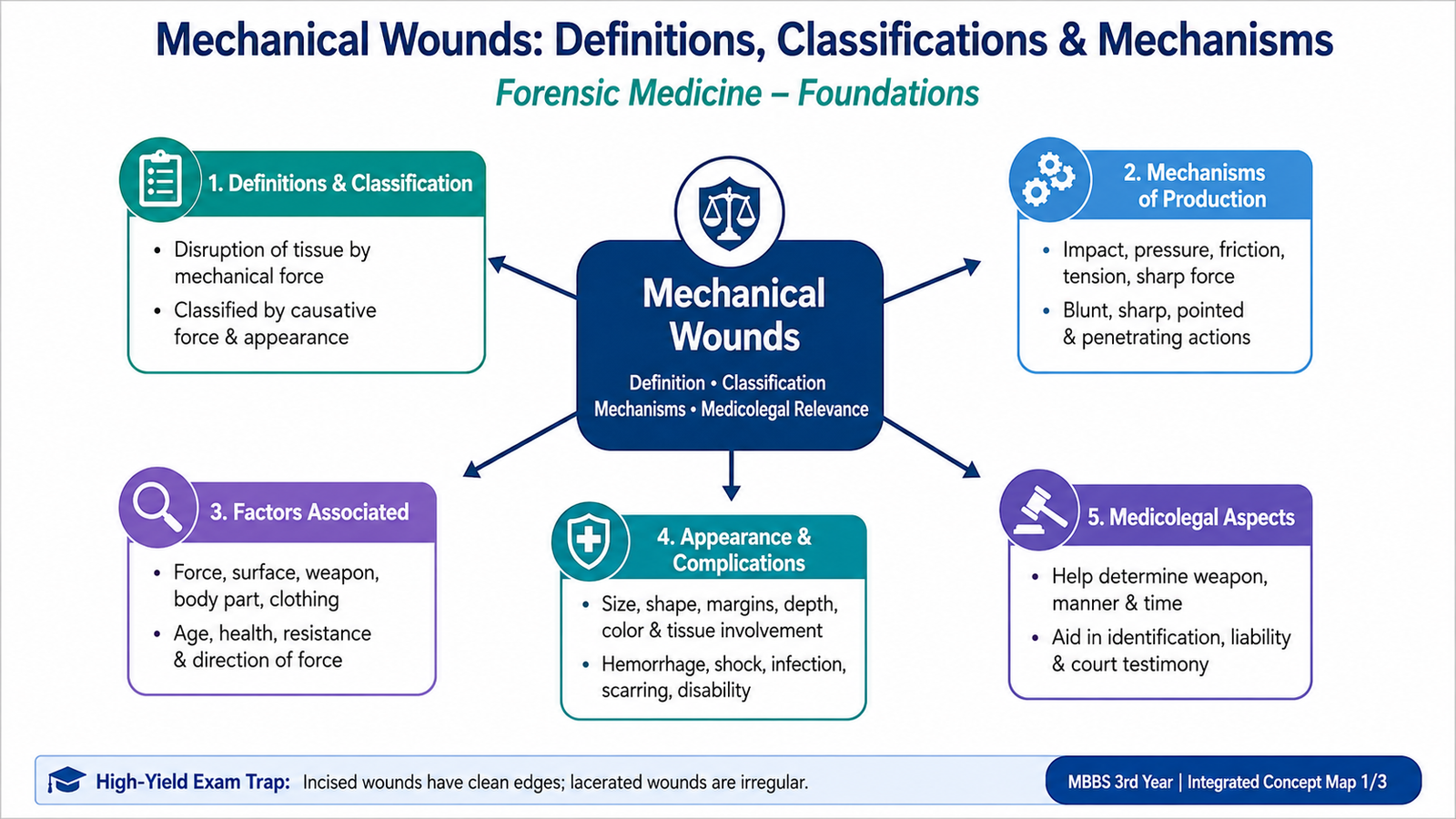
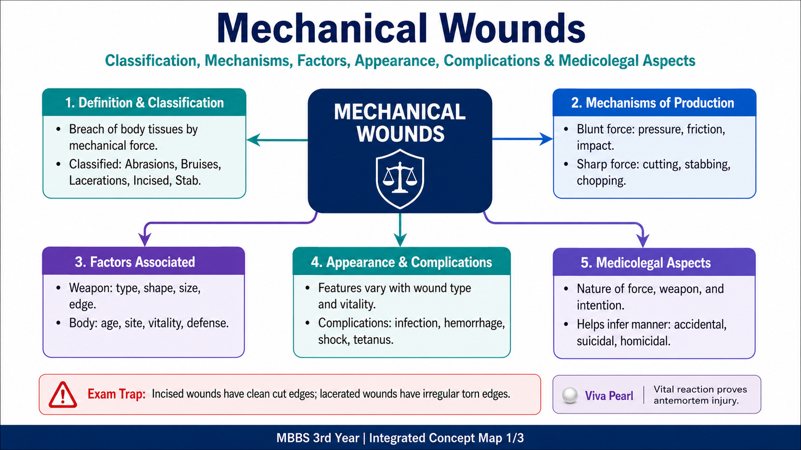
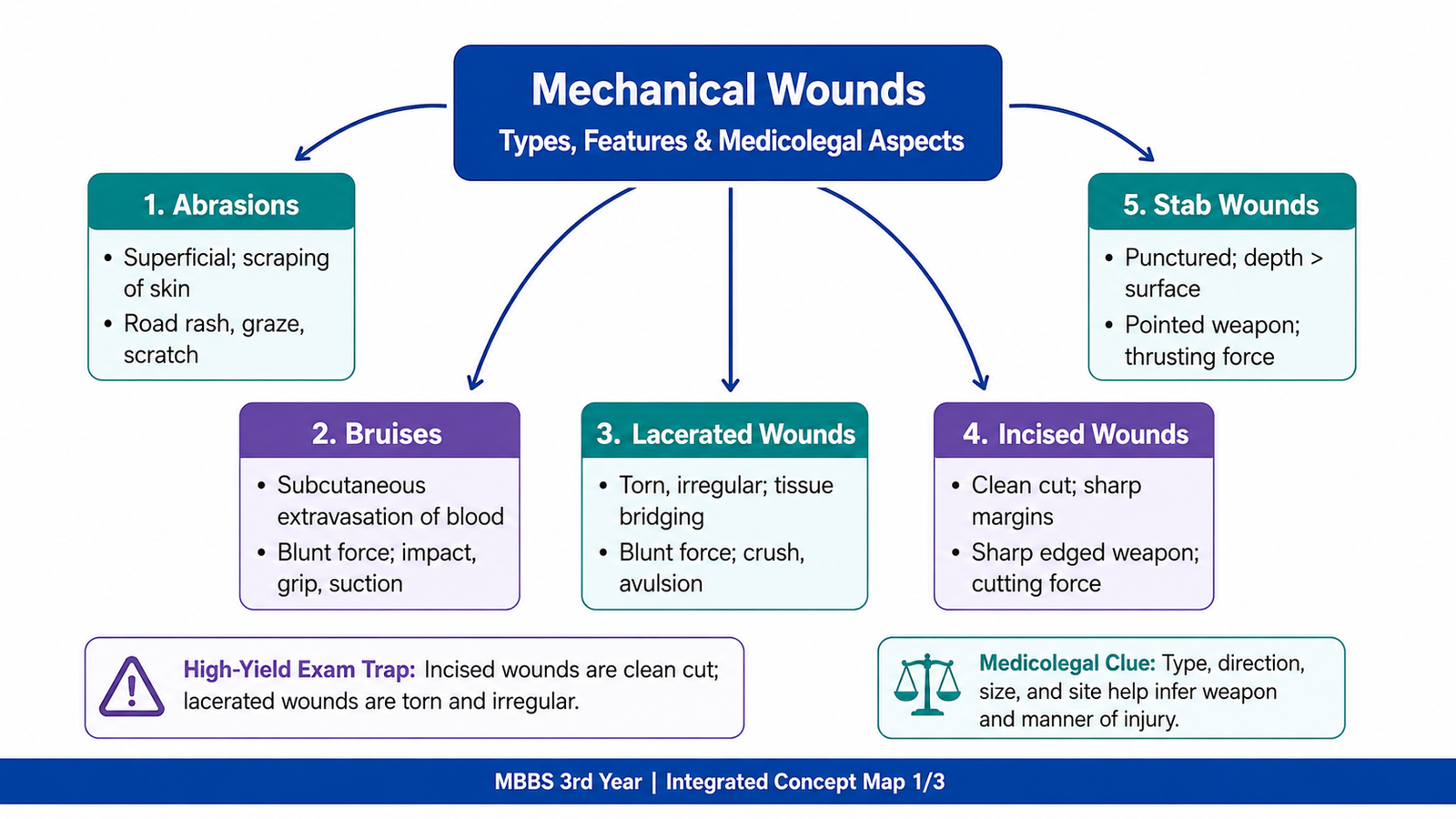
Mechanical wounds are injuries produced when physical force acts on the body. The same force does not produce the same appearance in every case because the final wound depends on the object, direction and strength of force, body site, tissue support and movement during contact. Forensic examination therefore begins by identifying the wound type and then explaining how its features could have developed.
This chapter explains abrasions, bruises, lacerations, incised wounds and stab wounds, including their classification, appearance, complications, vitality and medico-legal importance. It also covers the warning features of battered baby syndrome, the difference between empathy and sympathy, and the basic structure of a research proposal.
A. Mechanical Wounds: Definition, Classification and General Principles
A mechanical wound is best understood by linking the applied force with the tissue response. Classification provides the starting point, but interpretation also depends on the object, body site, direction of force and biological reaction.
Definition and core concept
A wound is a disruption of the normal continuity of living tissue produced by an external or internal force. In forensic practice, the visible lesion is not merely a diagnosis; it is also a record of interaction between a force, an object, the body surface, underlying tissues and the circumstances of the event.
Classification
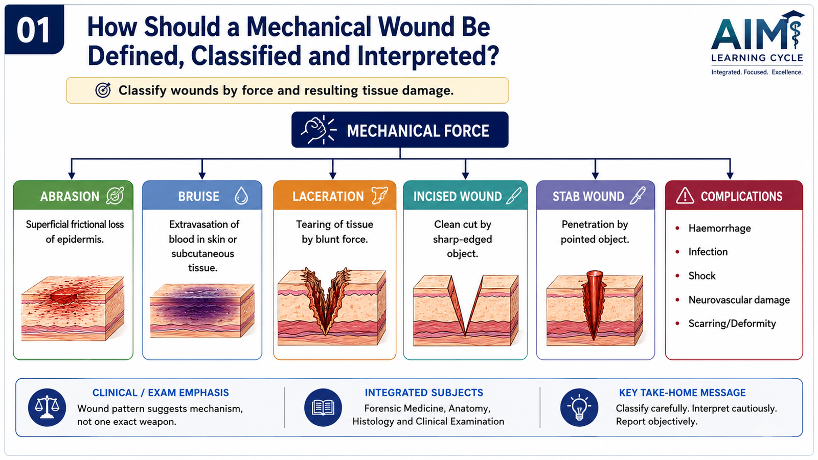
Mechanical wounds may be classified according to the dominant force and resulting tissue response:
- Abrasion: superficial loss or damage of epidermis, usually by friction, pressure or impact.
- Bruise or contusion: extravasation of blood into tissues following rupture of vessels, usually without loss of skin continuity.
- Laceration: tearing or splitting of tissue caused by blunt force.
- Incised wound: wound produced predominantly by the sharp cutting edge of an object, with surface length usually greater than depth.
- Stab or puncture wound: wound produced by penetration of a pointed or sharp-pointed object, with depth usually greater than surface dimensions.
Mechanisms of production
Factors affecting wound production and appearance
- Type, shape, size, edge and surface of the object.
- Magnitude, velocity, direction and duration of force.
- Angle of impact or penetration.
- Movement of the body or object during contact.
- Site, thickness, elasticity and vascularity of tissue.
- Presence of underlying bone or a loose mobile tissue plane.
- Clothing or intervening material.
- Age, nutritional state, disease, medications and bleeding tendency.
- Post-injury treatment, healing, infection and postmortem change.
Complications common to mechanical wounds
- Hemorrhage and shock.
- Damage to nerves, vessels, tendons, muscles, bones or internal organs.
- Infection, cellulitis, abscess formation or tetanus risk.
- Scarring, deformity, contracture or functional loss.
- Foreign-body retention.
- Air or fat embolic consequences in selected deep injuries.
- Death from blood loss, organ damage or secondary complications.
B. Abrasions
Abrasions mainly involve the epidermis. Their surface appearance may help explain friction, pressure, impact or movement, but patterns and direction must always be interpreted cautiously.
Definition
An abrasion is a superficial injury involving damage or removal of the epidermis, usually caused by friction, pressure or impact against a rough or patterned surface. Limited dermal involvement may produce slight bleeding or exudation.
Types and mechanisms
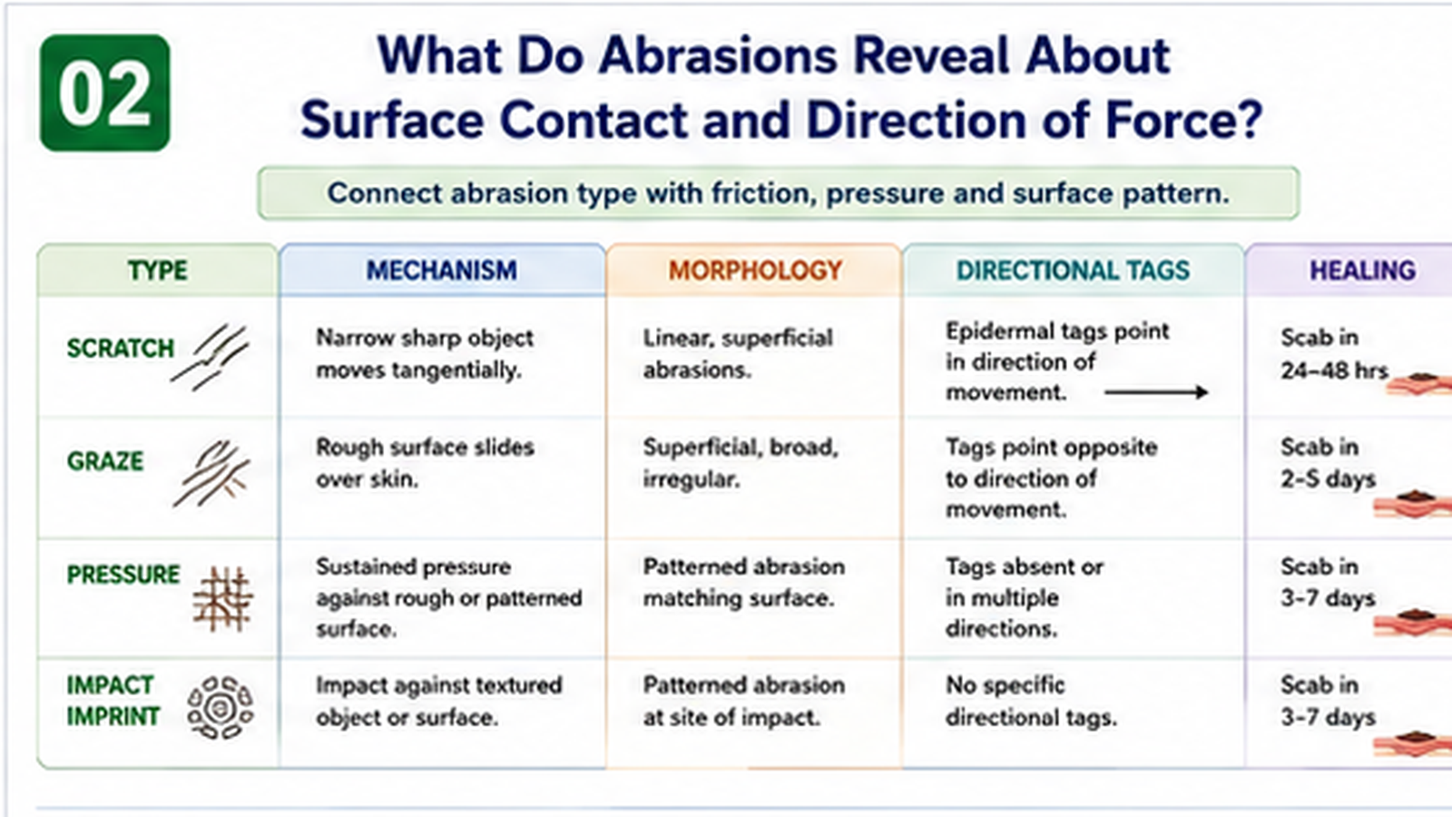
- Scratch abrasion: produced by a pointed object moving across skin, such as a fingernail or thorn.
- Graze or sliding abrasion: produced when skin rubs tangentially against a rough surface.
- Pressure abrasion: produced by compression of skin between an object and underlying tissue; it may reproduce part of the object’s pattern.
- Impact or imprint abrasion: caused by forceful perpendicular contact, sometimes displaying the shape or texture of the contacting surface.
Appearance and associated factors
- Fresh abrasions are often reddish and moist, with possible pinpoint bleeding.
- Drying and exudate produce a scab during healing.
- In a graze abrasion, epidermal tags may accumulate toward the terminal end and help suggest direction, although interpretation must be cautious.
- Patterned abrasions may reproduce a weave, edge, ligature or textured surface.
- Superficial abrasions usually heal without a permanent scar unless the dermis is damaged or infection occurs.
Antemortem versus postmortem abrasion
| Feature | Antemortem abrasion | Postmortem abrasion |
|---|---|---|
| Vital reaction | May show bleeding, exudation, inflammation and healing. | No true vital inflammatory response. |
| Surface | Initially moist or blood-stained; later forms a scab. | Often dry, yellow-brown and parchment-like. |
| Interpretation | Supports injury during life when vital reactions are present. | May result from handling, dragging, insects or environmental contact after death. |
Important limitation: Very early antemortem abrasions may have minimal visible reaction, and decomposition may obscure vitality. No single feature should be interpreted in isolation.
Medicolegal importance
- May indicate the site of impact or friction.
- May suggest direction of movement in suitable graze abrasions.
- May reproduce the pattern of an object, surface, ligature or fingernail.
- May support restraint, struggle, dragging, assault or sexual violence when interpreted with history and distribution.
- May provide trace material such as dirt, fibers or biological material.
- May help assess whether the alleged mechanism is plausible.
C. Bruises
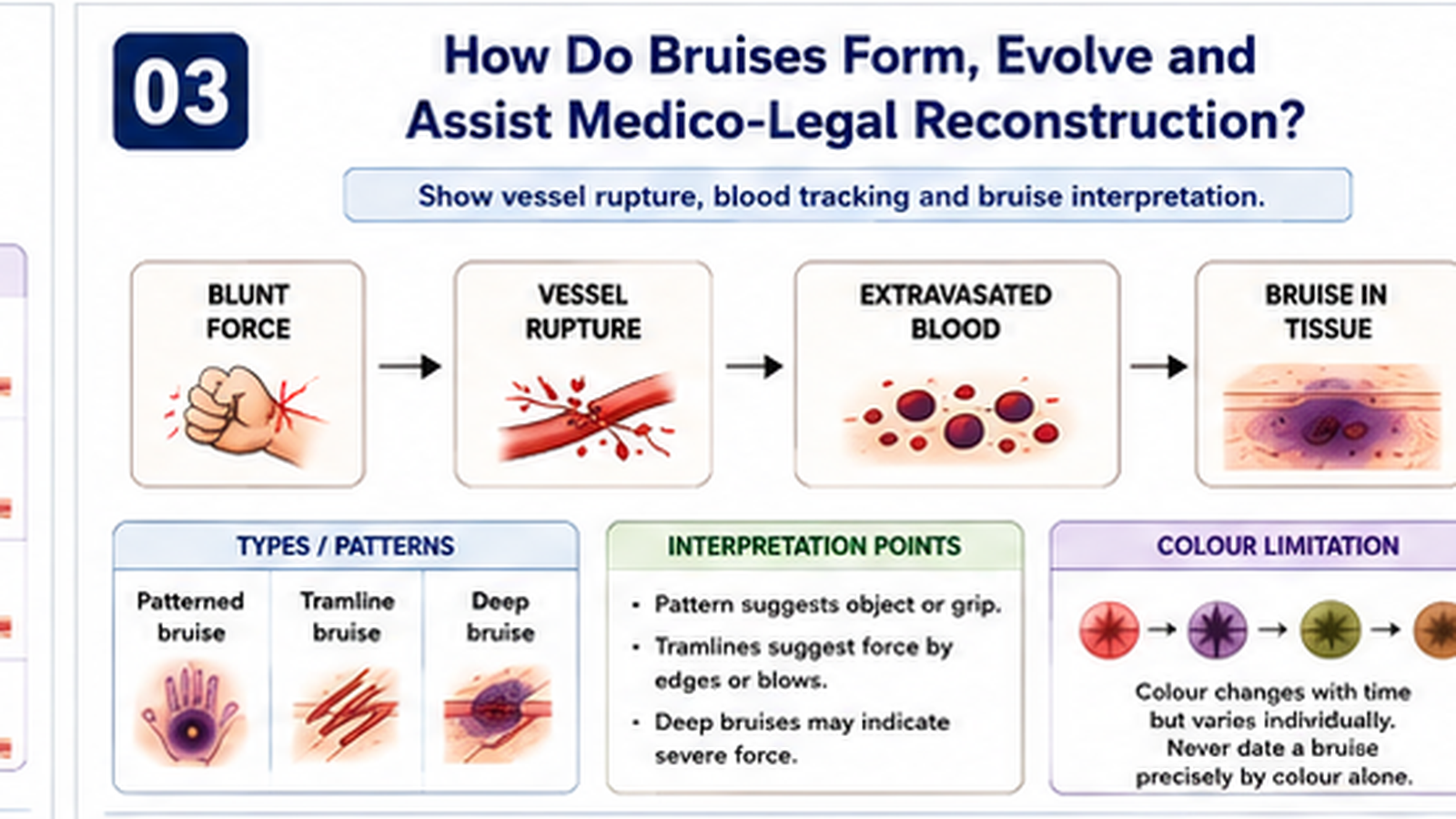
Bruises form when blunt force ruptures vessels and blood escapes into tissues. Because blood can spread, track and change with time, their site, pattern and colour require careful interpretation.
Definition
A bruise or contusion is extravasation of blood into tissues following rupture of small vessels, usually from blunt-force compression, impact or shearing, while the overlying skin may remain intact.
Types and mechanisms
- Simple bruise: localized blood extravasation after blunt impact.
- Patterned bruise: reflects part of the shape or surface of the causative object.
- Tramline bruise: two parallel lines of bruising caused by a rod-like object compressing tissue at the margins while the center is relatively spared.
- Deep bruise: forms in muscles or deeper tissues and may become externally visible only after blood tracks toward the skin.
- Ectopic or migratory bruise: appears at a site distant from the original injury due to gravitational or tissue-plane tracking of blood.
- Intraneural, intramuscular or visceral contusion: may cause significant dysfunction despite limited external discoloration.
Factors affecting appearance
- Force and surface area of impact.
- Vascularity and looseness of underlying tissue.
- Depth of the injured vessel.
- Age, body composition and skin pigmentation.
- Bleeding disorders, liver disease, malnutrition and vascular fragility.
- Use of anticoagulant or antiplatelet medicines.
- Gravity, movement and tissue-plane tracking.
- Time elapsed and environmental conditions.
Colour change and age estimation
A bruise may change from red or purple to blue, green, yellow and gradually fade as hemoglobin is broken down and cleared. However, the sequence and timing vary markedly. Colour alone cannot provide an exact injury age, particularly when assessed from photographs, in different skin tones or in deep injuries.
Antemortem versus postmortem bruising
| Feature | Antemortem bruise | Postmortem discoloration or artifact |
|---|---|---|
| Blood distribution | Blood infiltrates and may be incorporated into tissue planes. | Hypostasis remains mainly within vessels unless tissue is damaged. |
| Incision of area | Shows extravasated blood infiltrating tissue and not easily washed away. | Lividity shows dark fluid blood mainly within vessels and may wash from a cut surface. |
| Vital response | May show inflammatory or repair reaction if survival occurred. | No true vital inflammatory response. |
Complications and medico-legal importance
- Pain, swelling and temporary functional impairment.
- Hematoma formation, pressure effects or compartment syndrome.
- Underlying fracture or internal-organ injury despite minor external change.
- Pattern may support contact with a particular class of object.
- Distribution may support defense, restraint, gripping or repeated assault.
- Deep or concealed bruises are particularly important in children.
- Bruises at protected sites or in clusters may conflict with an accidental history.
D. Lacerated Wounds
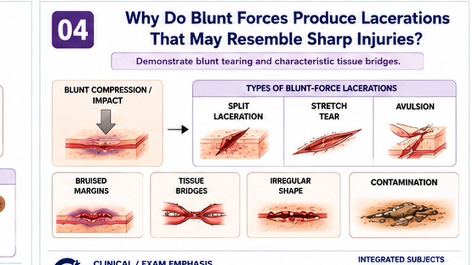
Lacerations are produced by blunt force that tears, splits or crushes tissues. The blunt mechanism explains their irregular margins, surrounding bruising, contamination and retained tissue bridges.
Definition
A laceration is a tear, split or crushing disruption of skin or deeper tissue caused predominantly by blunt force. It results from compression, shearing, stretching or crushing rather than clean cutting.
Types and mechanisms
- Split laceration: skin is compressed against underlying bone and splits; common over the scalp and bony prominences.
- Stretch laceration: excessive stretching or shearing tears skin and soft tissue.
- Tear laceration: irregular tearing by blunt projections or moving machinery.
- Avulsion: tissue is forcibly detached or reflected from its attachments.
- Crush laceration: heavy compression produces extensive tissue devitalization.
- Cut laceration: a relatively sharp but blunt-edged heavy object combines crushing with cutting characteristics.
Characteristic appearance
- Margins are commonly irregular, bruised and abraded.
- Hair bulbs, vessels, nerves and fibrous strands may remain intact across the depth as tissue bridges.
- Surrounding swelling, contamination and crushed tissue may be present.
- Bleeding may be less externally dramatic than in an incised wound because vessels are crushed and irregularly torn.
- Shape may be influenced by skin tension lines, site and underlying bone.
- A scalp split laceration may appear linear and deceptively resemble an incised wound.
Antemortem versus postmortem laceration
- Antemortem lacerations may show hemorrhage into margins, swelling, clotting, inflammation and healing.
- Postmortem tears generally lack vital tissue reaction, although gravity and manipulation can cause limited oozing.
- Very early death after injury may result in little inflammatory response; interpretation should combine gross findings, internal examination and histology where available.
Complications
- Contamination and infection.
- Retained foreign material.
- Tissue necrosis due to crushing.
- Damage to deeper vessels, nerves, tendons or bone.
- Prominent scarring and functional impairment.
- Major blood loss or internal injury depending on site.
Medico-legal importance
The morphology usually supports blunt-force causation. Associated abrasions, bruising, tissue bridges, embedded debris and underlying fractures may help reconstruct the event. However, the shape alone does not always reproduce the object, and scalp lacerations require careful inspection before being classified.
E. Incised Wounds and Their Difference from Lacerations
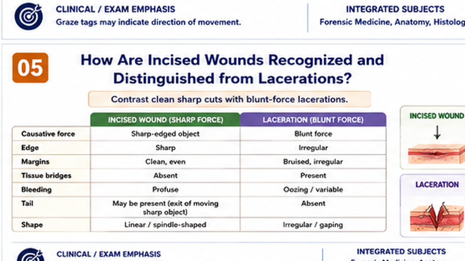
Incised wounds result from clean cutting by a sharp edge. Their regular margins and absence of tissue bridges usually separate them from lacerations, although scalp wounds and heavy sharp weapons can create difficulty.
Definition and mechanism
An incised wound is produced by the cutting action of a sharp-edged object. Its length on the skin surface is usually greater than its depth. The sharp edge divides tissue cleanly rather than crushing or tearing it.
Classification by circumstance and pattern
- Simple incised wound: a clean cut produced by one movement of a sharp edge.
- Slash wound: a relatively long incised wound produced by sweeping movement.
- Chop wound: caused by a heavy sharp-edged weapon and may combine clean cutting with crushing, bone injury and tissue destruction.
- Hesitation or tentative cuts: multiple superficial, parallel or nearby cuts that may occur in self-inflicted injury, but must be interpreted with the complete circumstances.
- Defense cuts: incised injuries on hands or forearms sustained while warding off a sharp weapon.
Appearance and associated factors
- Margins are clean, regular and usually not bruised or abraded.
- Tissue bridges are absent.
- Hair, vessels, nerves and tendons within the path are cleanly divided.
- Bleeding may be profuse because vessels are sharply cut.
- Gapping depends on relation to skin tension lines and depth.
- The ends may indicate direction only in selected wounds; a superficial “tail” may occur as the blade leaves the skin.
- Depth may vary along the wound according to pressure, blade movement and body contour.
Incised wound versus laceration
| Feature | Incised wound | Laceration |
|---|---|---|
| Dominant force | Sharp cutting. | Blunt tearing, splitting, stretching or crushing. |
| Margins | Clean and regular. | Irregular, bruised or abraded. |
| Tissue bridges | Absent. | Usually present. |
| Hair and vessels | Cleanly divided. | Crushed or irregularly torn. |
| Bleeding | Often free and profuse. | May be less externally profuse due to crushing. |
| Contamination | Usually less unless the object or environment is contaminated. | Often greater, with debris or devitalized tissue. |
Antemortem versus postmortem incised wounds
Antemortem incised wounds may show active hemorrhage, blood infiltration, clot formation, vessel retraction and later inflammation or repair. Postmortem cuts usually show little pressure-driven bleeding, limited tissue infiltration and no true vital reaction. Nevertheless, a cut inflicted soon after death may show some passive blood escape, and rapid death after injury may limit visible inflammation.
Complications and medico-legal significance
- Severe external hemorrhage.
- Division of tendons, nerves or major vessels.
- Air embolic risk in selected injuries to large neck veins.
- Functional impairment and scarring.
- Wound location and associated marks may assist interpretation of accident, assault or self-infliction.
- Defense injuries and tentative cuts are supportive patterns, not individually conclusive proof of manner.
F. Stab Wounds
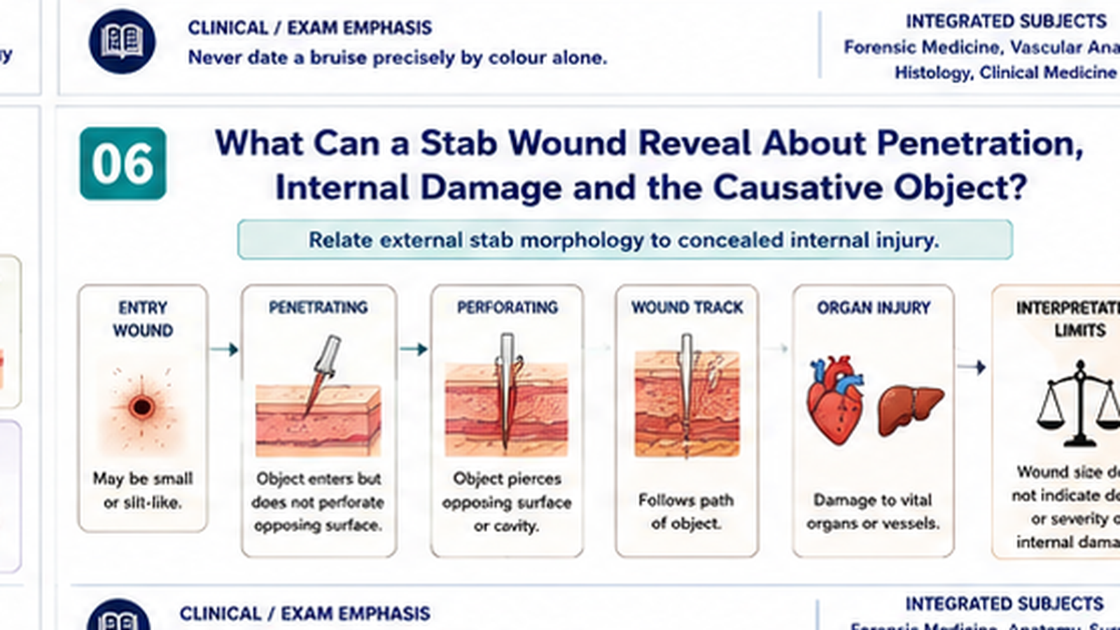
Stab wounds concentrate force through a pointed object and create a track deeper than the visible surface wound. Internal examination is therefore more important than the small external opening may suggest.
Definition
A stab wound is produced when a pointed or sharp-pointed object penetrates the body. Its depth is usually greater than the surface length or width. The external opening may be small while the internal damage is extensive.
Classification
- Penetrating wound: the object enters the body but does not emerge through another surface.
- Perforating wound: the object passes through and produces an entry and an exit wound.
- Puncture wound: produced by a pointed object, sometimes without a sharp cutting edge.
- Incised-stab wound: penetration by an object with one or more sharp edges.
External appearance
- The wound may be slit-like, elliptical, triangular or irregular depending on blade shape, skin tension and movement.
- A single-edged blade may produce one sharper angle and one more rounded or split angle, but this feature is not always preserved.
- A double-edged blade may produce two relatively sharp angles.
- Abrasions or bruises around the opening may result from the hilt, guard or forceful impact.
- Twisting, withdrawal and body movement can alter the surface appearance.
- Skin elasticity may make wound length smaller or occasionally different from the blade width.
Wound track and internal examination
The wound track should be assessed in relation to body position, tissue movement and organ displacement. Its apparent direction may change when the body is repositioned. The path may involve skin, subcutaneous tissue, muscle, vessels, pleura, lungs, heart, abdomen or solid organs.
- Depth of the track does not necessarily equal blade length because tissues may be compressed during forceful penetration.
- Blade length cannot be precisely determined from track depth alone.
- The track may be longer than the measured blade penetration if the body wall was compressed.
- Multiple tracks can result from repeated movements through one skin opening.
Antemortem versus postmortem stab wounds
Antemortem stab wounds may show hemorrhage into the track, blood aspiration into airways when respiratory activity persists, blood pressure effects and later inflammatory changes. Postmortem stab wounds lack true vital reaction and usually show limited passive bleeding. Interpretation is difficult when death is immediate or the wound is inflicted near the time of death.
Complications
- External or concealed internal hemorrhage.
- Hemothorax, pneumothorax or cardiac tamponade.
- Perforation of hollow organs with contamination and peritonitis.
- Damage to major vessels, spinal structures, nerves or solid organs.
- Infection and retained foreign material.
- Death despite a small external wound.
Medico-legal importance
- May suggest whether a pointed, single-edged, double-edged or blunt-pointed object was involved.
- Can help assess direction and depth of penetration, with stated limitations.
- Location, accessibility, clothing defects and associated injuries may contribute to assessment of circumstances.
- Defense wounds may be present on hands and forearms.
- Weapon comparison requires correlation with dimensions, shape, clothing damage and internal track; it cannot rely solely on skin appearance.
G. Wound Vitality, Age and Medico-Legal Assessment
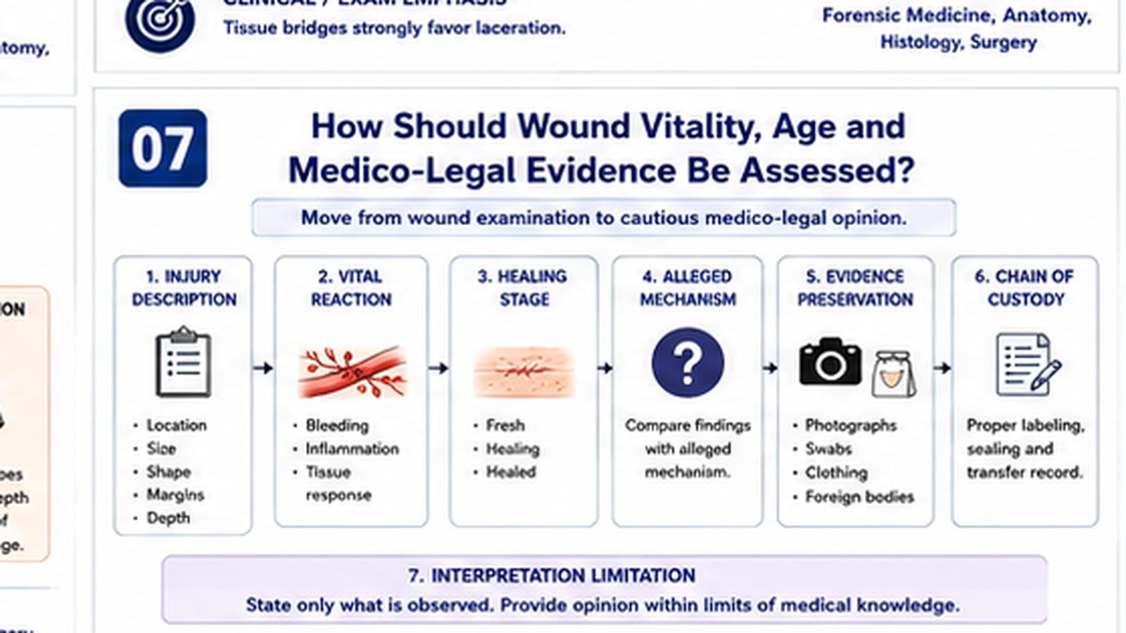
Medico-legal assessment combines careful description with cautious interpretation. No single sign reliably proves exact timing, exact weapon or manner of injury, so observations and conclusions must be clearly separated.
Systematic injury examination
Each wound should be examined and documented independently before a combined interpretation is made. The doctor should record:
- Type of injury.
- Exact anatomical site and side.
- Number, size, shape and orientation.
- Margins, ends, depth and surrounding changes.
- Swelling, tenderness, bleeding, scab, discharge or healing.
- Associated bruising, abrasion, fracture or internal injury.
- Foreign material, contamination or clothing damage.
- The patient’s account and alleged mechanism, documented in neutral language.
Evidence of vitality
Vitality means that an injury was sustained during life and elicited a biological response. Relevant findings include:
- Active hemorrhage or blood infiltration into tissues.
- Clot formation and vessel response.
- Swelling and inflammatory reaction.
- Leukocyte infiltration and tissue repair on microscopy.
- Scab formation, granulation tissue or scar formation.
- Physiological consequences such as aspiration of blood or vital organ response in appropriate circumstances.
Age estimation
The age of a wound is estimated from the combined appearance, healing stage, symptoms, histological reaction and clinical history. Exact timing is often impossible. Environmental conditions, infection, treatment, age, nutrition and disease alter healing. Reports should use cautious expressions such as “recent,” “healing,” “older than another injury” or “compatible with the stated interval” when supported.
Medico-legal questions commonly addressed
- What type of wound is present?
- What class of force or object could produce it?
- Is the alleged mechanism medically plausible?
- Could the injury be self-inflicted, accidental or inflicted by another person?
- Is the injury recent, healing or of uncertain age?
- Was it produced during life?
- What complications, functional effects or danger to life may result?
- Is specialist referral, imaging, surgery or safeguarding required?
Objective evidence handling
- Confirm identity and obtain appropriate consent according to the clinical and legal context.
- Prioritize emergency care before detailed medico-legal procedures.
- Record date, time, history source and relevant circumstances.
- Use body diagrams, measurements and clinically appropriate photography where authorized.
- Preserve clothing, foreign material and specimens when relevant.
- Label, seal and document transferred evidence.
- Maintain chain of custody from collection to authorized handover.
- State observations separately from interpretations.
- Acknowledge limitations and avoid determining guilt.
H. Battered Baby Syndrome
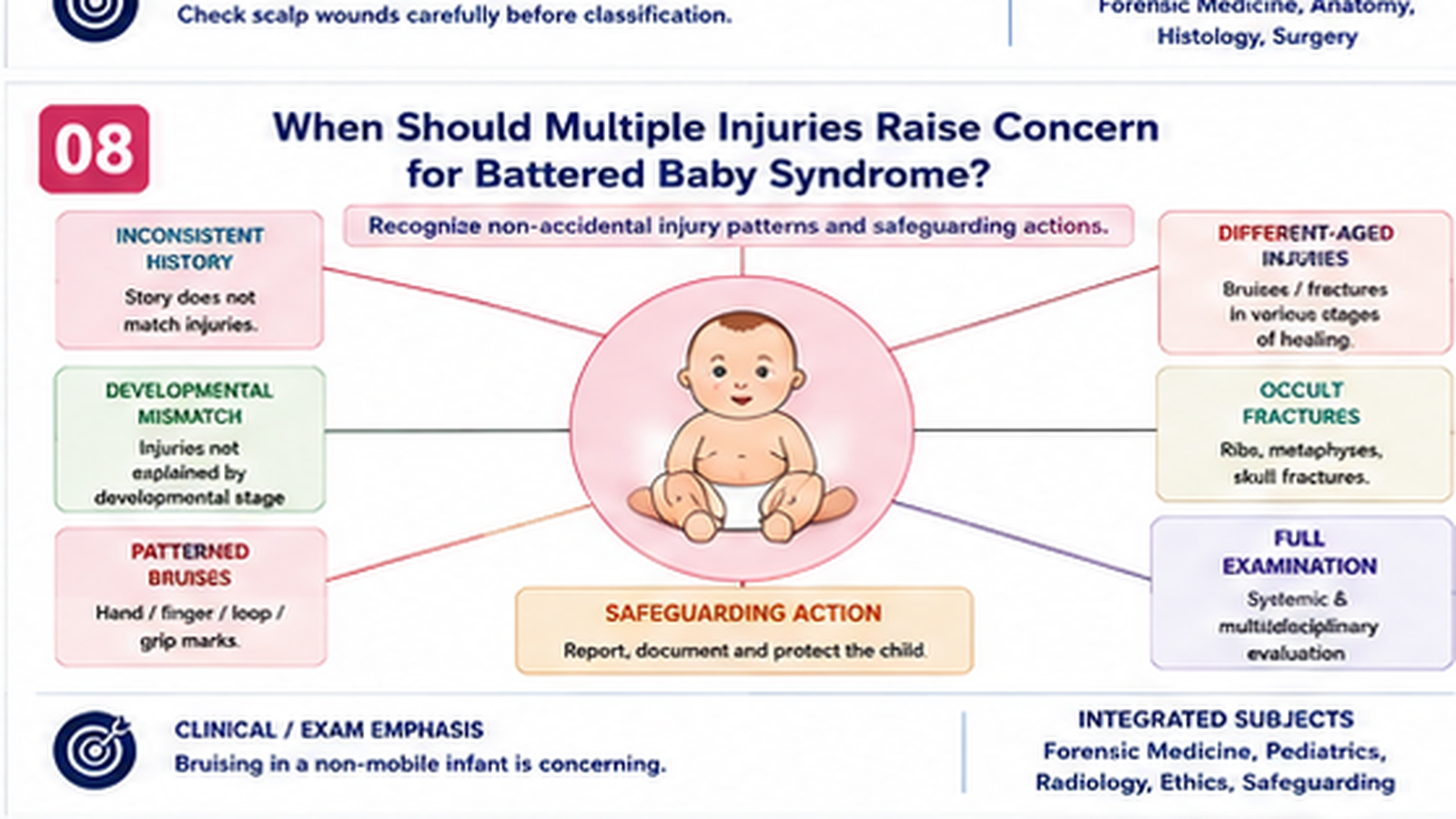
Battered baby syndrome is recognized from a pattern of concern rather than one isolated injury. The history, developmental ability, distribution and age of injuries, investigations and child behaviour must be considered together.
Core concept
Battered baby syndrome describes a pattern of non-accidental physical injury in an infant or young child caused by a caregiver or another person. Modern practice considers it within the broader framework of child physical abuse and safeguarding. Diagnosis is not based on one bruise or fracture alone; it depends on the consistency of the history, developmental ability, injury pattern, examination, investigations and multidisciplinary assessment.
Features that should raise suspicion
- History that is absent, vague, changing or inconsistent between caregivers.
- Mechanism incompatible with the child’s developmental abilities.
- Delay in seeking medical care without a reasonable explanation.
- Multiple injuries of different apparent ages.
- Repeated presentations for unexplained injuries.
- Bruises in a non-mobile infant.
- Injuries at protected sites such as the trunk, ears, neck, inner thighs, buttocks or genital region.
- Patterned bruises, bite marks, loop marks or grip marks.
- Fractures with different healing stages or unusual patterns for the alleged event.
- Burns with clear margins or patterned contact appearance.
- Intracranial injury, retinal findings or unexplained neurological deterioration.
- Visceral injury without a plausible high-energy accidental mechanism.
- Fearful behavior, withdrawal or marked interactional changes around a particular caregiver.
Clinical and forensic reasoning
The doctor should consider differential explanations including accidental injury, bleeding disorders, bone fragility, dermatological lesions, birth-related injury and cultural practices. However, investigation of alternatives must not delay protection when the child may remain at risk.
Examination and investigations
- Assess airway, breathing, circulation, neurological status and pain.
- Undress and examine the child respectfully from head to toe.
- Record growth, nourishment, hygiene and general condition.
- Document every visible lesion with site, dimensions, colour, shape and surrounding findings.
- Assess oral cavity, scalp, ears, neck, trunk, limbs and protected areas.
- Perform imaging and laboratory evaluation according to clinical findings and specialist guidance.
- Consider occult fractures, intracranial injury, retinal injury, abdominal trauma and bleeding disorders.
Immediate responsibilities
- Treat urgent injuries and relieve pain.
- Ensure the child is not discharged into an unsafe situation.
- Document statements verbatim where important.
- Use neutral, non-accusatory communication.
- Involve senior clinicians, pediatric specialists and the appropriate safeguarding or medico-legal pathway.
- Preserve relevant evidence and maintain confidentiality within lawful professional limits.
I. Empathy, Communication and Research-Proposal Writing
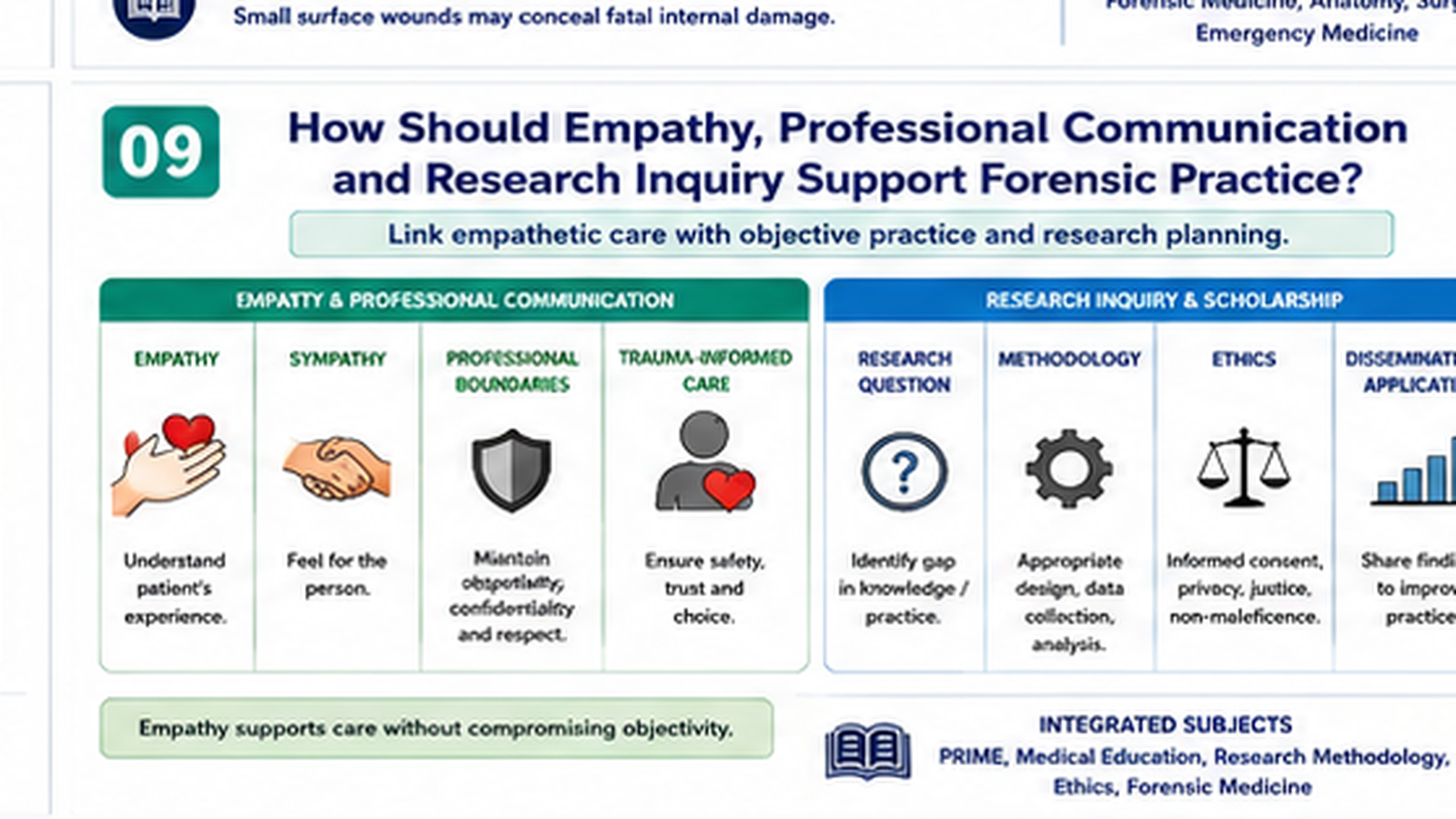
Professional forensic practice requires both humane communication and objective reasoning. The same disciplined approach also supports a clear, ethical and feasible research proposal.
Empathy versus sympathy
| Aspect | Empathy | Sympathy |
|---|---|---|
| Meaning | Understanding the person’s feelings and perspective while maintaining professional boundaries. | Feeling sorrow, pity or concern for another person. |
| Clinical response | “This examination may feel frightening. I will explain each step and stop if you need a pause.” | “I feel very sorry for you.” |
| Professional value | Supports trust, disclosure, consent, dignity and trauma-informed care. | May communicate concern but can become pitying or emotionally over-involved. |
Empathy does not mean accepting every account uncritically. A forensic clinician can understand distress, avoid judgment and maintain dignity while still documenting inconsistencies and interpreting findings objectively.
Trauma-informed communication
- Introduce yourself and explain your role.
- Use age-appropriate language and an appropriate caregiver or support person when safe.
- Explain the purpose and steps of examination.
- Seek consent and assent where applicable.
- Limit unnecessary repetition of distressing questions.
- Avoid blame, leading questions and promises that cannot be guaranteed.
- Respect privacy while explaining any professional duty to escalate safety concerns.
Developing a research proposal
A research proposal is a structured plan explaining what will be studied, why it matters and how the study will be conducted ethically and scientifically. A topic related to this chapter could examine documentation quality, awareness of non-accidental injury indicators or patterns of mechanical wounds presenting to a healthcare facility.
- Title: concise, specific and reflective of population, setting and main variable.
- Introduction: background, magnitude and clinical or medico-legal importance of the problem.
- Rationale: the knowledge or practice gap the study will address.
- Research question: a clear, answerable question.
- Objectives: one general objective and focused specific objectives.
- Operational definitions: clear definitions of key variables, injury patterns or documentation standards.
- Methodology: study design, setting, duration, population, inclusion and exclusion criteria, sampling method, sample-size basis and data-collection procedure.
- Data analysis plan: variables, descriptive analysis and any planned comparison or association.
- Ethical considerations: approval, confidentiality, consent, safeguarding and management of sensitive medico-legal information.
- Limitations: anticipated sources of bias or restricted generalizability.
- Work plan and budget: practical sequence and resource requirements where requested.
- References and annexures: standard citation style, data tool, consent information and relevant permissions.
Example research framework
Proposed title: Completeness of medico-legal documentation of mechanical wounds in patients presenting to a tertiary-care emergency department.
Research question: What proportion of medico-legal injury records contain the essential descriptive elements required for objective wound interpretation?
General objective: To assess the completeness of documentation of mechanical wounds.
Possible specific objectives: To assess recording of site, size, type, margins, surrounding changes, possible mechanism, treatment and interpretation limitations; and to identify common documentation gaps.
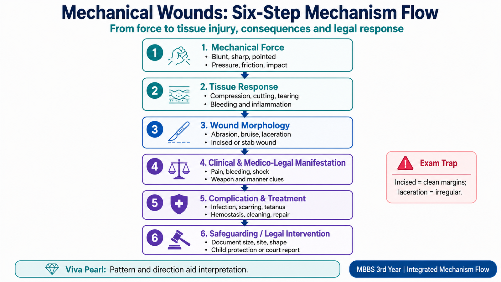
- ⭐ Mechanical wounds are classified by the dominant force and tissue response: abrasion, bruise, laceration, incised wound and stab wound.
- An abrasion is a superficial epidermal injury. Graze abrasions may show epidermal tags toward the terminal end, while pressure or impact abrasions may reproduce a pattern.
- A bruise is blood extravasation into tissue after vessel rupture. Its appearance depends on depth, site, vascularity, health and medicines; colour alone cannot date it precisely.
- A laceration is caused by blunt tearing, splitting or crushing. Irregular bruised margins and tissue bridges are key differentiating features.
- An incised wound is produced by a sharp cutting edge. It usually has clean regular margins, no tissue bridges and surface length greater than depth.
- A stab wound usually has greater depth than surface dimensions. A small skin opening may conceal severe internal bleeding or organ injury.
- Antemortem wounds may show hemorrhage, tissue infiltration, inflammation and healing. Very recent injuries or rapid death may show only limited vital reaction.
- Exact wound age and exact weapon identification are often impossible; conclusions should use cautious terms such as compatible, consistent or possible.
- Incised wounds and lacerations are distinguished mainly by margins, tissue bridges, clean division of structures, bruising and contamination.
- Objective documentation includes site, size, shape, orientation, margins, depth, surrounding changes, associated injuries and the patient’s account in neutral language.
- Bruising in a non-mobile infant, injuries at protected sites, different injury ages and a history inconsistent with development are major warning features of battered baby syndrome.
- Empathy means understanding the patient’s perspective while maintaining professional boundaries; sympathy mainly means feeling sorrow or pity.
- A research proposal should include title, background, rationale, question, objectives, methods, analysis, ethics, limitations, work plan and references.
Mechanical Wounds and Medicolegal Assessment
Watch the main video after completing the chapter. Focus on the mechanism, appearance and medicolegal importance of abrasions, bruises, lacerations, incised wounds and stab wounds.
“`
Mechanical Injuries: Blunt Force, Sharp Trauma and Stab Wounds
Covers the major mechanical injuries and helps connect the type of force with the resulting wound pattern.
Battered Baby Syndrome
Reinforces the important history, injury patterns and diagnostic warning signs associated with child physical abuse.
“`