
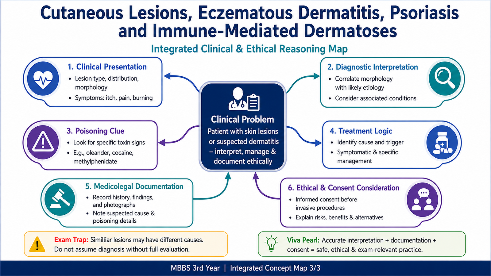
This chapter follows the KMU learning outcomes and builds the topic in a logical sequence. First understand what each skin lesion represents and why it develops, then use the AIM High-Yield Review for revision.
Cutaneous Lesions, Eczematous Dermatitis, Psoriasis and Immune-Mediated Dermatoses
MSK Module — recognition of important skin lesions, inflammatory dermatoses, vesiculobullous disorders, psoriasis, warts and juvenile dermatomyositis.
Topic Introduction
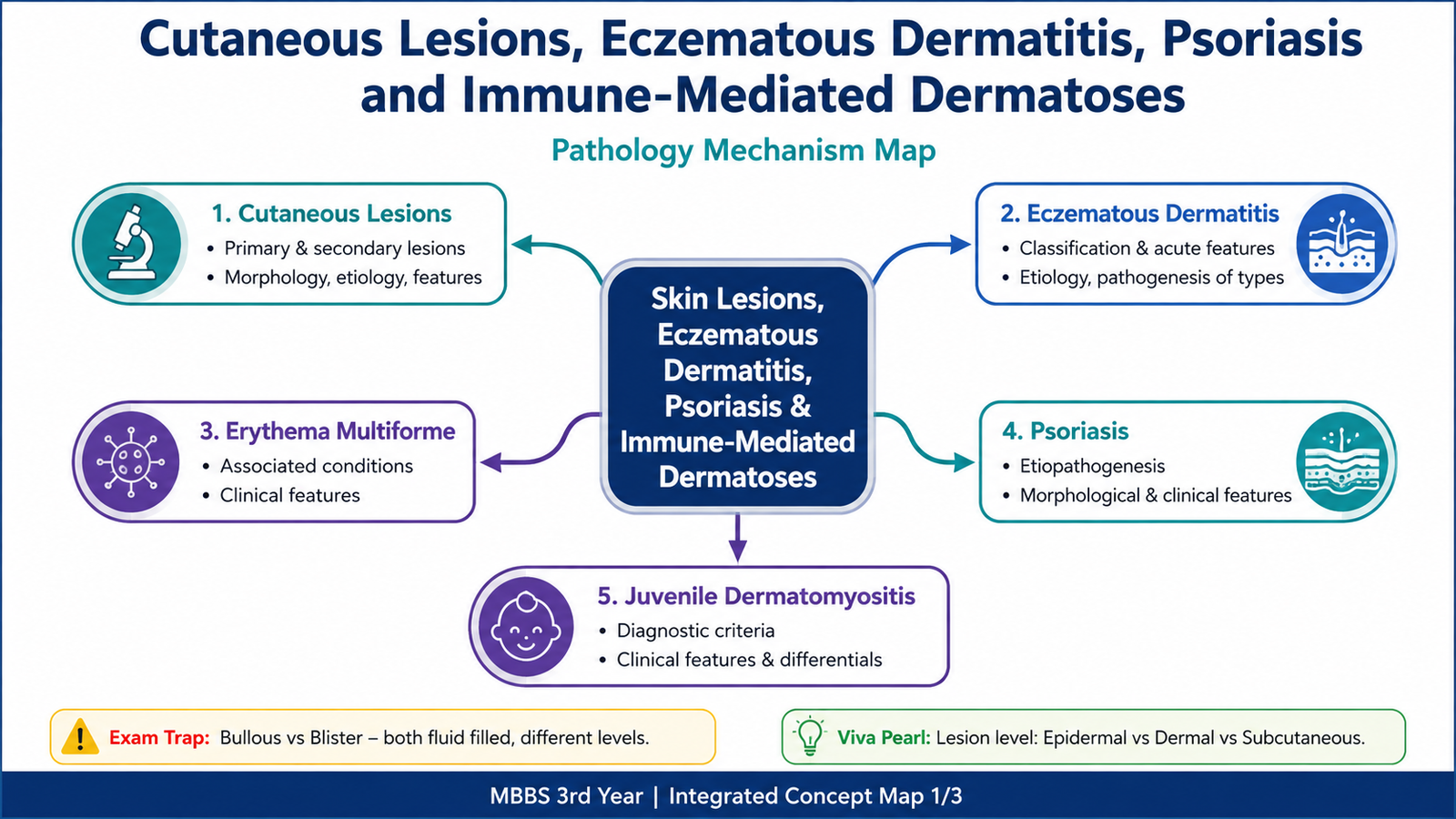
Skin diseases are often recognized by the type of lesion they produce and by the way that lesion is distributed or changes with time. For this reason, learning the language of dermatology is the first step toward understanding disorders such as eczema, psoriasis and blistering diseases. In this chapter, you will first learn the important primary and secondary skin lesions and the pathological terms used to describe them. You will then study eczematous dermatitis, erythema multiforme, psoriasis, urticaria, pemphigus, bullous pemphigoid and warts. The final section covers juvenile dermatomyositis, with emphasis on its diagnostic features and important differential diagnoses.
A. Language of Cutaneous Lesions and Important Morphological Terms
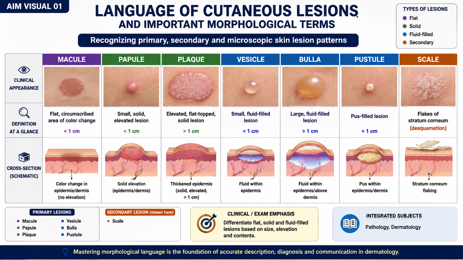
Accurate description of a skin lesion begins by identifying what type of lesion is present. Some terms describe lesions visible to the eye, such as a macule or vesicle, while others describe microscopic changes in the epidermis, such as acanthosis or parakeratosis. Understanding both levels helps connect clinical appearance with underlying pathology.
Primary skin lesions
Primary lesions are the initial visible changes produced directly by the disease process.
- Macule: a flat, circumscribed area of altered skin color without elevation or depression. Examples include areas of hyperpigmentation or erythema.
- Papule: a small, solid, raised lesion produced by epidermal thickening, dermal inflammation or deposition of material.
- Nodule: a deeper and usually larger solid lesion that extends into the dermis or subcutaneous tissue.
- Plaque: a broad, raised, relatively flat-topped lesion. It may develop by enlargement or coalescence of papules, as commonly seen in psoriasis.
- Vesicle: a small, fluid-filled elevation caused by separation within or beneath the epidermis.
- Bulla: a larger fluid-filled blister. The level of separation within the skin influences whether the roof is fragile or tense.
- Blister: a general clinical term for a fluid-filled lesion; a small blister is a vesicle and a larger one is termed a bulla.
- Pustule: a raised lesion containing purulent material composed mainly of inflammatory cells and cellular debris.
Secondary surface changes
- Scale: visible flakes of keratin produced by abnormal or accelerated shedding of the stratum corneum.
- Lichenification: thickening of skin with exaggerated normal skin markings, usually caused by repeated rubbing or scratching.
- Excoriation: superficial loss of epidermis caused by scratching or mechanical trauma.
Important microscopic terms
- Hyperkeratosis: thickening of the stratum corneum.
- Parakeratosis: retention of nuclei within cells of the stratum corneum. It usually reflects accelerated epidermal turnover.
- Acanthosis: thickening of the epidermis, particularly the stratum spinosum.
- Dyskeratosis: abnormal or premature keratinization of individual epidermal cells.
- Acantholysis: loss of adhesion between keratinocytes, causing separation of epidermal cells and formation of an intraepidermal cleft or blister.
- Papillomatosis: surface elevation caused by elongation or irregular prominence of dermal papillae and overlying epidermis.
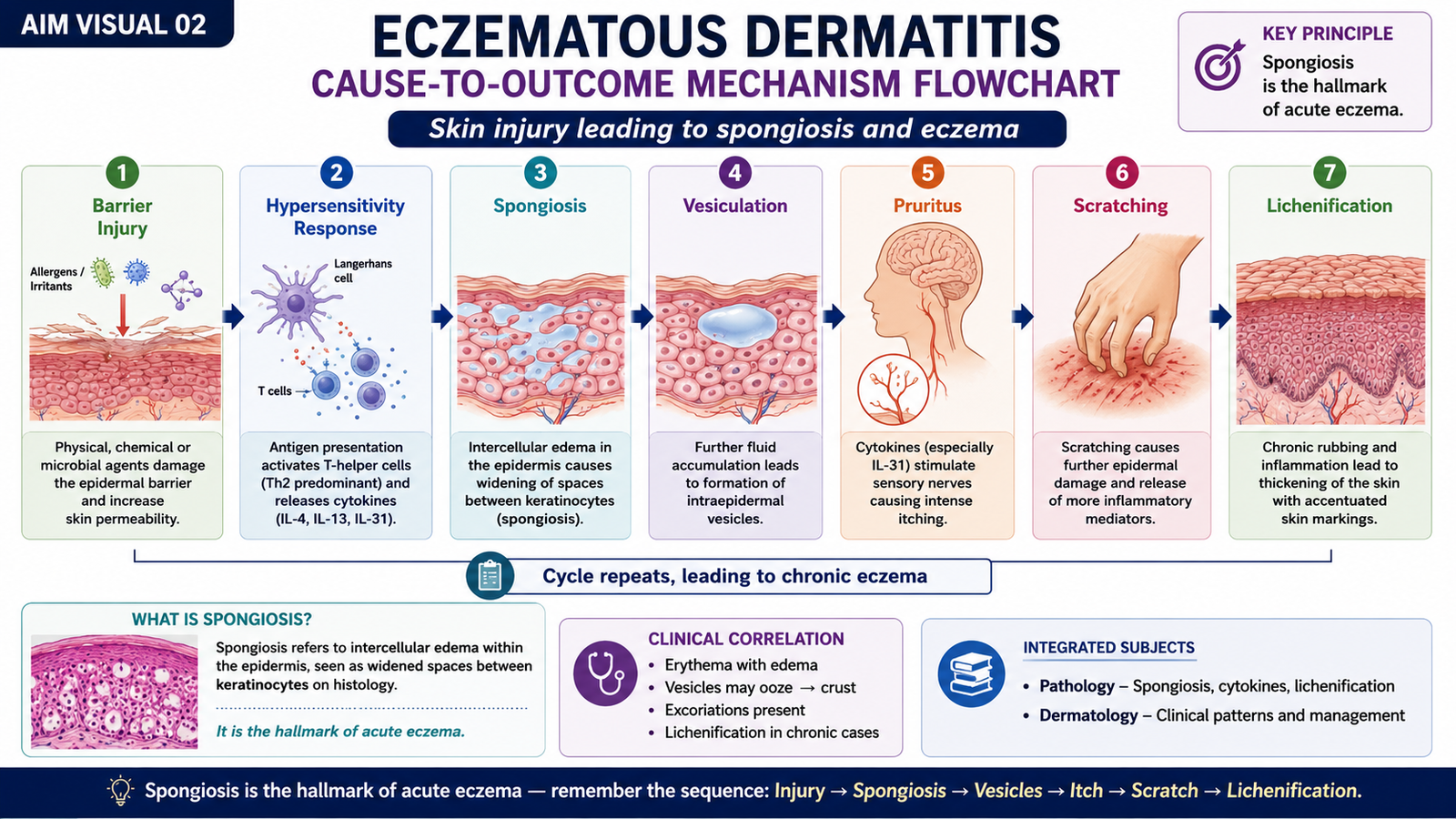
- Spongiosis: accumulation of intercellular edema within the epidermis. Widening between keratinocytes may become marked enough to produce vesicles in eczematous dermatitis.
- Lentiginous pattern: increased numbers or prominence of cells arranged along the basal layer of the epidermis. When the supplied term is used with spongiosis, it refers descriptively to epidermal alteration in which basal-zone change and intercellular edema are appreciated histologically.
B. Eczematous Dermatitis
Eczematous dermatitis is a pattern of inflammatory skin reaction characterized clinically by itching, erythema and variable vesiculation, oozing, crusting or scaling. Its characteristic microscopic feature in the acute stage is spongiosis, which means intercellular edema within the epidermis. Different causes can produce the same basic eczematous reaction pattern.
Classification and major types
Eczematous dermatitis may develop through immune-mediated hypersensitivity, inherited barrier dysfunction, direct chemical irritation, drug reactions or light-induced injury.
- Allergic contact dermatitis
- Atopic dermatitis
- Drug-related eczematous dermatitis
- Photoeczematous eruption
- Primary irritant dermatitis
Acute eczematous dermatitis: morphology
In acute eczema, inflammation increases epidermal intercellular fluid. This separates keratinocytes without immediately destroying their attachments. The resulting spongiosis can progress to small intraepidermal vesicles. Microscopic morphology:
- Prominent epidermal spongiosis.
- Formation of small intraepidermal vesicles when edema becomes marked.
- Variable inflammatory cell infiltration within the epidermis and superficial dermis.
- Superficial dermal vascular dilatation and inflammatory infiltrates may accompany the epidermal reaction.
Clinical and gross appearance:
- Erythematous and pruritic lesions.
- Papules and small vesicles may develop.
- Vesicles may rupture, causing oozing and crusting.
- With persistence, scaling and epidermal thickening become more prominent.
- Repeated scratching may produce excoriation and lichenification.
Allergic contact dermatitis
Allergic contact dermatitis occurs when a sensitized individual develops a delayed immune reaction after the skin is re-exposed to a particular contact allergen. The substance penetrates the epidermis, is presented to T lymphocytes and produces an inflammatory response on subsequent exposure.
The distribution usually corresponds to areas exposed to the offending substance, although inflammation may extend beyond the exact point of contact.
Atopic dermatitis
Atopic dermatitis is a chronic, relapsing, intensely pruritic inflammatory skin disorder in which impaired epidermal barrier function and abnormal immune responses contribute to inflammation. Barrier dysfunction allows increased water loss and easier entry of irritants and environmental antigens. Itching promotes scratching, and repeated scratching further damages the barrier, creating a continuing itch-scratch cycle.
- Pruritus is a dominant clinical feature.
- Acute lesions may show erythema, papules, vesicles and oozing.
- Chronic lesions become dry, scaly and lichenified.
Drug-related eczematous dermatitis
Some medications can trigger an inflammatory skin reaction with an eczematous pattern. The eruption results from an adverse immune or hypersensitivity response to the drug or its metabolites. Clinically, it may resemble other forms of eczema, so the temporal relationship with medication exposure is important.
Photoeczematous eruption
A photoeczematous eruption develops when light exposure contributes to a cutaneous inflammatory reaction. The eruption therefore tends to involve sun-exposed areas. The inflammatory reaction produces the same basic eczematous pattern of erythema, itching and spongiosis.
Primary irritant dermatitis
Primary irritant dermatitis is caused by direct injury to the epidermal barrier rather than a specific sensitized immune response. Strong chemicals, repeated exposure to detergents or other irritating substances may directly damage keratinocytes and disturb barrier function. Inflammation then develops at the exposed site.
C. Erythema Multiforme
Erythema multiforme is an acute, immune-mediated skin reaction characterized by distinctive erythematous lesions, particularly the classic target or iris lesion. It is commonly triggered by an infection and less often by medications or other stimuli. The immune response damages epidermal keratinocytes, producing the characteristic clinical pattern.
Associated conditions
- Infections, especially herpes simplex infection.
- Other infectious triggers may occasionally be associated.
- Drugs can trigger an erythema-multiforme-like reaction in some patients.
Clinical features
The hallmark lesion is a target-shaped lesion with concentric zones of color change. The center may appear dusky or damaged, surrounded by paler and then erythematous zones. Lesions commonly develop symmetrically on the extremities and may spread centrally.
- Acute onset.
- Symmetrical erythematous papules that evolve into target lesions.
- Extremities are commonly involved.
- Mucosal involvement may occur in more significant disease.

D. Psoriasis
Psoriasis is a chronic immune-mediated inflammatory disorder characterized by excessive and abnormal epidermal proliferation. The epidermis turns over much more rapidly than normal, so keratinocytes reach the surface before completing normal maturation. This explains the characteristic combination of epidermal thickening, parakeratosis and surface scaling.
Etiopathogenesis
Psoriasis develops in genetically susceptible individuals through dysregulated immune activation, particularly involving T lymphocytes and inflammatory cytokine pathways. These signals stimulate keratinocyte proliferation and maintain chronic inflammation.
Gross and clinical morphology
Psoriatic lesions are usually well-demarcated erythematous plaques covered by silvery-white scale. The plaque is raised because of epidermal hyperplasia, while the scale reflects rapid and incomplete keratinization.
- Sharply demarcated erythematous plaques.
- Silvery-white surface scale.
- Common involvement of extensor surfaces such as elbows and knees.
- Scalp involvement is common.
- Nail changes may occur.
Microscopic morphology
- Acanthosis: regular epidermal thickening due to increased keratinocyte proliferation.
- Parakeratosis: retained nuclei in the stratum corneum because epidermal maturation is accelerated.
- Elongation of rete ridges contributes to the thickened epidermal appearance.
- Neutrophils may accumulate within the superficial epidermis or stratum corneum.
- Superficial dermal vessels are dilated and lie close to the epidermal surface.
The proximity of dilated superficial vessels to a thinned suprapapillary epidermis helps explain why pinpoint bleeding may occur when scale is removed.

E. Urticaria, Pemphigus and Bullous Pemphigoid
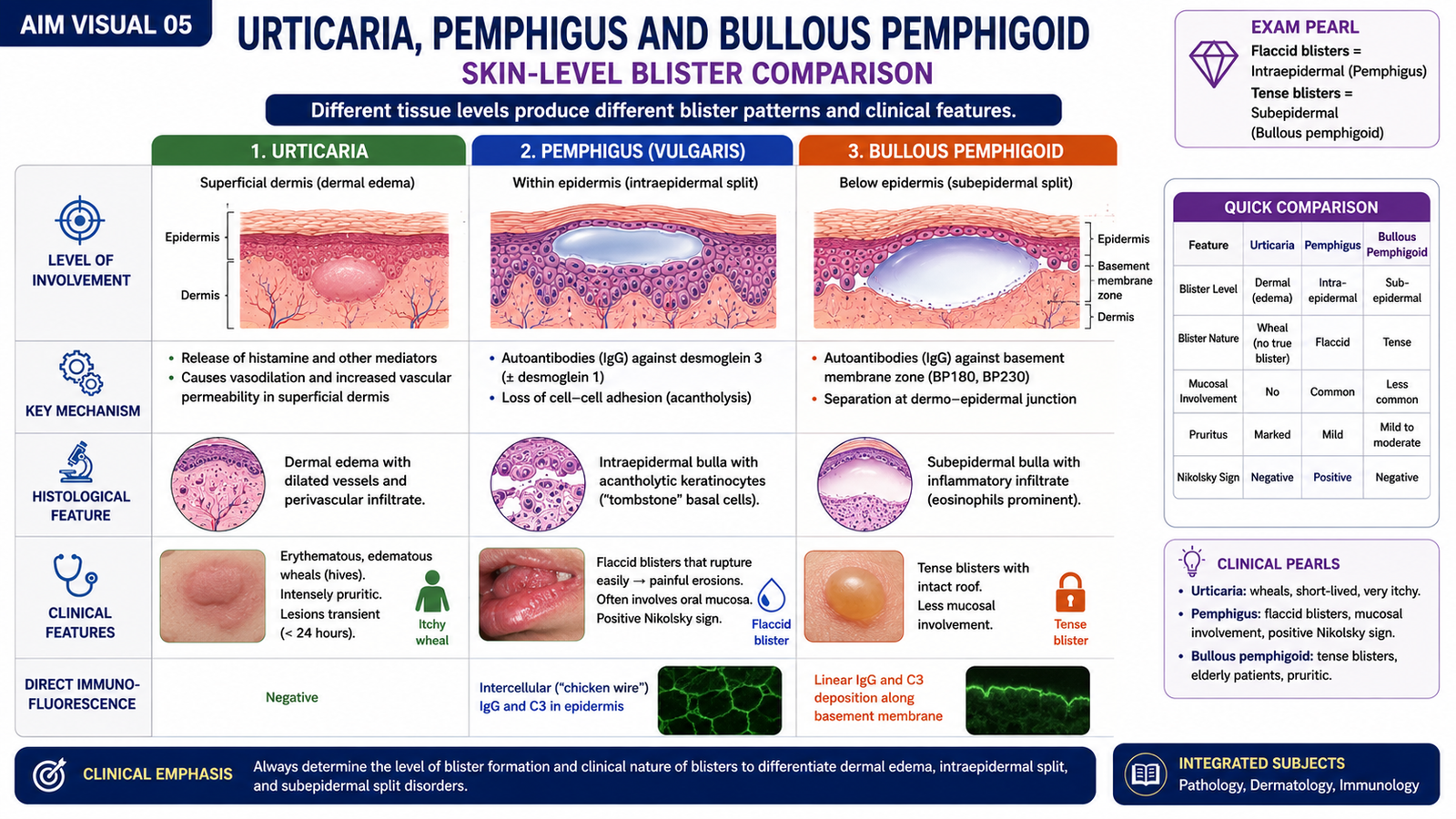
Urticaria, pemphigus and bullous pemphigoid are clinically different disorders, but each illustrates how the level and mechanism of tissue injury determine the appearance of a skin lesion. Urticaria is mainly caused by dermal edema, whereas pemphigus and bullous pemphigoid produce true blisters through separation at different levels of the epidermis.
Urticaria
Urticaria is characterized by transient, raised, erythematous and usually intensely pruritic wheals. It develops when mast-cell mediators increase vascular permeability in the superficial dermis. Plasma then moves into the tissue and produces localized dermal edema.
Pemphigus
Pemphigus is an autoimmune blistering disorder in which antibodies interfere with adhesion between epidermal keratinocytes. Loss of intercellular attachment is called acantholysis. Because the split occurs within the epidermis, the blister roof is relatively thin and fragile.
- Flaccid, easily ruptured blisters.
- Erosions may be more obvious than intact blisters.
- Mucosal involvement may occur.
- Histology demonstrates intraepidermal separation with acantholysis.
Bullous pemphigoid
Bullous pemphigoid is also an autoimmune blistering disorder, but the immune attack is directed against components involved in attachment of the epidermis to the underlying basement membrane zone. The epidermis therefore separates from the dermis, producing a subepidermal blister.
- Blisters are typically tense because the full epidermis forms the blister roof.
- The split is subepidermal rather than intraepidermal.
- The stronger blister roof makes lesions less fragile than those of pemphigus.
| Feature | Pemphigus | Bullous Pemphigoid |
|---|---|---|
| Main abnormality | Loss of keratinocyte adhesion | Loss of epidermal attachment to basement membrane zone |
| Level of split | Intraepidermal | Subepidermal |
| Acantholysis | Present | Absent |
| Blister character | Flaccid and fragile | Tense |
F. Warts
Warts are benign epidermal proliferations caused by infection with human papillomavirus. Viral infection stimulates proliferation and altered maturation of epidermal keratinocytes, producing a localized rough or papillomatous lesion. Their appearance therefore reflects both increased epidermal growth and surface keratinization.
Morphological features
- Well-defined papules or plaques with a rough or verrucous surface.
- Hyperkeratosis contributes to the thick surface.
- Acanthosis reflects epidermal thickening.
- Papillomatosis produces the characteristic undulating or finger-like surface configuration.
Clinically, warts may vary in shape and site, but their common underlying feature is a benign HPV-associated proliferation of epidermis.

G. Juvenile Dermatomyositis
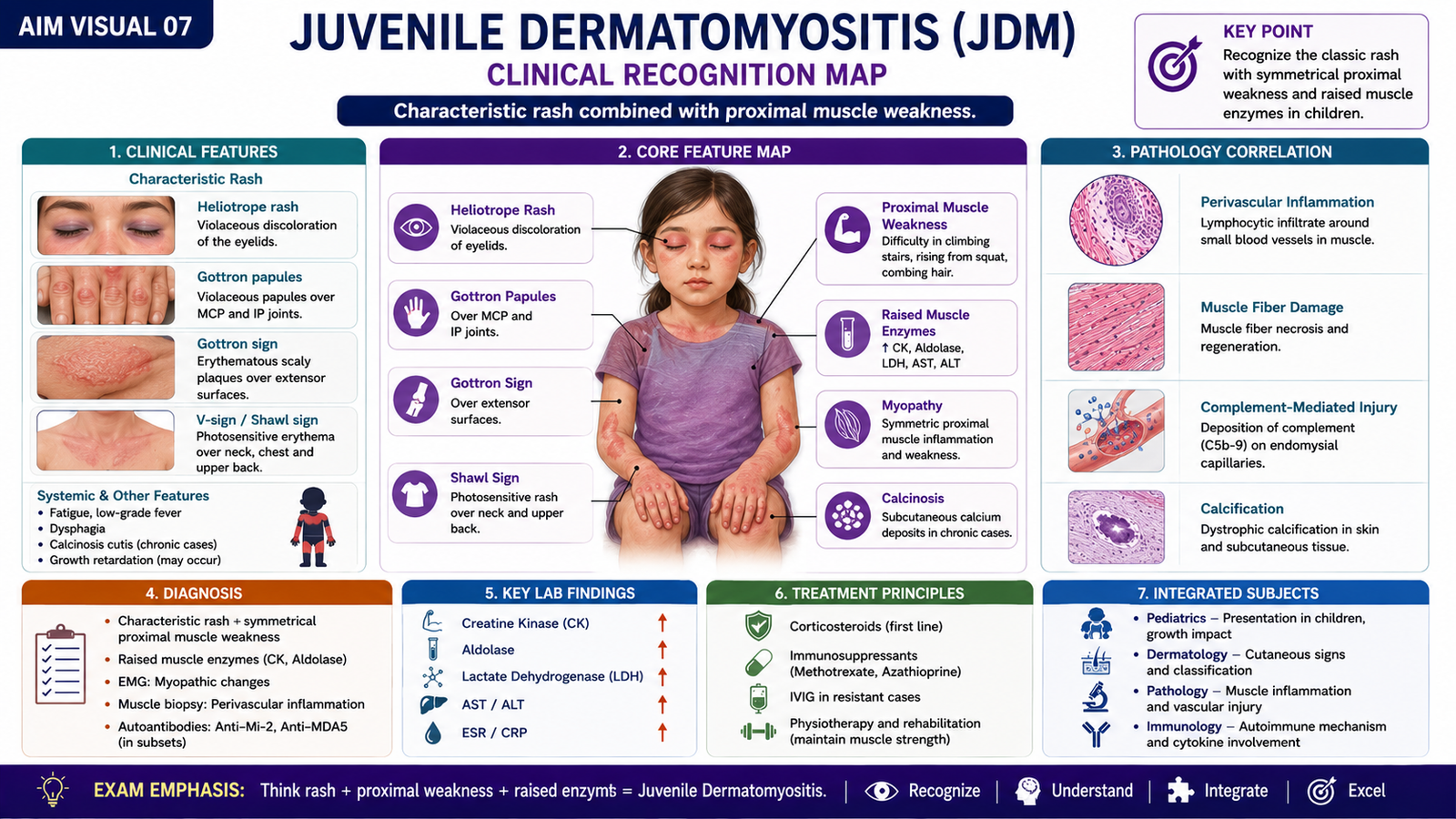
Juvenile dermatomyositis is an inflammatory disease of childhood involving both skeletal muscle and skin. Its key clinical pattern is a characteristic cutaneous eruption together with symmetrical proximal muscle weakness. Diagnosis is based on the combination of these clinical findings with evidence of muscle inflammation or injury.
Clinical features
Muscle involvement mainly affects proximal muscle groups. Children may therefore struggle with activities that require shoulder or hip strength.
- Symmetrical proximal muscle weakness.
- Difficulty climbing stairs, rising from a seated position or lifting the arms.
- Heliotrope rash: violaceous discoloration around the eyelids, often associated with periorbital swelling.
- Gottron papules: violaceous or erythematous papules over extensor surfaces of joints, particularly the knuckles.
- Other photosensitive erythematous eruptions may occur.
- Calcification within soft tissues may occur in juvenile disease.
Diagnostic criteria and supportive evidence
The diagnosis is supported when characteristic skin changes occur together with evidence of inflammatory myopathy. No single finding should be interpreted in isolation.
- Characteristic cutaneous manifestations such as heliotrope rash or Gottron papules.
- Symmetrical proximal muscle weakness.
- Raised serum muscle enzymes indicating muscle injury.
- Electrophysiological evidence compatible with myopathy when assessed.
- Muscle biopsy may demonstrate inflammatory muscle injury when required for diagnostic confirmation.
Differential diagnosis
The differential diagnosis depends on whether muscle weakness, skin findings or both dominate the presentation. The most useful distinction is whether the child has true inflammatory proximal muscle weakness together with the characteristic rash.
- Other inflammatory myopathies.
- Muscular dystrophies and other primary muscle disorders.
- Systemic autoimmune diseases with muscle or skin manifestations.
- Other dermatological conditions that mimic the rash without producing inflammatory myopathy.
Important Comparison — Common Cutaneous Reaction Patterns
| Condition | Key Process | Characteristic Morphology | Clinical Clue |
|---|---|---|---|
| Eczema | Spongiosis | Intercellular epidermal edema ± vesicles | Pruritic erythematous, oozing or scaly eruption |
| Psoriasis | Accelerated epidermal proliferation | Acanthosis + parakeratosis | Well-demarcated plaque with silvery scale |
| Pemphigus | Acantholysis | Intraepidermal blister | Fragile, flaccid blister |
| Bullous pemphigoid | Subepidermal separation | Subepidermal blister | Tense blister |
⭐ AIM High-Yield Review
- A macule is flat; a papule is solid and raised; a vesicle or bulla contains fluid.
- Spongiosis means intercellular epidermal edema and is the key microscopic feature of acute eczematous dermatitis.
- Repeated scratching in chronic eczema produces lichenification.
- Allergic contact dermatitis is immune mediated and requires sensitization; irritant dermatitis results from direct epidermal injury.
- ⭐ The classic lesion of erythema multiforme is the target lesion.
- Psoriasis results from immune-driven acceleration of epidermal proliferation and abnormal keratinocyte maturation.
- Acanthosis + parakeratosis + silvery scaling strongly support psoriasis.
- Urticaria is caused mainly by mediator-induced superficial dermal edema.
- Acantholysis is the loss of adhesion between keratinocytes and is characteristic of pemphigus.
- ⭐ Pemphigus produces an intraepidermal, fragile blister, whereas bullous pemphigoid produces a subepidermal, tense blister.
- Warts are HPV-associated benign epidermal proliferations showing hyperkeratosis, acanthosis and papillomatosis.
- In juvenile dermatomyositis, symmetrical proximal muscle weakness occurs with characteristic skin findings.
- Gottron papules and a heliotrope rash are particularly important diagnostic clues for juvenile dermatomyositis.
Topic 16 — Cutaneous Lesions, Eczematous Dermatitis, Psoriasis and Immune-Mediated Dermatoses
3rd Year MBBS • MSK Module • Pathology, Dermatology & Pediatrics
Focus on identifying the morphology of common skin lesions first, then connect these patterns with eczema, psoriasis, blistering disorders, warts and juvenile dermatomyositis.
▶️ Watch the complete explanation before attempting the related AIM MCQs and post-test.
🎯 What to Focus on During the Video
- Differentiate macules, papules, nodules, plaques, vesicles, bullae, pustules and scales.
- Understand hyperkeratosis, parakeratosis, acanthosis, dyskeratosis, acantholysis and papillomatosis.
- Recognize spongiosis as the key pathological change in acute eczematous dermatitis.
- Differentiate allergic contact dermatitis from primary irritant dermatitis.
- Recognize the classical target lesion of erythema multiforme.
- Connect accelerated epidermal turnover with the morphology of psoriasis.
- Differentiate pemphigus from bullous pemphigoid according to the level of blister formation.
- Relate HPV infection to the morphology of warts.
- Recognize proximal muscle weakness, Gottron papules and heliotrope rash in juvenile dermatomyositis.
Pay special attention to spongiosis in eczema, acanthosis and parakeratosis in psoriasis, acantholysis in pemphigus, subepidermal blistering in bullous pemphigoid, and the characteristic Gottron papules and heliotrope rash of juvenile dermatomyositis.