
📖 AIM Learning Material
Topic Introduction
Bone and cartilage tumours are best understood by linking four clues: the patient’s age, the exact part of the bone involved, the matrix produced by the lesion and its radiological behaviour. Most primary bone lesions are benign, while primary malignant bone tumours are uncommon but important because delayed recognition can allow local destruction or metastasis. This chapter explains classification, frequency, common clinical features, the morphology of major bone-forming and cartilage-forming tumours, Ewing sarcoma, giant-cell tumour and fibrous tumour-like lesions. It also shows how plain radiography, CT, MRI and planned biopsy are used to recognize and investigate suspicious lesions.
A. Classification, Frequency and General Recognition of Bone Tumours
Core Classification Principle
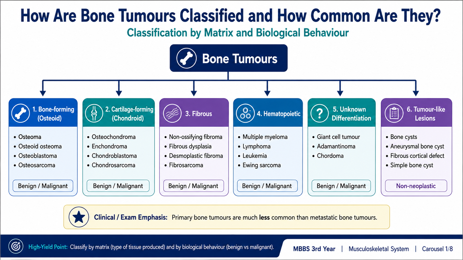
Bone tumours are primarily classified according to the type of tissue or matrix produced by the neoplastic cells. Classification must then be combined with biological behaviour, patient age and skeletal location. A lesion that produces osteoid belongs to the bone-forming group, while one producing cartilage matrix belongs to the cartilage-forming group.
| Tumour Group | Common Benign Examples | Important Malignant Examples | Defining Product or Pattern |
|---|---|---|---|
| Bone-forming | Osteoid osteoma, osteoblastoma | Osteosarcoma | Osteoid or immature bone |
| Cartilage-forming | Osteochondroma, enchondroma, chondroblastoma, chondromyxoid fibroma | Chondrosarcoma | Chondroid matrix |
| Fibrous | Fibrous cortical defect, non-ossifying fibroma | Fibrosarcoma of bone | Spindle cells and collagen |
| Hematopoietic | Uncommon | Plasma-cell myeloma, primary bone lymphoma | Marrow-derived cells |
| Unknown or uncertain differentiation | Giant-cell tumour is usually locally aggressive rather than simply benign | Ewing sarcoma | Distinct clinicomolecular and cellular patterns |
| Tumour-like lesions | Fibrous cortical defect, non-ossifying fibroma and related reactive or developmental lesions | Not true malignant neoplasms | May simulate neoplasia clinically or radiologically |
Frequency in the General Population
Primary bone tumours are much less common than metastatic deposits in bone. Among primary lesions, benign tumours and tumour-like lesions are encountered more frequently than primary malignant tumours. Osteochondroma is among the most common benign bone tumours, while osteosarcoma is the most common primary malignant bone-forming tumour. Chondrosarcoma is an important malignant cartilage-forming tumour and occurs mainly in adults. Ewing sarcoma is uncommon but is a major primary malignant bone tumour of children and adolescents.
Common Clinical Features
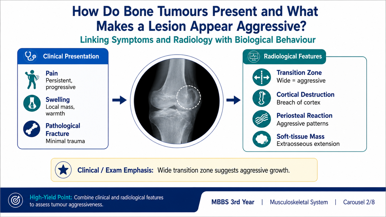
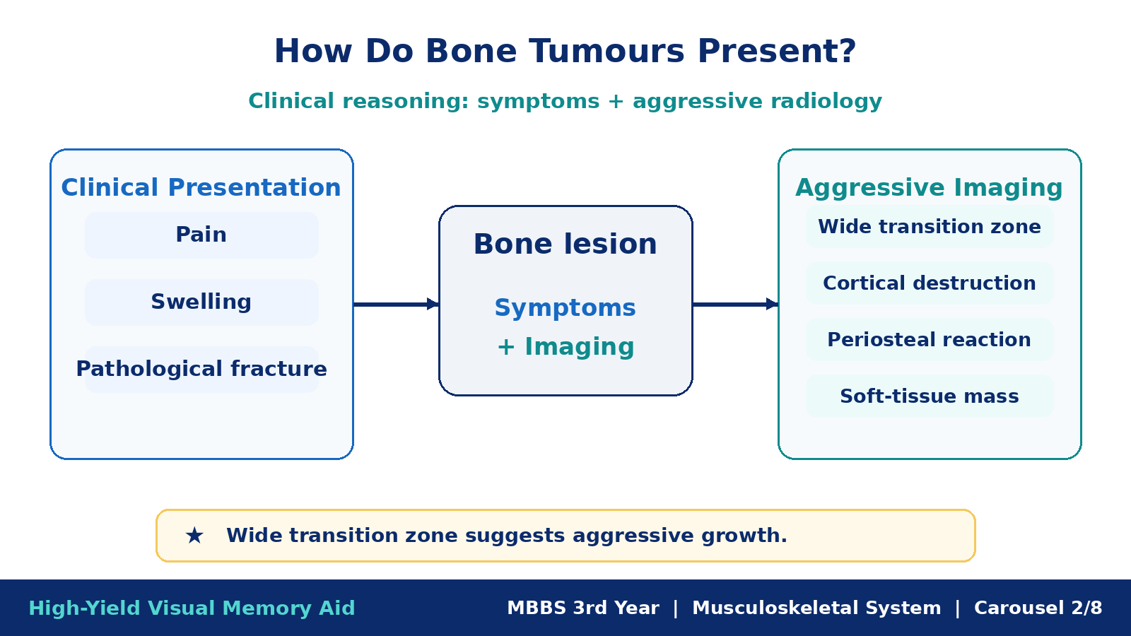
The clinical presentation reflects tumour growth, pressure within bone, periosteal irritation, cortical destruction and involvement of nearby soft tissue or joints. Small benign lesions may be asymptomatic and discovered incidentally, whereas aggressive tumours more commonly cause progressive symptoms.
- Pain: may result from expansion, microfracture, periosteal irritation or inflammatory mediator release.
- Swelling or palpable mass: suggests expansion beyond normal bone contour or soft-tissue extension.
- Restricted joint movement: occurs when a lesion lies near a joint or produces pain and mechanical limitation.
- Pathological fracture: develops when tumour-induced bone destruction weakens structural integrity.
- Neurological or vascular symptoms: may occur when an expanding mass compresses adjacent structures.
- Systemic manifestations: fever, elevated inflammatory markers or malaise may occur in Ewing sarcoma and can mimic infection.
Radiological Assessment of Biological Behaviour
| Feature | Usually Less Aggressive | Usually Aggressive |
|---|---|---|
| Margin | Well-defined, often sclerotic | Poorly defined |
| Zone of transition | Narrow | Wide |
| Cortex | Smooth expansion or thinning | Destruction or breakthrough |
| Periosteal reaction | Absent or solid | Interrupted, laminated, spiculated or elevated |
| Soft-tissue mass | Usually absent | May be present |
Radiology suggests biological behaviour but does not replace histopathological diagnosis. A benign-appearing lesion can occasionally be clinically important, while some inflammatory lesions may imitate malignancy.
B. Osteosarcoma: Pathogenesis, Morphology and Recognition
Definition and Core Concept
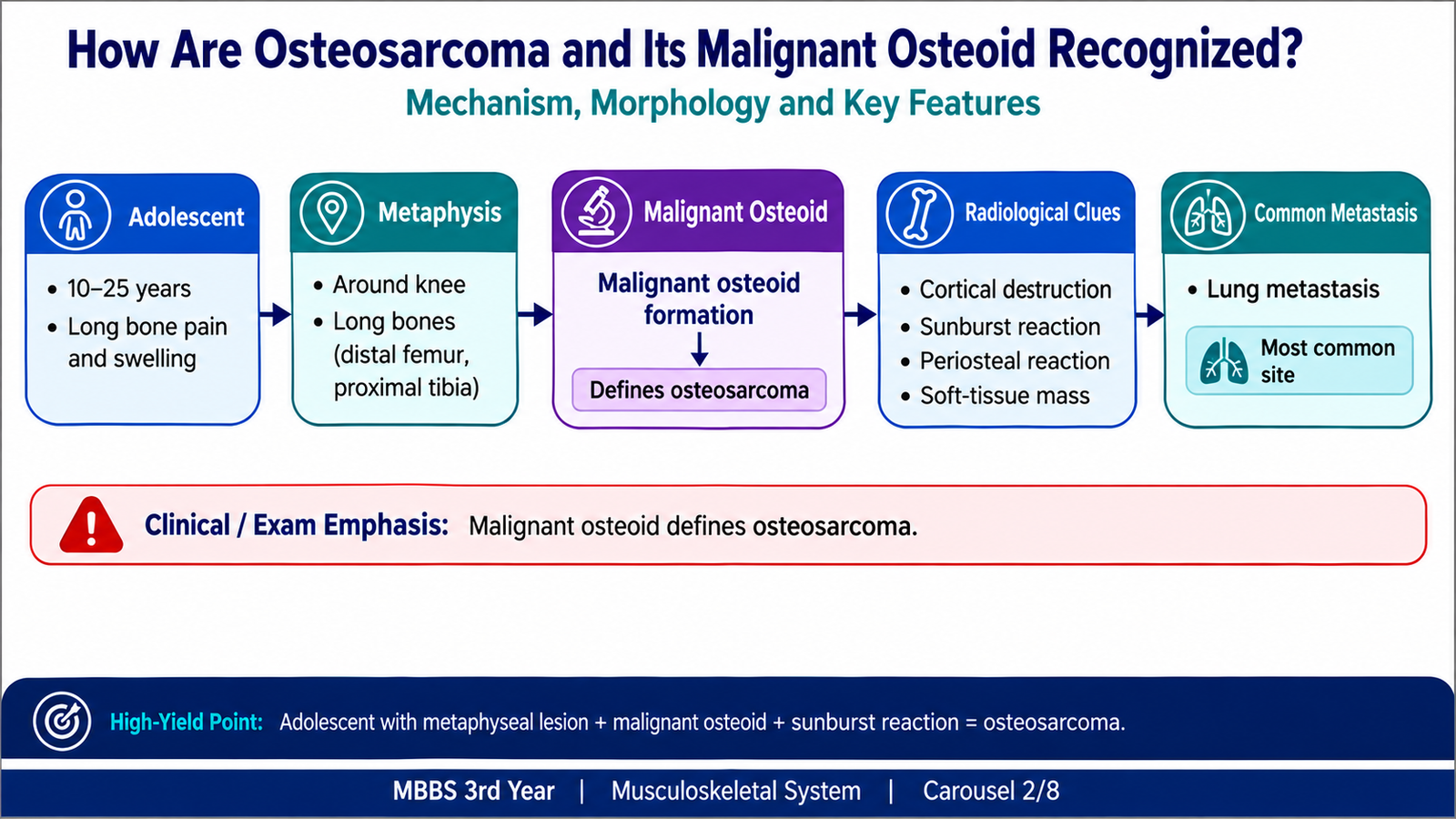
Osteosarcoma is a malignant mesenchymal tumour in which the neoplastic cells directly produce osteoid or immature bone. The presence of malignant osteoid is essential for diagnosis; radiological bone formation alone is not sufficient.
Frequency, Age and Site
- It is the most important primary malignant bone-forming tumour.
- Conventional primary osteosarcoma most often affects adolescents and young adults.
- It commonly arises in the metaphysis of long bones.
- The region around the knee, especially the distal femur and proximal tibia, is a classic location.
- A second age peak may occur in older adults, often in association with pre-existing bone abnormalities.
Etiology and Pathogenesis
Many cases are sporadic. Molecular abnormalities affecting tumour-suppressor pathways permit primitive osteogenic cells to proliferate without normal control. Important associations include hereditary abnormalities involving the retinoblastoma pathway, Li-Fraumeni syndrome, Paget disease of bone and prior irradiation.
Gross Morphology
- Large, bulky, destructive mass arising within the metaphysis.
- Cut surface is often gritty, grey-white and variegated.
- Hemorrhage, necrosis and cystic change may be present.
- The tumour may destroy the cortex and form an extraosseous soft-tissue mass.
- Joint cartilage may temporarily resist direct penetration, although nearby structures can be extensively involved.
Microscopic Morphology
- Markedly pleomorphic malignant stromal cells.
- Hyperchromatic nuclei and atypical mitotic figures.
- Irregular, lace-like osteoid deposited directly by malignant cells.
- Variable areas of cartilage, fibrous tissue, hemorrhage and necrosis may coexist.
Clinical and Radiological Correlation
Pain and progressive swelling result from rapidly expanding tumour, cortical destruction and periosteal irritation. Radiographs commonly show a mixed lytic and sclerotic metaphyseal lesion. Tumour osteoid may create a dense cloud-like pattern. Perpendicular new bone formation can produce a sunburst-like appearance, while periosteal elevation may form a triangular shadow at the tumour margin.
C. Osteoid Osteoma and Osteoblastoma
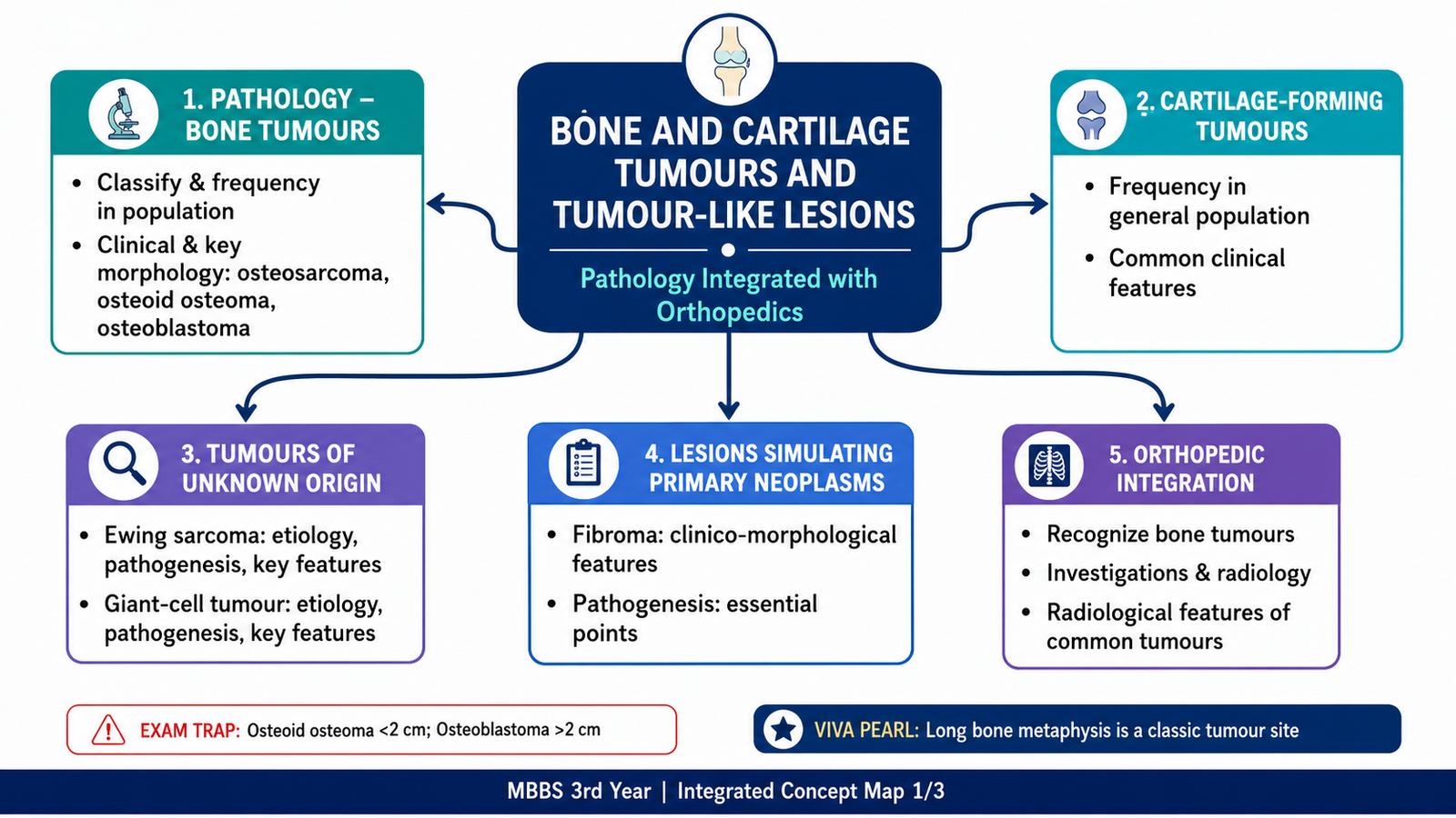
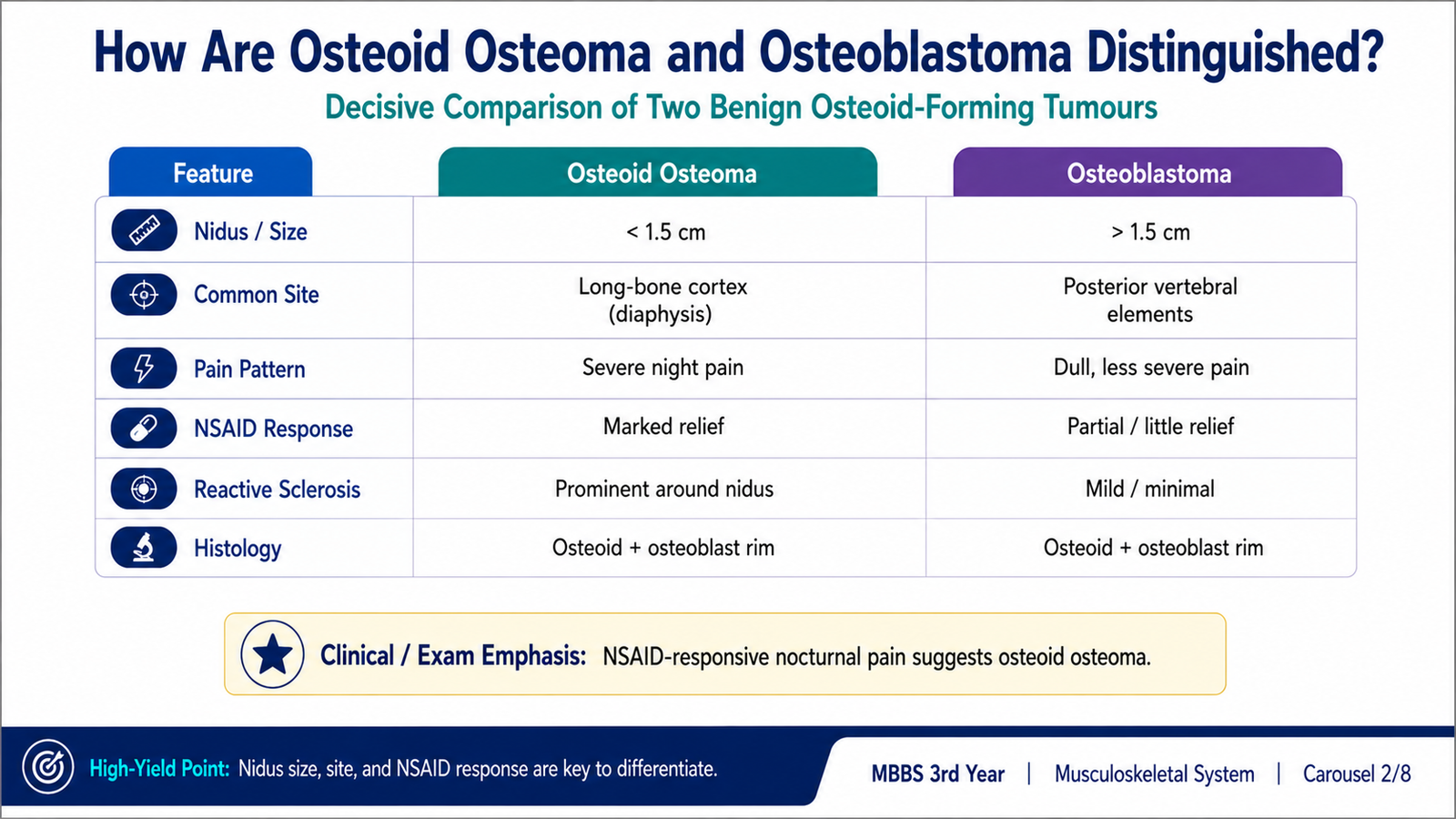
Osteoid osteoma and osteoblastoma are benign bone-forming tumours composed of osteoid and woven bone rimmed by osteoblasts. Their histological appearance can overlap, so clinical behaviour, size, site and radiological pattern are essential for distinction.
| Feature | Osteoid Osteoma | Osteoblastoma |
|---|---|---|
| Usual patient | Children and young adults | Adolescents and young adults |
| Typical site | Cortex of long bones | Posterior elements of vertebrae and long bones |
| Size | Small nidus, classically less than about 2 cm | Usually larger |
| Pain pattern | Severe nocturnal pain, characteristically relieved by NSAIDs | Pain may be less responsive to NSAIDs |
| Radiology | Small radiolucent nidus with prominent surrounding reactive sclerosis | Larger expansile, mixed or lucent lesion with less striking sclerosis |
| Histology | Interlacing woven bone and osteoid rimmed by osteoblasts in vascular stroma | Similar pattern, but larger and sometimes more locally aggressive |
Why Osteoid Osteoma Causes Characteristic Pain
The nidus produces high levels of prostaglandins, which stimulate pain-sensitive nerve endings and contribute to intense nocturnal pain. Cyclooxygenase inhibition explains the characteristic relief obtained with NSAIDs.
Gross and Microscopic Features
Both lesions contain gritty red-brown tissue composed of woven bone and osteoid trabeculae lined by prominent osteoblasts. The intervening stroma is loose and richly vascular. The osteoblasts are cytologically bland, distinguishing these lesions from osteosarcoma.
D. Common Cartilage-Forming Tumours
Frequency and General Clinical Pattern
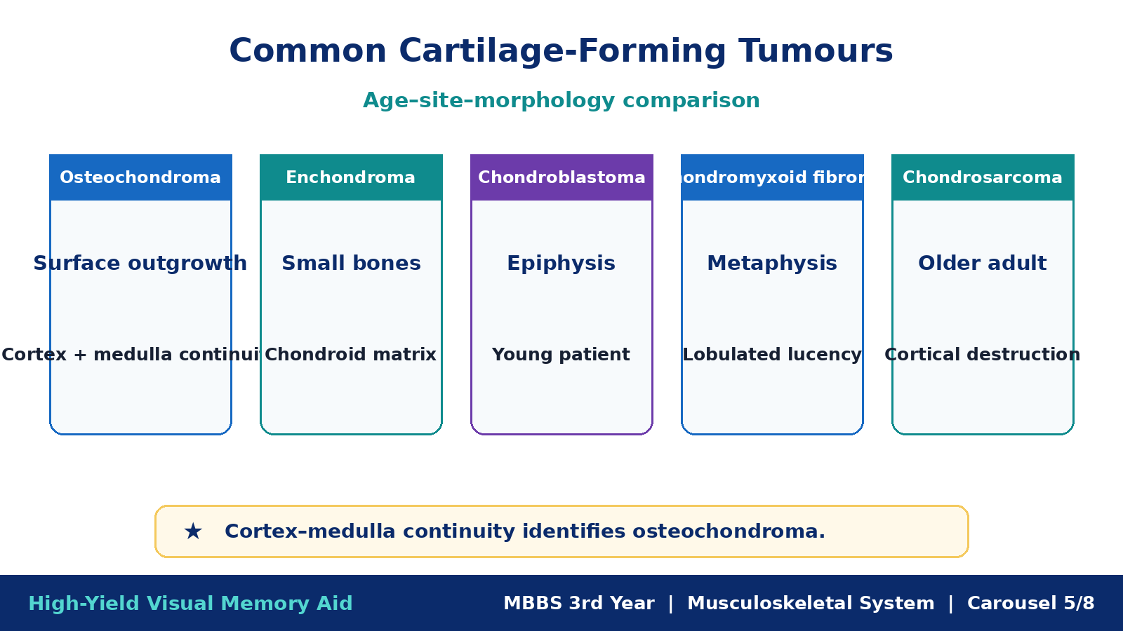
Cartilage-forming tumours range from common benign exostoses and intramedullary lesions to malignant chondrosarcoma. Benign cartilaginous tumours are more frequent than primary malignant cartilage tumours. Their presentation varies with location: some are incidental, while others cause deformity, pain, restricted movement or pathological fracture.
| Tumour | Typical Age and Site | Clinical Clue | Radiological or Morphological Clue |
|---|---|---|---|
| Osteochondroma | Usually develops during skeletal growth; metaphysis near growth plates | Painless bony mass, deformity or mechanical symptoms | Cartilage-capped bony projection with cortex and medulla continuous with the parent bone |
| Enchondroma | Often young adults; medullary cavity, especially small bones of hands and feet | Often incidental or presents with pathological fracture | Well-circumscribed intramedullary cartilaginous lesion with chondroid calcification |
| Chondroblastoma | Adolescents and young adults; epiphysis | Joint pain, stiffness and localized tenderness | Chondroblasts, giant cells and delicate “chicken-wire” calcification |
| Chondromyxoid fibroma | Young patients; metaphysis of long bones | Pain and swelling | Lobulated lesion containing chondroid, myxoid and fibrous tissue |
| Chondrosarcoma | Mainly adults; pelvis, shoulder girdle, ribs and proximal long bones | Progressive deep pain and enlarging mass | Destructive cartilage-producing tumour with lobulated growth and ring-and-arc-type calcification |
Morphological Logic
Benign cartilaginous tumours usually show orderly lobules of hyaline cartilage with relatively uniform chondrocytes. Features suggesting malignancy include permeative growth, cortical destruction, soft-tissue extension, increasing cellularity, nuclear atypia and entrapment of pre-existing bone. Pain in a previously stable cartilage lesion, renewed growth after skeletal maturity or increasing cartilage-cap thickness in an osteochondroma should raise concern for malignant transformation and prompt further assessment.

E. Ewing Sarcoma
Definition and Etiology
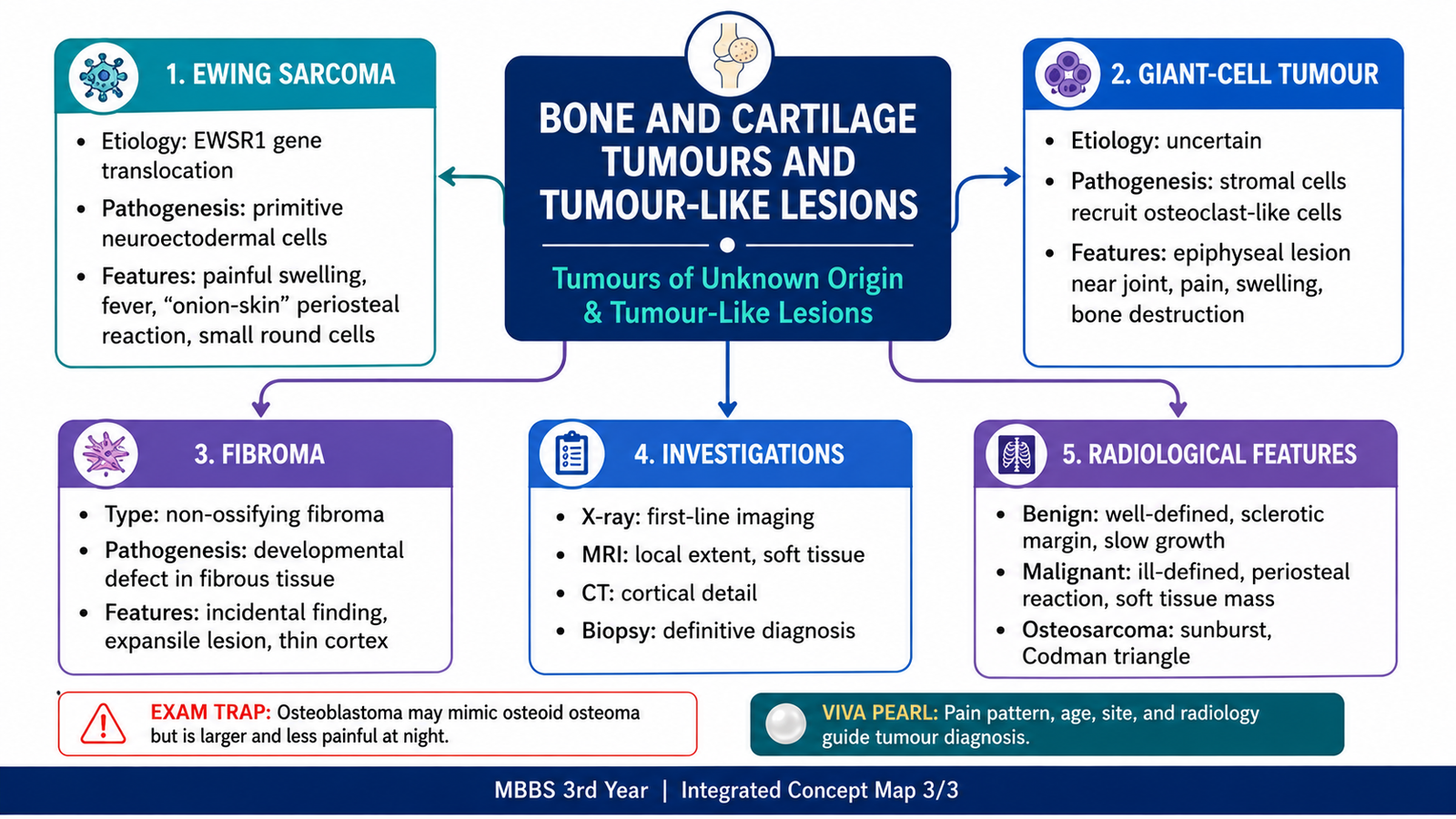
Ewing sarcoma is a highly malignant small round-cell tumour of bone and soft tissue, most often affecting children and adolescents. It is associated with a characteristic chromosomal translocation involving the EWSR1 gene, most commonly producing an EWS–FLI1 fusion transcription factor.
Pathogenesis
Clinical Features
- Localized pain, swelling and tenderness.
- Frequently involves the diaphysis or metadiaphysis of long bones and flat bones.
- Fever, malaise, leukocytosis and raised inflammatory markers may be present.
- The inflammatory presentation may resemble osteomyelitis.
- Metastasis may involve lungs, bone and bone marrow.
Morphology
Grossly, the tumour is soft, grey-white and often contains hemorrhage and necrosis. Microscopically, it consists of uniform small round cells arranged in sheets. The cells have scant clear or pale cytoplasm that contains glycogen, round nuclei and inconspicuous nucleoli. Necrosis may be extensive.
Radiology and Diagnostic Correlation
Radiographs may show a destructive permeative lesion with layered periosteal new bone, often described as an onion-skin pattern. A large soft-tissue component may be present. Diagnosis requires biopsy with histopathology and supportive immunohistochemical and molecular testing.
F. Giant-Cell Tumour of Bone
Definition and Usual Setting
Giant-cell tumour of bone is a locally aggressive neoplasm usually occurring in skeletally mature young adults. It characteristically arises in the epiphysis of a long bone and commonly extends toward the articular surface. The region around the knee is frequently involved.
Pathogenesis
The true neoplastic component is the mononuclear stromal cell population, not the multinucleated giant cells. Stromal cells express signals that recruit and activate osteoclast-type giant cells, producing marked bone resorption.
Clinical and Radiological Features
- Localized pain, swelling and reduced movement near a joint.
- Usually occurs after closure of the growth plate.
- Radiographs show an eccentric, expansile, purely lytic epiphyseal lesion.
- The lesion may extend to subarticular bone and produce a soap-bubble appearance.
- Cortical thinning, cortical breach and soft-tissue extension may occur.
Gross and Microscopic Morphology
Grossly, the lesion is soft, red-brown and may contain areas of hemorrhage, necrosis and cystic change. Microscopically, numerous osteoclast-type multinucleated giant cells are distributed relatively evenly among mononuclear stromal cells. The nuclei of the giant cells resemble those of the stromal cells.
Outcome and Complications
Giant-cell tumour can recur after local treatment because of its infiltrative local behaviour. Although histologically benign in many cases, it can rarely metastasize to the lungs or undergo malignant transformation. Its classification therefore requires more nuance than the simple label “benign.”
G. Fibrous Tumour-Like Lesions and Investigation of Bone Tumours
Fibrous Cortical Defect and Non-Ossifying Fibroma
In the context of lesions simulating primary neoplasms, “fibroma” commonly refers to fibrous cortical defect and its larger counterpart, non-ossifying fibroma. These are developmental or reactive fibrous lesions rather than true aggressive neoplasms. They occur mainly in children and adolescents and are frequently discovered incidentally.
Essential Pathogenesis
The lesion is thought to represent a localized disturbance in bone development in which normal cortical bone is replaced by fibrous tissue containing fibroblasts, histiocytes and giant cells. Most lesions stabilize and regress as skeletal growth proceeds.
Clinico-Morphological Features
- Usually affects the metaphysis of long bones in children or adolescents.
- Most lesions are asymptomatic.
- Radiographs show an eccentric, well-demarcated, radiolucent cortical lesion with a sclerotic rim.
- Larger lesions may weaken bone and predispose to pathological fracture.
- Microscopy shows spindle fibroblasts in a storiform pattern with histiocytes, foam cells and scattered giant cells.
Orthopedic Diagnostic Pathway
Why Biopsy Planning Matters
A poorly placed biopsy tract may contaminate uninvolved tissue compartments and complicate limb-sparing surgery. Imaging should therefore precede biopsy, and the biopsy route should be selected so that the tract can be removed during definitive surgery.
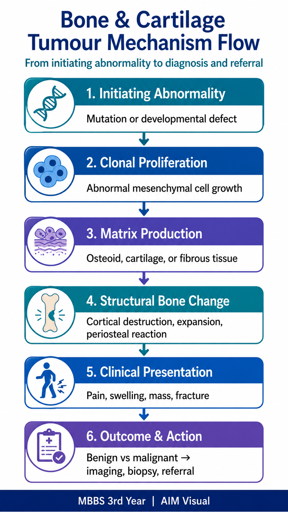
Integrated Mechanism Flow
AIM High-Yield Review
🎥 AIM Recommended Video
Watch this short overview after completing the chapter. Focus on how patient age, tumour location, radiological appearance and tumour type are combined to recognize common benign and malignant bone tumours.