
📖 AIM Learning Material
Topic Introduction
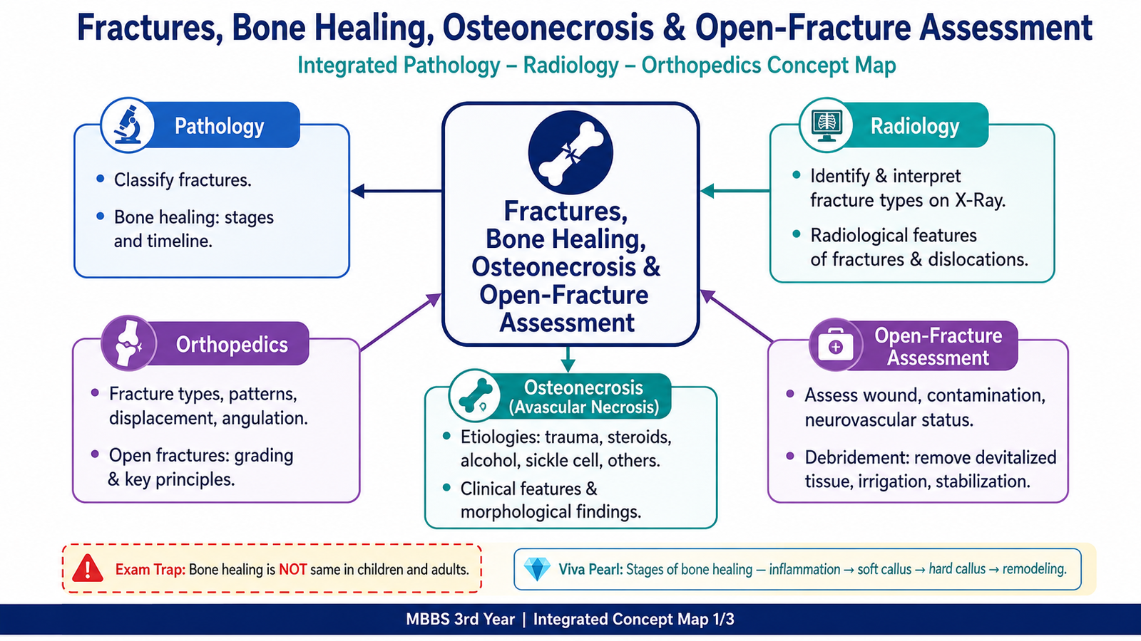
A fracture is a complete or incomplete break in the continuity of bone. Understanding fractures requires more than recognizing a fracture line: the student must be able to classify the injury, describe displacement and angulation, examine the patient safely and interpret the X-ray systematically. Bone usually repairs itself through an organized sequence of inflammation, callus formation and remodeling, but healing may fail when blood supply, stability or tissue viability is poor. Open fractures are especially important because the wound communicates with the fracture and introduces contamination. This chapter also explains osteonecrosis, in which loss of blood supply causes death of bone and marrow and may eventually lead to collapse of the affected joint surface.
Core Learning Material
A. Fracture Definition and Classification
Definition and core concept
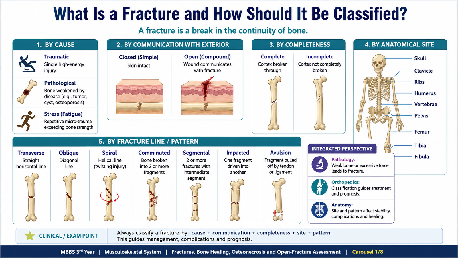
A fracture is a complete or incomplete interruption in the continuity of a bone or its cartilage. The break may follow a single major force, repeated submaximal stress or weakening of bone by an underlying disease. Classification is clinically important because it predicts stability, soft-tissue damage, contamination, healing potential and possible complications.
Classification by cause
- Traumatic fracture: normal bone breaks after force exceeding its strength.
- Pathological fracture: diseased or structurally weakened bone breaks after minor or normal physiological force.
- Stress fracture: repetitive loading causes accumulated microdamage.
- Insufficiency fracture: normal loading acts on bone with reduced mechanical strength.
Classification by communication with the exterior
- Closed fracture: no communication between the fracture and external environment.
- Open fracture: a wound communicates with the fracture or fracture hematoma, even when the wound is small or distant from the apparent fracture line.
Classification by completeness and pattern
| Pattern | Description | Usual mechanical implication |
|---|---|---|
| Transverse | Fracture line approximately perpendicular to the long axis. | Often produced by direct force or bending. |
| Oblique | Diagonal fracture line across the bone. | May shear and shorten under loading. |
| Spiral | Fracture line winds around the shaft. | Suggests a torsional mechanism. |
| Comminuted | Bone is broken into more than two fragments. | Usually reflects greater energy and instability. |
| Segmental | Two separate fracture levels isolate a bone segment. | Blood supply to the isolated segment may be compromised. |
| Impacted | One fragment is driven into another. | May appear shortened and relatively stable. |
| Avulsion | A tendon or ligament pulls away a bone fragment. | Traction force at an attachment site. |
| Compression | Trabecular bone collapses under axial loading. | Commonly recognized in vertebral bodies. |
| Depressed | A bone fragment is displaced inward. | Classically described in flat bones such as the skull. |
Classification by anatomical site
- Epiphyseal, physeal, metaphyseal or diaphyseal.
- Intra-articular or extra-articular.
- Proximal, middle or distal part of the bone.
B. Clinical Assessment and Systematic X-Ray Interpretation
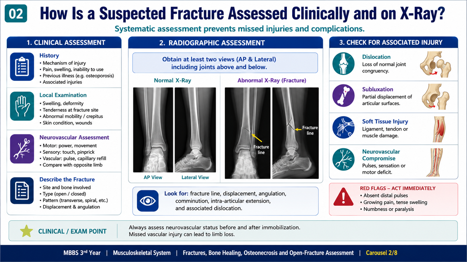
Focused musculoskeletal history
History should establish what happened, what force acted on the limb and whether the patient had normal bone before the event. The mechanism often predicts the fracture pattern and associated soft-tissue injury.
- Time and mechanism of injury: direct blow, fall, twisting, crush or high-energy collision.
- Site, severity and onset of pain; inability to use or bear weight.
- Audible crack, deformity, bleeding or visible wound.
- Numbness, weakness, coldness or altered color distal to the injury.
- Previous fractures, bone disease, malignancy, infection or prolonged medicine exposure affecting bone.
- In children, ask about the exact mechanism and whether it is developmentally plausible.
Focused examination
Examination begins with the patient’s general condition and proceeds to the injured region without repeatedly manipulating an unstable limb.
Systematic X-ray interpretation
Radiographs should usually include at least two orthogonal views. The image should include the relevant joints above and below when a long-bone injury is assessed. A systematic approach reduces missed fractures and prevents vague descriptions.
- Confirm: patient, side, date, view and image quality.
- Identify the bone and exact location: proximal, shaft or distal; epiphyseal, metaphyseal or diaphyseal.
- Describe completeness and pattern: transverse, oblique, spiral, comminuted or other recognized pattern.
- Describe displacement: direction of the distal fragment relative to the proximal fragment.
- Describe angulation: direction in which the apex points.
- Assess shortening, distraction, rotation and impaction.
- Examine cortex and trabeculae: follow each cortical margin continuously.
- Assess joints and growth plates: alignment, congruity and physeal involvement.
- Inspect soft tissues: swelling, gas or foreign material.
Normal X-ray versus structural deformity
| Feature | Normal | Abnormal clue |
|---|---|---|
| Cortex | Smooth and continuous. | Break, step, buckle or depression. |
| Trabeculae | Expected continuous internal pattern. | Disruption, compression or abnormal lucency. |
| Alignment | Anatomical axes and joint relationships preserved. | Translation, angulation, shortening or rotation. |
| Joint | Congruent articular surfaces. | Loss of congruity, widening or associated fracture. |
| Soft tissue | No unexpected swelling or gas. | Swelling, gas, foreign body or displaced fat plane. |
Radiological characteristics of dislocation
A dislocation produces complete loss of normal joint congruity. The examiner should identify the displaced bone, describe its direction relative to the adjoining bone and search carefully for an associated fracture. In subluxation, some articular contact remains. Comparison of joint alignment on both views is essential because a dislocation may be overlooked on a single projection.
C. Fracture Patterns in Children and Adults
Why fracture behavior differs
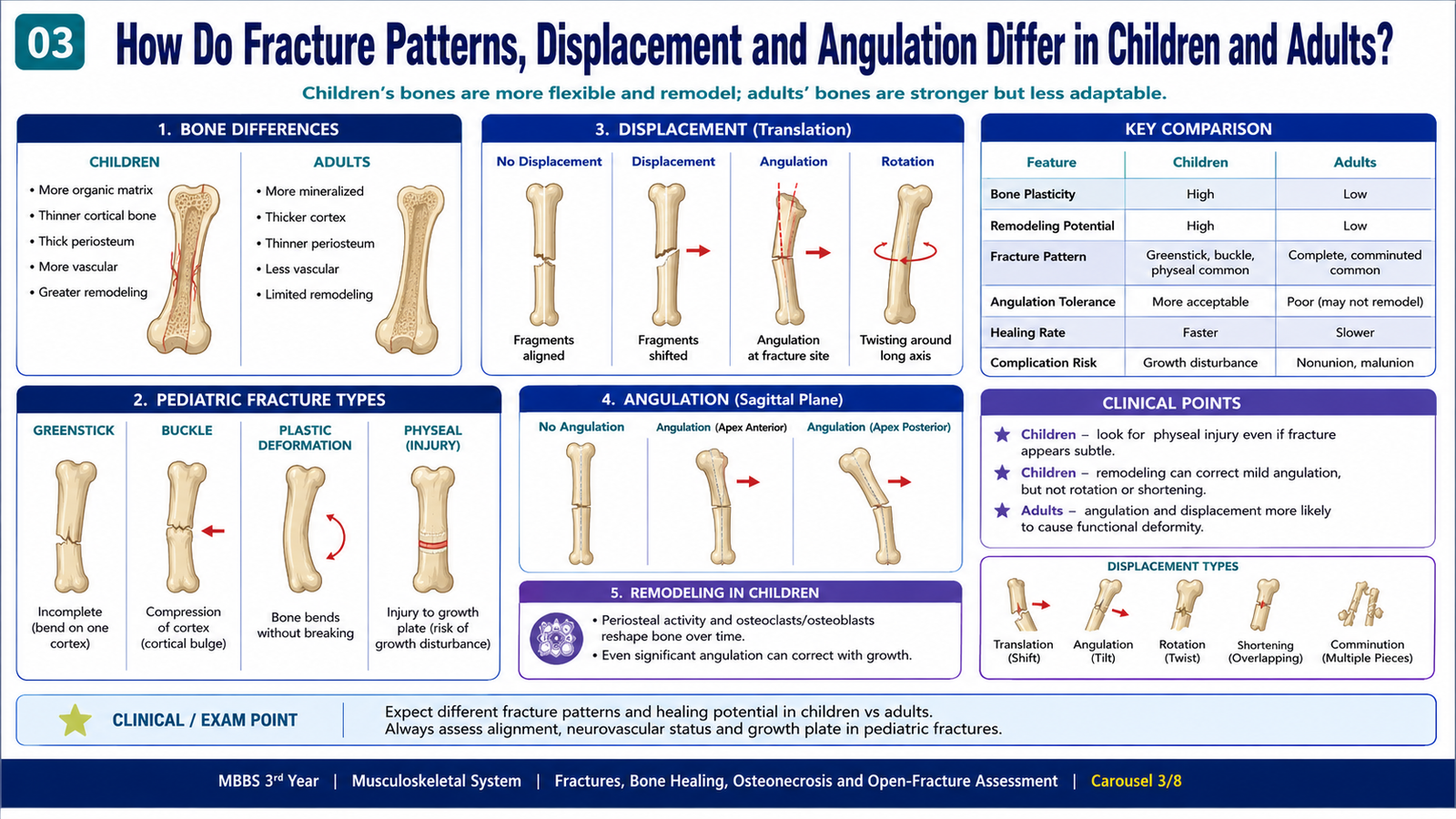
Adult bone is relatively rigid and more likely to fail completely once its elastic limit is exceeded. Pediatric bone is more porous and flexible, and its periosteum is thicker and biologically active. Consequently, children may sustain incomplete fractures and often retain a periosteal hinge that limits displacement.
Important pediatric patterns
- Greenstick fracture: one cortex breaks while the opposite cortex bends; angulation occurs around the intact periosteal hinge.
- Torus or buckle fracture: cortical compression causes localized bulging without a complete fracture line.
- Plastic deformation: bone bends beyond its elastic limit without an obvious complete cortical break.
- Physeal fracture: injury extends through or near the growth plate and may affect future growth.
Displacement and angulation
Muscle pull, gravity, the direction of injury and the stability of the periosteum determine the final position of fragments. Displacement may be described as translation, shortening or distraction. Angulation describes deviation between the axes of the fragments. Rotation is particularly important because it may not remodel adequately and can produce persistent functional malalignment.
| Feature | Children | Adults |
|---|---|---|
| Bone behavior | More flexible; incomplete failure is common. | More rigid; complete fracture is more common. |
| Periosteum | Thick and may remain partly intact. | Thinner and less likely to act as a stable hinge. |
| Characteristic patterns | Greenstick, buckle, plastic deformation and physeal injury. | Complete transverse, oblique, spiral and comminuted patterns. |
| Remodeling | Greater potential, especially near active growth plates and in the plane of joint motion. | Limited remodeling; accurate alignment is generally more important. |
| Special concern | Growth-plate injury and growth disturbance. | Comorbidity, poor bone quality and reduced healing reserve. |
D. Fracture Healing
Core pathological response
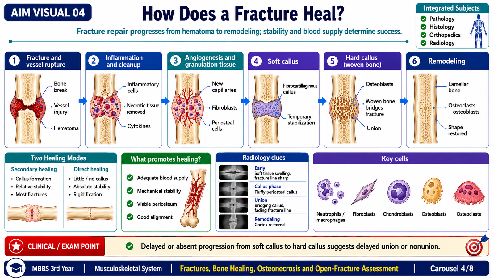
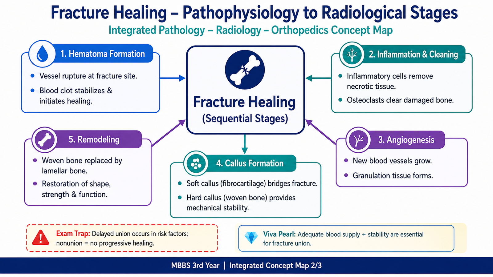
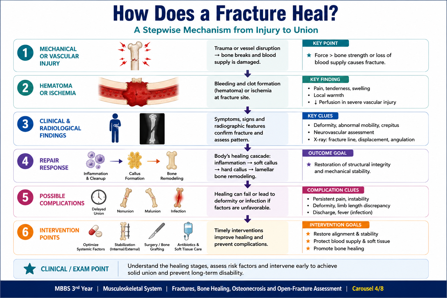
Fracture healing is a coordinated regenerative process involving hematoma formation, inflammation, removal of damaged tissue, production of a temporary stabilizing callus, formation of woven bone and eventual remodeling into mechanically organized lamellar bone. Successful repair requires adequate blood supply, viable osteogenic cells and sufficient mechanical stability.
Secondary or indirect healing
Secondary healing is the usual pattern when relative stability permits limited controlled movement and callus formation.
Primary or direct healing
Direct healing occurs when fracture ends are closely apposed and held with near-absolute stability. There is little visible external callus. Osteoclast-led cutting cones cross the fracture line, followed by vascular channels and osteoblasts that form new lamellar bone.
Radiological progression
- The initial fracture line may be sharply visible.
- Early resorption at fragment margins can temporarily make the line more apparent.
- Callus gradually appears and bridges the fracture.
- The fracture line becomes less distinct as union progresses.
- Remodeling restores a more normal contour over time.
E. Problems and Complications of Fracture Healing
Local and systemic determinants
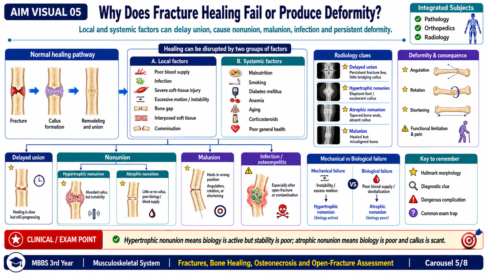
Healing depends on biology and mechanics. A well-vascularized fracture with viable tissues and appropriate stability can form bridging bone. Severe vascular injury, infection, loss of tissue or excessive movement disrupts the repair sequence. Conversely, extreme rigidity with a persistent gap may also prevent effective bridging.
- Poor blood supply or extensive periosteal stripping
- Severe soft-tissue damage
- Infection
- Large fracture gap or bone loss
- Interposition of soft tissue
- Excessive movement or inadequate stabilization
- Highly comminuted or open injury
- Poor nutritional state
- Advanced age or impaired biological reserve
- Metabolic or systemic disease affecting bone repair
- Smoking
- Medicines or exposures that impair bone formation or vascularity
Major outcomes
| Outcome | Meaning | Clinical or radiological clue |
|---|---|---|
| Delayed union | Healing progresses more slowly than expected but retains the potential to unite. | Persistent fracture line with incomplete bridging over an unexpectedly prolonged course. |
| Nonunion | The fracture fails to unite without further intervention. | Persistent mobility, pain and absent effective bony continuity. |
| Malunion | The fracture unites in an unsatisfactory position. | Residual angulation, rotation, translation or shortening. |
| Infection-related failure | Microbial contamination and tissue necrosis interfere with repair. | Persistent wound drainage, systemic or local inflammatory findings and destructive bone change. |
Hypertrophic versus atrophic nonunion
Hypertrophic nonunion shows biological activity and abundant callus, suggesting that instability is the major problem. Atrophic nonunion shows little callus and poor biological activity, commonly reflecting impaired blood supply, tissue loss or loss of viable bone.
F. Open Fractures: Assessment and Risks
Definition
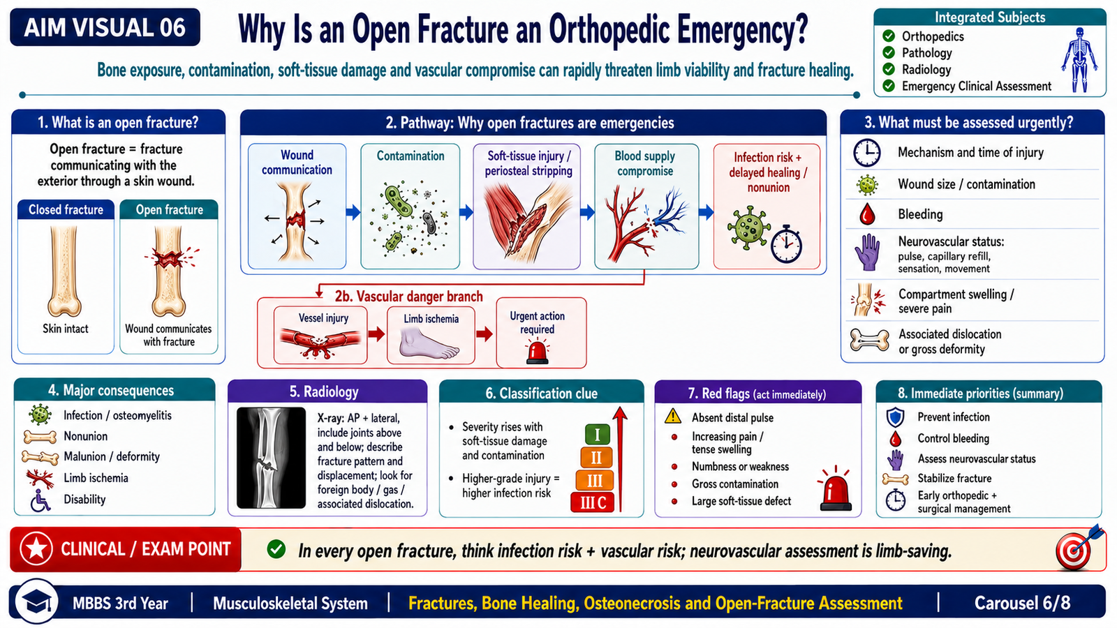
An open fracture is a fracture associated with a wound that communicates with the fracture, fracture hematoma or surrounding deep tissues. The wound may be created by an external object entering the limb or by a sharp bone fragment piercing the skin from within.
Why the injury is dangerous
The severity of an open fracture depends not only on wound size but also on the energy of injury, contamination, soft-tissue stripping, vascular damage, bone loss and the ability to achieve durable coverage.
Basic severity framework
The commonly taught Gustilo–Anderson framework groups open fractures by increasing wound severity, contamination, comminution and soft-tissue or vascular injury. It is most reliable after operative inspection rather than from a brief initial view alone.
| General category | Typical principle | Increasing concern |
|---|---|---|
| Lower-grade injury | Relatively limited wound and soft-tissue damage. | Contamination and communication still require urgent care. |
| Intermediate injury | Larger wound with greater soft-tissue trauma. | Higher infection and healing risk. |
| Severe high-energy injury | Extensive contamination, comminution, soft-tissue loss or vascular injury. | Complex reconstruction, infection, nonunion and limb-threatening ischemia. |
Initial assessment priorities
- Address life-threatening injuries before focusing exclusively on the limb.
- Inspect the wound without repeated probing or unnecessary manipulation.
- Record contamination, visible tissue damage and possible bone exposure.
- Document distal pulses, capillary refill, temperature, sensation and motor function.
- Cover the wound with an appropriate sterile dressing and immobilize the limb.
- Arrange urgent orthopedic assessment for debridement, stabilization and soft-tissue planning.
- Use appropriate infection-prevention measures and assess tetanus protection according to clinical protocol.
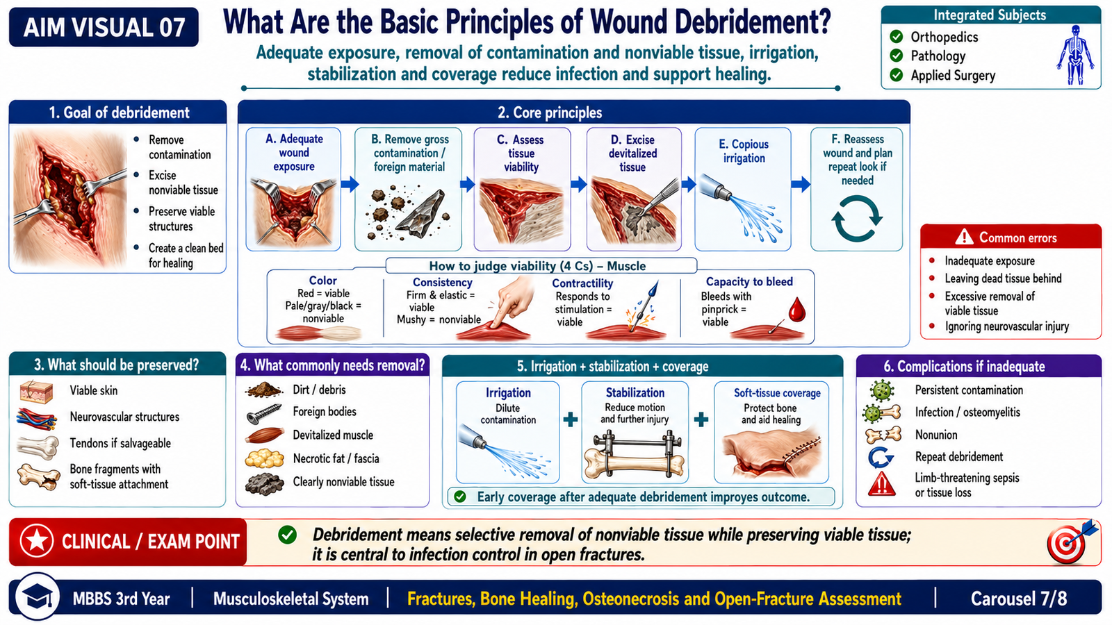
G. Basic Principles of Wound Debridement
Purpose
Debridement converts a contaminated traumatic wound into the cleanest and most biologically viable wound reasonably achievable. Its purpose is to remove contamination and nonviable tissue while preserving structures that can survive and contribute to function, healing and reconstruction.
Core principles
Assessment of tissue viability
Muscle viability is judged from its appearance, consistency, ability to contract and capacity to bleed when appropriately assessed. Skin, subcutaneous tissue, fascia, tendon, bone and neurovascular structures are evaluated separately. The decision is biological and functional rather than based on wound appearance alone.
Relationship to stabilization and coverage
Debridement is not an isolated act. The wound, fracture stability, blood supply and soft-tissue coverage must be considered together. Stable bone supports healing, while viable vascularized coverage protects the fracture from further contamination and tissue desiccation.

H. Osteonecrosis (Avascular Necrosis)
Definition
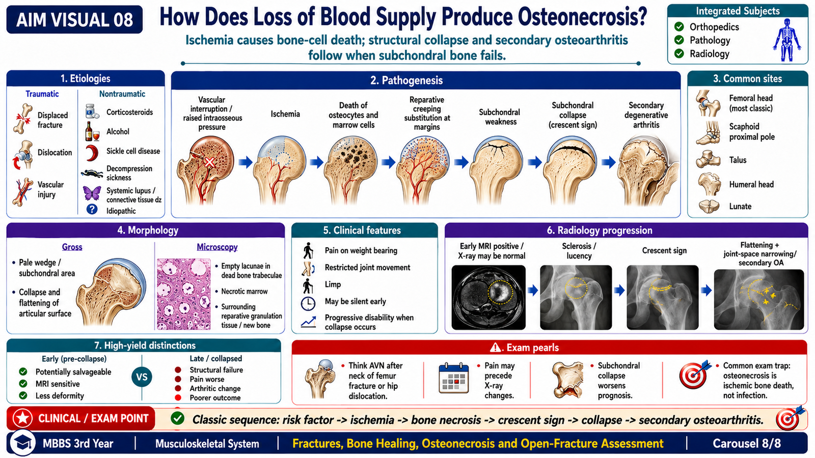
Osteonecrosis, also called avascular necrosis, is ischemic death of bone and marrow elements caused by interruption or severe reduction of blood supply. The consequences depend on the site, extent of necrosis and mechanical load placed on the affected bone.
Major etiologies
- Fracture disrupting critical vessels
- Dislocation compromising blood supply
- Direct vascular injury
- Prolonged or high cumulative corticosteroid exposure
- Excessive alcohol exposure
- Sickle cell disease and other vaso-occlusive states
- Decompression-related vascular injury
- Radiation injury
- Systemic disorders affecting vessels or marrow
- Idiopathic cases
Pathogenesis
Morphology
- Often a wedge-shaped or segmental area of necrosis.
- Subchondral involvement is particularly important in weight-bearing joints.
- Advanced lesions may show flattening, fragmentation or collapse of the articular surface.
- Secondary degenerative changes may develop in the adjoining joint.
- Empty osteocyte lacunae in dead trabeculae.
- Necrosis of marrow fat and hematopoietic cells.
- Repair at viable margins with osteoclastic resorption and osteoblastic new bone deposition.
- Microfractures and structural collapse when repair cannot match mechanical stress.
Clinical features
- Early disease may be asymptomatic.
- Deep, gradually progressive pain, often worsened by weight bearing.
- Reduced joint movement and functional limitation as collapse develops.
- Symptoms may follow trauma or occur in a patient with a recognized systemic risk factor.
Radiological findings
Early plain radiographs may be normal. With progression, imaging may show altered density, sclerosis, cystic change, a subchondral fracture line, flattening or collapse of the articular surface and secondary degenerative changes. More sensitive imaging may identify early marrow abnormalities before collapse becomes visible on plain X-ray.
Integrated Mechanism Flow
AIM High-Yield Review
- Fracture classification: describe the cause, communication with the exterior, completeness, pattern, anatomical site and fragment position.
- Open fracture: any wound communicating with the fracture or fracture hematoma makes the injury open, even when bone is not visible.
- X-ray description: state the bone and site, fracture pattern, displacement of the distal fragment, apex of angulation, shortening, rotation, joint alignment and soft-tissue findings.
- Dislocation: complete loss of joint congruity; subluxation means partial loss of congruity.
- Pediatric fractures: flexible bone and a thick periosteum explain greenstick, buckle and plastic-deformation patterns.
- Secondary healing: hematoma and inflammation → soft callus → woven-bone hard callus → lamellar-bone remodeling.
- Primary healing: occurs with close apposition and near-absolute stability, with little external callus.
- Healing failure: poor blood supply, infection, tissue loss and instability may cause delayed union or nonunion; malunion means union in an abnormal position.
- Nonunion clue: abundant callus suggests active biology but inadequate stability, while little callus suggests poor biological activity or blood supply.
- Open-fracture priority: document neurovascular status, protect the wound, reduce contamination and obtain urgent orthopedic assessment.
- Debridement: remove foreign material and nonviable tissue while preserving viable structures needed for healing and coverage.
- Osteonecrosis: interruption of blood supply causes death of marrow and osteocytes; empty lacunae are an important microscopic feature.
- Radiological progression of osteonecrosis: early films may be normal, followed by sclerosis, subchondral fracture and collapse.
- Major outcome: collapse of subchondral bone damages the articular surface and may produce permanent joint deformity.
Watch this video after reading the fracture-healing section. Focus on the sequence from fracture hematoma and inflammation to soft callus, hard callus and bone remodeling.
Concept Not Clear? Ask AIM AI
Use the AIM Learning Assistant to clarify difficult concepts from this topic before proceeding to assessment.
You may ask AIM AI to:
- Explain a difficult concept in simpler words.
- Describe a mechanism step by step.
- Compare two confusing diseases, drugs or findings.
- Provide a clinically relevant example.
- Highlight a high-yield KMU examination point.
- Explain a common MCQ trap related to the topic.
Best use: Ask one focused question at a time. AIM AI supports your understanding but does not replace the complete learning material.