
This chapter follows the supplied KMU learning outcomes and explains the infections in a logical sequence. First understand how each organism produces disease and how the lesions differ; then use the final high-yield review for revision.
Topic 17 — Viral, Fungal, Bacterial and Parasitic Skin Infections
MSK Module — Etiopathogenesis, diagnosis and antimicrobial treatment of important skin infections, with primary-care recognition and management of cutaneous leishmaniasis.
Topic Introduction
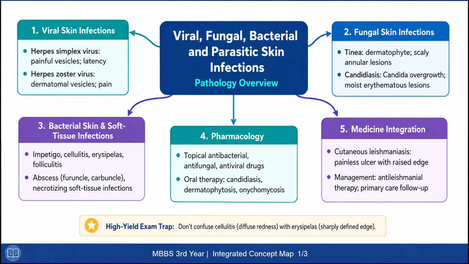
Skin infections may be caused by viruses, fungi, bacteria or parasites. Although all involve the skin, the way they enter, the tissue they affect and the lesions they produce are different. This chapter first explains herpes simplex and herpes zoster, followed by tinea and candidiasis. It then covers common bacterial skin and soft-tissue infections, including dangerous necrotizing infections. The antimicrobial preparations used on the skin and important oral antifungal treatments are explained through their mechanisms, uses and adverse effects. Finally, cutaneous leishmaniasis is discussed from the practical viewpoint of recognition and basic management in primary healthcare. The aim is to connect the causative organism with tissue injury, clinical appearance, diagnosis and appropriate treatment.
A. Viral Skin Infections — Herpes Simplex and Herpes Zoster
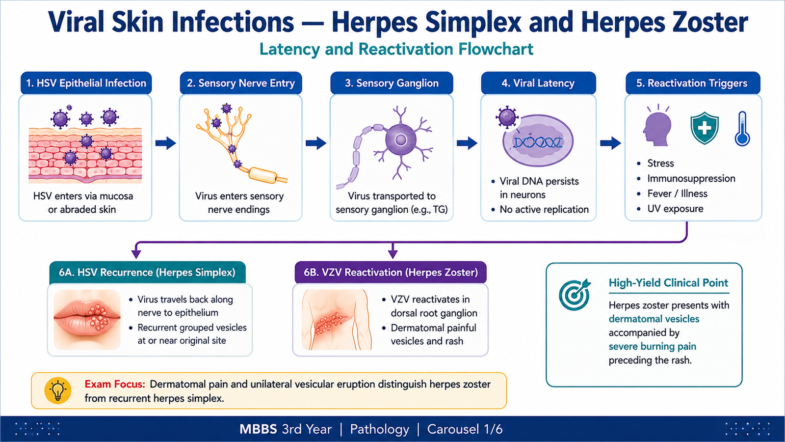
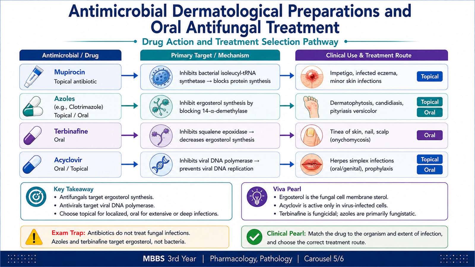
Herpes simplex virus and varicella-zoster virus belong to the herpesvirus group. An important property of these viruses is latency: after the initial infection, the virus remains inside sensory neurons and may reactivate later. Their characteristic skin lesions are vesicles, but the distribution and clinical pattern differ because the viruses reactivate in different ways.
Herpes Simplex Virus Infection
Herpes simplex virus infection is caused mainly by HSV-1 or HSV-2. HSV-1 commonly produces orolabial infection, whereas HSV-2 commonly involves the genital region, although either type can infect either site.
During primary infection, the virus enters through skin or mucosal epithelium and replicates in epithelial cells. Viral replication damages these cells and produces small intraepidermal vesicles. The virus then enters sensory nerve endings and travels along the nerve to a sensory ganglion, where its genome persists in a latent state.
Viral entry through skin or mucosa → replication in epithelial cells → epithelial cell injury and vesicle formation → entry into sensory nerve endings → transport to sensory ganglion → latency → later reactivation → recurrent localized vesicular lesions
Reactivation may follow conditions such as fever, local trauma, emotional stress or ultraviolet exposure. When reactivated, the virus travels back along the sensory nerve toward the skin or mucosa. Because immunity already exists, recurrent disease is usually more localized than the primary infection.
Important pathological features include ballooning degeneration of infected epithelial cells, formation of vesicles and characteristic viral nuclear changes. Multinucleated giant cells may be present in infected epithelium.
Herpes Zoster
Herpes zoster results from reactivation of latent varicella-zoster virus. The primary infection with this virus causes varicella, after which the virus remains latent in sensory ganglia. When cell-mediated immunity against the virus decreases, it may reactivate.
The reactivated virus travels along a sensory nerve to the skin. Because a sensory ganglion supplies a particular dermatome, the resulting vesicular eruption usually follows a dermatomal distribution. Inflammation involving the sensory nerve also explains the characteristic pain.
Previous varicella infection → latency in sensory ganglion → viral reactivation → inflammation of sensory nerve → spread along the nerve to skin → painful grouped vesicles in the corresponding dermatome
The rash is usually unilateral and normally does not cross the midline because an individual sensory ganglion supplies one side of the body. Vesicles may rupture and crust. Persistent pain after disappearance of the skin lesions may occur because the sensory nerve has been damaged.
| Feature | Herpes Simplex | Herpes Zoster |
|---|---|---|
| Virus | HSV-1 or HSV-2 | Varicella-zoster virus |
| Latency | Sensory ganglia | Sensory ganglia |
| Typical recurrence | Localized grouped vesicles | Dermatomal vesicular eruption |
| Pain | May cause local burning or pain | Often prominent due to sensory nerve inflammation |
B. Fungal Skin Infections — Tinea and Candidiasis
Fungal skin infections commonly involve superficial keratinized tissues or moist skin surfaces. Dermatophytes cause tinea by using keratin and therefore mainly infect the stratum corneum, hair and nails. Candida, in contrast, is an opportunistic yeast that may normally colonize the body but produces disease when local or host conditions allow excessive growth.
Tinea or Dermatophytosis
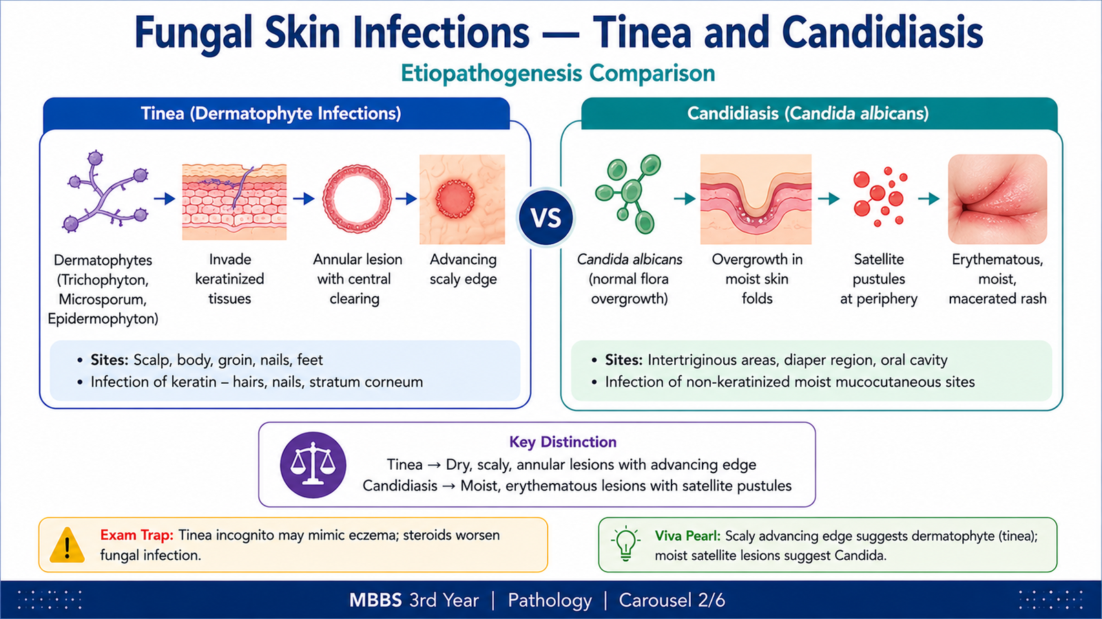
Tinea is a superficial fungal infection caused by dermatophytes. Important dermatophyte genera include Trichophyton, Microsporum and Epidermophyton. These organisms have the ability to utilize keratin, explaining why they primarily infect keratinized structures.
The fungus enters superficial keratinized tissue and grows within the stratum corneum. The host inflammatory response is often more active at the advancing edge of the infection. This produces the characteristic expanding, scaly margin seen in many forms of tinea.
Dermatophyte exposure → adherence to keratinized tissue → growth within stratum corneum, hair or nail → keratin degradation → local inflammatory response → scaling, pruritus and characteristic superficial lesions
Tinea is named according to the anatomical site involved:
- Tinea corporis: infection of glabrous body skin.
- Tinea cruris: infection of the groin.
- Tinea pedis: infection of the feet.
- Tinea capitis: involvement of scalp and hair.
- Tinea unguium: dermatophyte infection of nails.
Skin lesions often show erythema, scaling and an advancing margin. Nail infection may produce thickening, discoloration, brittleness and separation of the nail from the underlying nail bed.
Candidiasis
Candidiasis is most commonly caused by Candida albicans. Candida may normally exist as part of the microbial flora of the mouth, gastrointestinal tract, genital tract and skin. Disease develops when the normal balance between the organism and host defenses is disturbed.
Predisposing conditions may include prolonged moisture, occlusion, disruption of normal flora, diabetes or impaired immunity. On moist skin, Candida can proliferate and invade the superficial epithelium, producing inflammation.
Cutaneous candidiasis commonly affects warm, moist opposing skin surfaces. The affected area may be red and macerated, and small peripheral satellite lesions may occur around the main area of inflammation.
Candida colonization → favorable local or host condition → excessive fungal growth → superficial epithelial invasion → inflammatory response → erythematous, moist and sometimes satellite lesions
| Feature | Tinea | Candidiasis |
|---|---|---|
| Organism | Dermatophytes | Usually Candida albicans |
| Preferred tissue | Keratinized skin, hair and nails | Moist skin and mucosal surfaces |
| Typical clue | Scaly advancing edge | Maceration with satellite lesions |
C. Common Bacterial Skin and Soft-Tissue Infections
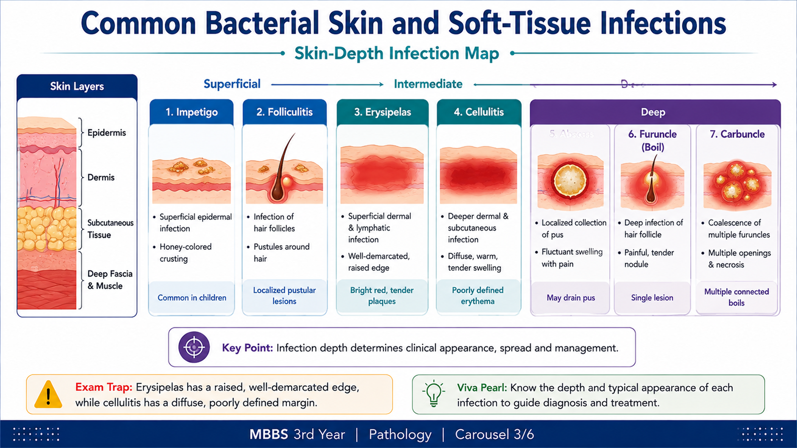
Bacterial infections may remain superficial or extend into progressively deeper tissues. The depth of infection helps explain the clinical appearance. Impetigo mainly involves superficial epidermis, folliculitis centers on hair follicles, erysipelas involves superficial dermis and lymphatics, cellulitis extends into deeper dermis and subcutaneous tissue, while abscesses produce localized collections of pus.
Impetigo
Impetigo is a superficial bacterial infection of the epidermis. It is commonly associated with Staphylococcus aureus and may also be produced by Streptococcus pyogenes.
Bacteria gain access through minor breaks in the skin and multiply within the superficial epidermis. Local tissue injury and inflammatory exudate produce superficial vesicles or pustules. When these rupture, the exudate dries on the surface and forms the characteristic crust.
A useful diagnostic clue is the presence of superficial erosions covered by honey-colored crusts.
Erysipelas and Cellulitis
Erysipelas is an acute infection involving the upper dermis and superficial lymphatics. The lesion is typically erythematous, warm and raised, with a relatively well-defined margin.
Cellulitis involves the deeper dermis and subcutaneous tissue. Because inflammation spreads through deeper tissue planes rather than remaining confined to superficial lymphatics, the border is usually less sharply defined.
Bacterial invasion → activation of acute inflammation → vasodilation and increased vascular permeability → redness, warmth and tissue edema → pain from inflammatory mediators and tissue pressure
Folliculitis
Folliculitis is inflammation and infection centered on a hair follicle. Staphylococcus aureus is a common bacterial cause. The infection is usually superficial and produces small erythematous papules or pustules centered around hair follicles.
The follicular location is an important diagnostic clue. When infection extends deeper into the follicle and surrounding tissue, a larger painful lesion can develop.
Abscess, Furuncle and Carbuncle
A skin abscess is a localized collection of pus within the dermis or subcutaneous tissue. Neutrophils accumulate at the site of bacterial infection, tissue undergoes liquefactive destruction and a cavity containing pus forms.
A furuncle is a deeper infection of a hair follicle that extends into surrounding dermis and forms a painful abscess. A carbuncle represents a larger, deeper inflammatory mass formed by several interconnected furuncles.
- Abscess: localized pus-filled cavity.
- Furuncle: deep follicular abscess.
- Carbuncle: interconnected deeper follicular abscesses.
Diagnosis
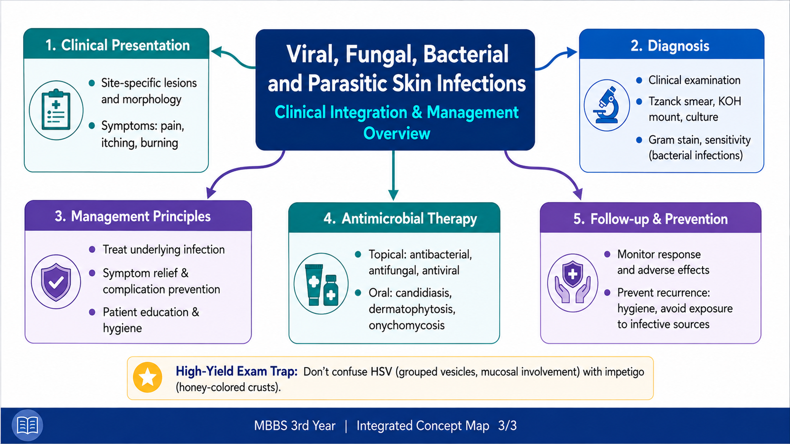
The diagnosis of common bacterial skin infections is often primarily clinical. The appearance of the lesion, its depth, margin, relation to a hair follicle, presence of pus and associated inflammatory changes help distinguish the different conditions.
- Honey-colored superficial crust: suggests impetigo.
- Raised, sharply defined erythematous plaque: favors erysipelas.
- Diffuse warm erythematous swelling with less-defined margins: favors cellulitis.
- Pustule centered on a hair follicle: suggests folliculitis.
- Localized fluctuant pus collection: suggests an abscess.
D. Necrotizing Soft-Tissue Infections
Necrotizing soft-tissue infections are rapidly progressive bacterial infections in which microorganisms spread through deep soft tissues and produce extensive tissue destruction. They differ from uncomplicated cellulitis because tissue necrosis, vascular damage and systemic toxicity can develop rapidly.
The infection may be caused by a single aggressive organism or by a mixture of aerobic and anaerobic bacteria. The exact organisms vary, but the key pathological process is rapid spread along tissue planes with severe inflammation and tissue ischemia.
Etiopathogenesis
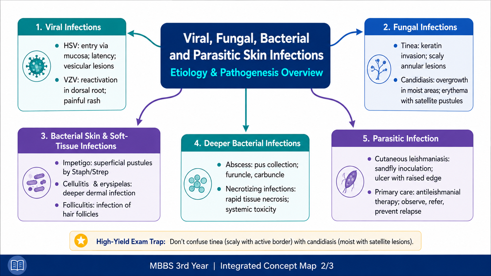
Bacteria enter through a break in the skin or deeper tissue. They multiply and release enzymes and toxins that promote tissue injury and spread. Severe inflammation damages small blood vessels and may produce thrombosis. As the local blood supply falls, tissue becomes ischemic and eventually necrotic.
Bacterial entry → rapid spread through soft tissue → toxin and enzyme-mediated injury → intense inflammation and vascular thrombosis → reduced blood supply → tissue necrosis → systemic toxicity
This vascular damage is especially important because necrotic tissue has poor perfusion. Tissue destruction can therefore progress despite an apparently limited abnormality on the skin surface during the early stage.
Important Diagnostic Clues
Early recognition is essential. A patient may initially appear to have cellulitis, but certain findings suggest a deeper and more destructive process.
- Severe pain that appears greater than expected from the visible skin lesion.
- Rapid progression of swelling and erythema.
- Increasing tenderness and tissue edema.
- Skin discoloration, bullae or areas of necrosis as disease advances.
- Marked systemic illness or toxicity.
Because this is a rapidly progressive and potentially life-threatening infection, clinical suspicion should lead to urgent hospital and surgical assessment. The important undergraduate principle is that necrotizing infection is not simply severe cellulitis; it represents destructive infection of deep soft tissues.
E. Antimicrobial Dermatological Preparations and Oral Antifungal Treatment
Antimicrobial dermatological preparations are drugs applied to the skin to control microorganisms causing superficial infection. They can be grouped according to the organism they target into topical antibacterial, antifungal and antiviral agents. Topical treatment is most useful when infection is limited to accessible superficial tissue. Oral therapy becomes more important when infection is extensive or involves structures such as hair or nails that are difficult to treat adequately with topical drugs alone.
Classification of Antimicrobial Dermatological Preparations
- Topical antibacterial agents: examples include mupirocin and fusidic acid.
- Topical antifungal agents: examples include azoles such as clotrimazole and imidazole-related agents, and allylamines such as terbinafine.
- Topical antiviral agents: examples include acyclovir and related antiviral preparations used in selected superficial herpes infections.
Topical Antibacterial Drugs
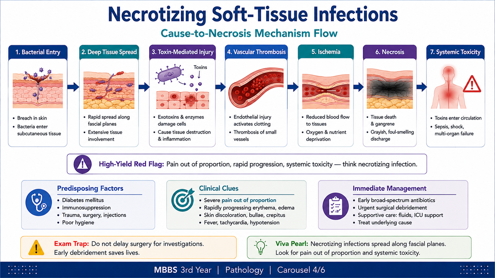
Mupirocin inhibits bacterial protein synthesis by blocking bacterial isoleucyl-transfer RNA synthetase. This prevents incorporation of isoleucine into bacterial proteins and suppresses bacterial growth.
Fusidic acid also interferes with bacterial protein synthesis and is used topically against susceptible organisms, particularly staphylococci.
Topical antibacterial agents are mainly used for localized superficial bacterial infections. Adverse effects are usually local and may include irritation, burning, itching or contact dermatitis. Unnecessary or prolonged use can also favor antimicrobial resistance.
Topical Antifungal Drugs
Topical antifungals are particularly useful for superficial dermatophytosis and cutaneous candidiasis.
Azole antifungals, such as clotrimazole, inhibit a fungal cytochrome P450-dependent enzyme needed for ergosterol synthesis. Ergosterol is an important component of the fungal cell membrane. Reduced ergosterol disrupts membrane structure and function.
Terbinafine inhibits squalene epoxidase, another enzyme in the fungal ergosterol pathway. The resulting disturbance of fungal membrane synthesis makes it particularly useful against dermatophytes.
Topical antifungals may cause local burning, irritation, erythema or itching. The appropriate agent is selected according to the likely fungal organism and anatomical site.
Topical Antiviral Drugs
Acyclovir and related antiviral agents interfere with viral DNA synthesis. Acyclovir is activated preferentially within infected cells and then inhibits viral DNA polymerase, limiting viral replication.
Topical antiviral preparations may be used in selected localized herpes infections. Local irritation or burning may occur.
Oral Treatment of Candidiasis
When candidiasis requires systemic treatment, an oral azole such as fluconazole may be used depending on the site and clinical situation. Like other azoles, fluconazole interferes with ergosterol synthesis and therefore disrupts fungal cell-membrane function.
Systemic azoles may produce gastrointestinal upset and liver-related adverse effects and can participate in clinically important drug interactions. The key principle is that systemic treatment is chosen when local therapy is insufficient or when the anatomical site requires oral treatment.
Oral Treatment of Dermatophytosis
Limited superficial dermatophytosis can often be treated topically. Oral therapy may be required when infection is extensive or involves hair-bearing areas or other sites where topical treatment is inadequate.
Terbinafine is an important systemic drug for dermatophyte infection because it inhibits squalene epoxidase. Azoles such as itraconazole are also active against dermatophytes.
Oral Treatment of Onychomycosis
Onychomycosis is fungal infection of the nail. Because the organism is located within the nail plate and nail bed, topical penetration may be inadequate in significant disease. Systemic therapy is therefore often required.
Terbinafine is an important oral option for dermatophyte onychomycosis. Itraconazole is another systemic antifungal option. Liver-related adverse effects and drug interactions are important considerations with systemic antifungal therapy.
F. Cutaneous Leishmaniasis — Clinical Features and Primary Healthcare Management
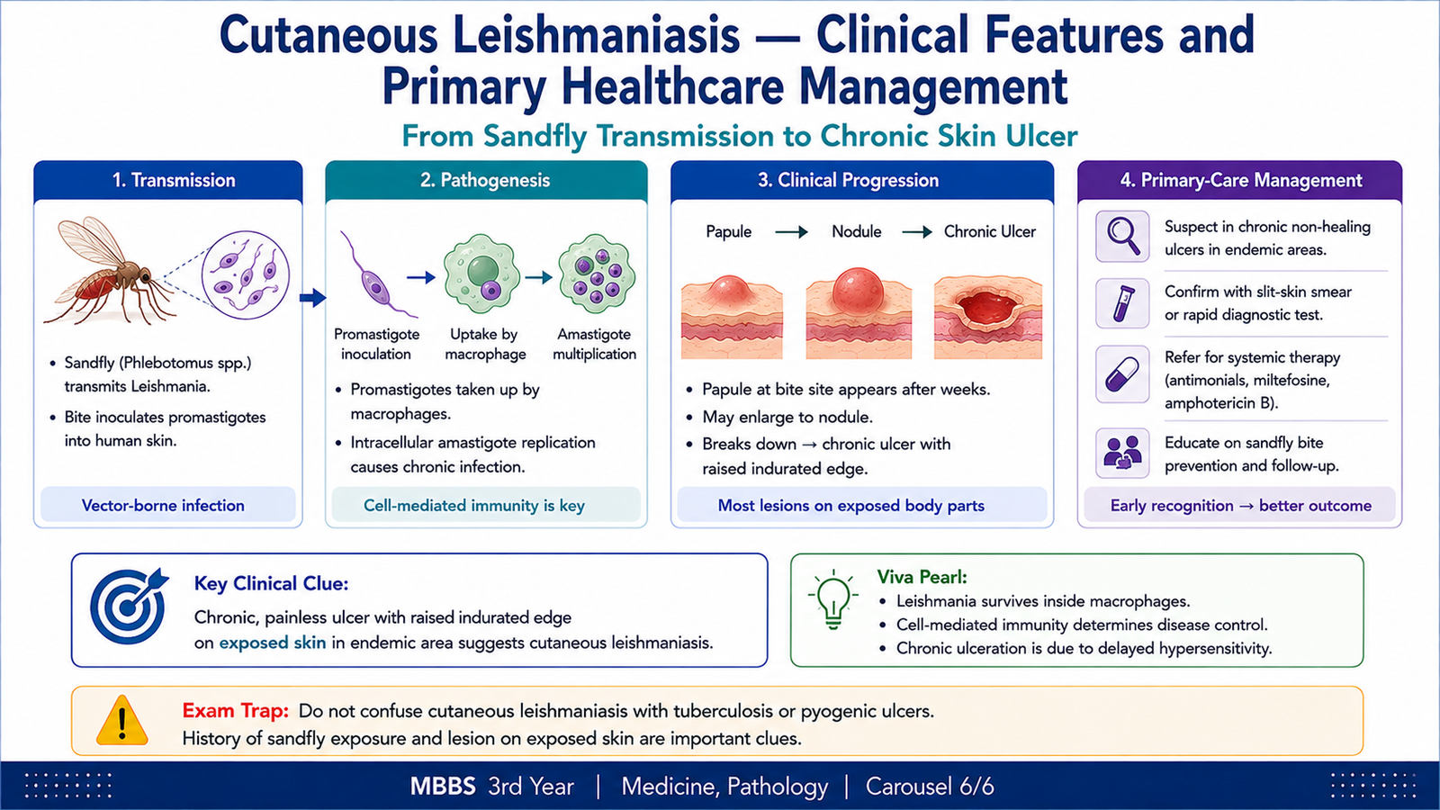
Cutaneous leishmaniasis is a parasitic infection of the skin caused by Leishmania species and transmitted by the bite of an infected sandfly. The parasite survives within macrophages in the skin, producing a chronic localized inflammatory lesion.
How the Skin Lesion Develops
During a sandfly bite, infective parasites are introduced into the skin. They are taken up by macrophages and transform into intracellular forms that multiply within these cells. Continued infection stimulates inflammation and produces progressive tissue damage.
Clinical Features
The lesion develops at or near the site of the infected sandfly bite. It may begin as a small papule or nodule and gradually enlarge. The lesion may subsequently ulcerate.
- Usually affects exposed skin.
- Begins as a papule or nodule.
- Enlarges gradually rather than behaving like an acute bacterial infection.
- May develop a central ulcer with an inflammatory or raised border.
- Lesions may persist for a prolonged period.
- Healing may leave a permanent scar.
The chronic nature of the lesion is an important clue. Unlike rapidly spreading bacterial cellulitis, cutaneous leishmaniasis commonly evolves gradually over time.
Diagnosis
Clinical suspicion is based on the appearance of a chronic skin lesion together with appropriate exposure. Confirmation involves demonstrating the parasite in material obtained from the lesion or through appropriate laboratory assessment.
The important diagnostic principle is to consider cutaneous leishmaniasis in a persistent papule, nodule or ulcer that does not behave like an ordinary bacterial skin infection.
Management in Primary Healthcare
Primary healthcare management begins with recognition of a suspicious lesion, assessment of the number and location of lesions, and identification of features that may require specialist assessment. Treatment selection depends on the parasite species, lesion characteristics, anatomical location and available local treatment pathways.
At primary-care level, the clinician should:
- Recognize the typical chronic cutaneous presentation.
- Assess the lesion site, number and extent.
- Arrange appropriate diagnostic confirmation where available.
- Provide local wound and skin care when required.
- Avoid unnecessary antibacterial treatment when bacterial infection is not responsible.
- Refer patients with extensive, atypical, complicated or difficult-site lesions for appropriate specialist management.
Specific antileishmanial treatment may be local or systemic depending on the clinical situation. At undergraduate primary-care level, the important principle is appropriate recognition, confirmation and referral or treatment selection rather than memorization of complex specialist regimens.
Integrated Mechanism Flow
↓
2. The organism targets a characteristic site: epithelium, keratinized tissue, hair follicle, dermis, deep soft tissue or macrophage.
↓
3. Microbial replication or persistence causes direct tissue injury and/or activates inflammation.
↓
4. The depth and mechanism of injury determine the lesion: vesicle, scale, crust, pustule, diffuse erythema, abscess, necrosis or chronic ulcer.
↓
5. Clinical appearance and tissue depth provide important diagnostic clues.
↓
6. Treatment is directed toward the responsible organism and the site or extent of infection.
↓
7. Rapidly destructive infection or difficult chronic disease requires timely referral and further management.
⭐ AIM High-Yield Review
- Herpesviruses establish latency in sensory ganglia after the initial infection.
- HSV recurrence produces localized grouped vesicular lesions, while herpes zoster characteristically follows a dermatome.
- Herpes zoster is painful because reactivation involves the sensory nerve and ganglion.
- Dermatophytes infect keratinized tissues: stratum corneum, hair and nails.
- Tinea commonly has a scaly advancing margin, whereas cutaneous candidiasis favors moist surfaces and may show satellite lesions.
- Honey-colored crusting is a classic diagnostic clue for impetigo.
- Erysipelas is more superficial and sharply demarcated; cellulitis involves deeper dermis and subcutaneous tissue and has less-defined margins.
- A furuncle is a deep follicular abscess; a carbuncle consists of interconnected deeper follicular abscesses.
- Severe pain out of proportion to the visible lesion and rapid progression should raise concern for a necrotizing soft-tissue infection.
- Necrotizing infection causes vascular injury and thrombosis, leading to ischemia and tissue necrosis.
- Azoles impair fungal ergosterol synthesis, while terbinafine inhibits squalene epoxidase.
- Acyclovir limits herpesvirus replication by inhibiting viral DNA synthesis.
- Localized superficial infection is generally more suitable for topical therapy; involvement of extensive skin, hair or nails may require systemic antifungal treatment.
- Cutaneous leishmaniasis results from intracellular survival of Leishmania within macrophages after transmission by a sandfly.
- A slowly evolving papule or nodule that becomes a chronic ulcer on exposed skin is an important clue to cutaneous leishmaniasis.
🎥 AIM Video Learning
Viral, Fungal and Bacterial Skin Infections
Watch this video after completing the learning material to reinforce the major clinical patterns and important differences between common skin infections.