
This chapter follows the KMU learning outcomes in a logical sequence. First understand how muscle fibres become weak or damaged, and then revise the distinguishing features in the AIM High-Yield Review.
Topic 12 — Skeletal-Muscle Atrophy, Myopathies and Muscular Dystrophies
MSK Module — Pathological muscle changes, inherited and acquired myopathies, Duchenne muscular dystrophy, disability concepts, poliomyelitis prevention and rehabilitation.
Topic Introduction
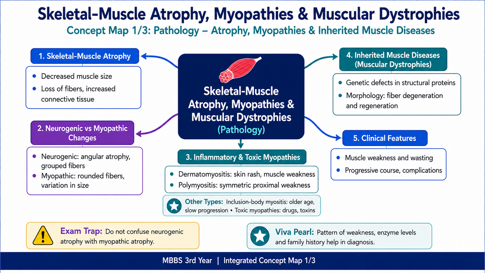
Skeletal-muscle weakness may result from loss of muscle fibres, injury within the muscle itself, inherited structural defects or interruption of its nerve supply. Muscle atrophy means a reduction in muscle-fibre size, while myopathy refers to a primary disorder of skeletal muscle. Muscular dystrophies are inherited diseases in which abnormal structural proteins cause progressive muscle-fibre degeneration. This chapter explains the pathological patterns of neurogenic and myopathic injury, inflammatory and toxic myopathies, inherited muscle diseases, the clinical investigation of muscle weakness, and Duchenne muscular dystrophy. It also connects poliomyelitis with disability, prevention, disease burden and rehabilitation.
A. Skeletal-Muscle Atrophy and Patterns of Muscle Injury
Skeletal muscle maintains its size through normal nerve stimulation, physical activity, adequate nutrition and balanced protein synthesis. When one of these supports is lost, muscle fibres become smaller. This process is called atrophy. The basic pattern of atrophy helps the pathologist determine whether weakness is caused mainly by loss of nerve supply or by a primary disease within the muscle.
Skeletal-Muscle Atrophy
Atrophy is a decrease in the size of individual muscle fibres. The number of fibres may remain relatively preserved at first, but prolonged disease can eventually cause fibre loss and replacement by fat or connective tissue.
Important causes include:
- Disuse: prolonged immobilization, bed rest or a limb kept in a cast.
- Denervation: interruption of the motor nerve supply.
- Reduced blood supply: chronic ischemia limits nutrient and oxygen delivery.
- Malnutrition or cachexia: muscle proteins are broken down to provide energy.
- Loss of hormonal stimulation: reduced anabolic support promotes muscle wasting.
- Aging: gradual loss of motor units and muscle mass produces sarcopenia.
Atrophic fibres contain fewer myofilaments and organelles. Protein breakdown increases through cellular protein-degradation pathways, while protein synthesis falls. The fibres therefore become smaller and weaker.
Neurogenic Change
Neurogenic atrophy occurs when muscle fibres lose their motor-nerve supply. Each motor neuron normally supplies a group of muscle fibres called a motor unit. Loss of that neuron removes stimulation from all fibres in its motor unit, producing a characteristic grouped pattern of atrophy.
Microscopic features include:
- Small, angular atrophic fibres.
- Groups of atrophic fibres corresponding to affected motor units.
- Relative preservation of initially unaffected fibres.
- Fibre-type grouping when surviving motor neurons reinnervate denervated fibres.
- Group atrophy when a previously enlarged reinnervated motor unit later loses its nerve supply.
Fibre-type grouping occurs because a surviving nerve branch may connect with nearby denervated fibres. These newly reinnervated fibres adopt the metabolic type of the new motor neuron and form groups of similar fibre type.
Myopathic Change
In myopathic disorders, the primary abnormality is within the muscle fibre. Individual fibres are damaged independently of their motor units, so the microscopic changes are usually scattered rather than grouped.
Typical myopathic changes include:
- Variation in muscle-fibre size.
- Rounded rather than sharply angular atrophic fibres.
- Muscle-fibre necrosis and regeneration.
- Internal or centrally placed nuclei.
- Splitting of fibres.
- Increased endomysial connective tissue in chronic disease.
- Replacement of muscle by fat in advanced disease.
| Feature | Neurogenic Pattern | Myopathic Pattern |
|---|---|---|
| Primary problem | Motor neuron or peripheral nerve | Muscle fibre |
| Atrophic fibres | Small and angular | Often rounded, with variable size |
| Distribution | Grouped according to motor units | Scattered individual fibres |
| Necrosis and regeneration | Usually not prominent initially | Common in active muscle disease |
| Special clue | Fibre-type grouping | Internal nuclei and fibre splitting |

B. Inflammatory Myopathies
Inflammatory myopathies are acquired muscle diseases in which immune-mediated inflammation damages skeletal-muscle fibres and causes weakness. The most important forms are dermatomyositis, polymyositis and inclusion-body myositis. They share muscle weakness but differ in age, distribution of weakness, immune mechanism and microscopic pattern.
General Clinical Pattern
These disorders commonly produce progressive weakness of proximal muscles, such as the shoulder and pelvic-girdle muscles. Patients may therefore have difficulty climbing stairs, rising from a chair, lifting objects or combing their hair. Pain may occur, but weakness is usually the main complaint.
Dermatomyositis
Dermatomyositis is an immune-mediated inflammatory myopathy involving both skeletal muscle and skin. It may occur in children or adults. The main injury affects small blood vessels in muscle, leading to reduced blood supply and damage that is most prominent around the edges of muscle fascicles.
Important morphological features include:
- Inflammatory cells mainly around blood vessels and within the perimysial connective tissue.
- Perifascicular atrophy, involving fibres at the edges of muscle fascicles.
- Muscle-fibre degeneration and regeneration.
- Reduced capillary density in affected areas.
Important clinical features include:
- Symmetrical proximal muscle weakness.
- A violaceous or dusky discoloration around the eyelids, called a heliotrope rash.
- Scaly erythematous papules over the knuckles, called Gottron papules.
- Skin changes over sun-exposed areas.
- Difficulty swallowing when pharyngeal muscles are involved.
Polymyositis
Polymyositis is an inflammatory myopathy that mainly affects adults and does not produce the characteristic skin rash of dermatomyositis. Cytotoxic T lymphocytes directly attack muscle fibres that express abnormal immune-related surface molecules.
Microscopic features include:
- Endomysial inflammatory infiltrates between individual muscle fibres.
- Predominantly cytotoxic T lymphocytes.
- Direct invasion and destruction of non-necrotic muscle fibres.
- Scattered fibre necrosis and regeneration.
Clinically, polymyositis causes symmetrical proximal muscle weakness. Skin manifestations are absent. Weakness of respiratory or swallowing muscles may occur in severe disease.
Inclusion-Body Myositis
Inclusion-body myositis is a slowly progressive inflammatory and degenerative muscle disease that usually affects older adults. Unlike dermatomyositis and polymyositis, weakness is often asymmetrical and may involve both proximal and distal muscles.
Important features include:
- Slow progression over several years.
- Weakness of quadriceps muscles and finger flexors.
- Endomysial inflammation.
- Muscle fibres containing rimmed vacuoles.
- Abnormal protein inclusions within affected fibres.
- Limited response to immunosuppressive treatment compared with other inflammatory myopathies.
| Feature | Dermatomyositis | Polymyositis | Inclusion-Body Myositis |
|---|---|---|---|
| Typical age | Children or adults | Mainly adults | Usually older adults |
| Weakness | Symmetrical and proximal | Symmetrical and proximal | Often asymmetrical; proximal and distal |
| Skin rash | Characteristic rash present | Absent | Absent |
| Inflammation | Perivascular and perimysial | Endomysial | Endomysial |
| Hallmark morphology | Perifascicular atrophy | T-cell invasion of muscle fibres | Rimmed vacuoles and inclusions |

C. Toxic and Other Acquired Myopathies
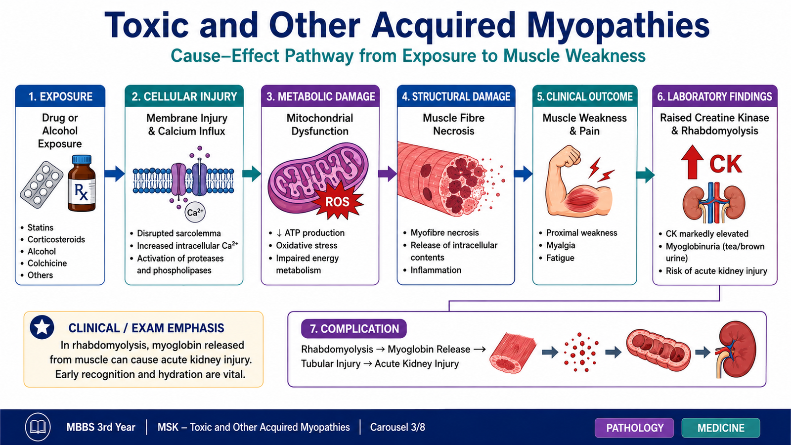
Toxic myopathies are muscle disorders caused by medicines, alcohol or other harmful substances. The toxic agent may directly injure muscle fibres, disturb energy production, alter electrolyte balance or trigger immune-mediated muscle damage. The severity ranges from mild muscle pain to extensive fibre necrosis and acute loss of muscle contents.
Mechanisms of Toxic Muscle Injury
Different toxic agents damage muscle through different pathways. Some disrupt the muscle-cell membrane, while others interfere with mitochondria and reduce energy production. Certain agents cause abnormal accumulation of lipids or cellular organelles inside fibres. Severe injury may cause widespread muscle-fibre necrosis.
Important causes include:
- Alcohol: may produce acute muscle necrosis or chronic proximal weakness.
- Lipid-lowering medicines: may cause muscle pain, weakness or, rarely, severe fibre breakdown.
- Glucocorticoids: prolonged exposure causes selective atrophy of particular muscle fibres and painless proximal weakness.
- Antimalarial medicines: may produce vacuolar changes within muscle fibres.
- Other medicines or toxins: may interfere with mitochondrial function, lysosomes or muscle-cell membranes.
Pathological Features
Morphology depends on the responsible agent. Muscle biopsy may show fibre necrosis, regeneration, vacuoles, lipid accumulation, mitochondrial abnormalities or selective fibre atrophy. In steroid-associated myopathy, inflammation is usually not prominent because weakness results mainly from protein breakdown and fibre atrophy rather than immune-mediated destruction.
Clinical Recognition
A temporal relationship between exposure and symptoms is an important clue. The patient may report muscle pain, tenderness, cramps or proximal weakness. Severe fibre breakdown releases intracellular substances into the blood and may produce dark urine and kidney injury.
Assessment therefore includes a careful medication and exposure history, examination of muscle strength, measurement of muscle enzymes and evaluation for complications when severe injury is suspected.
D. Inherited Muscle Diseases and Muscular Dystrophies
Inherited muscle diseases result from genetic abnormalities that disturb the structure, contraction, energy metabolism or development of muscle fibres. Muscular dystrophies are a major group of inherited disorders characterized by progressive muscle-fibre degeneration, repeated attempts at regeneration and eventual replacement of muscle by fat and fibrous tissue.
General Pathogenesis of Muscular Dystrophy
Muscle fibres repeatedly contract and relax throughout life. Structural proteins connect the internal contractile apparatus to the muscle-cell membrane and surrounding extracellular matrix. A genetic defect in one of these proteins makes the muscle fibre vulnerable to mechanical stress.
General Morphology
Early disease shows muscle-fibre necrosis together with regenerating fibres. Fibre size becomes variable, and nuclei may move from the edge of the fibre to a central position. As repeated injury exceeds the regenerative capacity of muscle, fibres are lost and replaced by adipose tissue and collagen.
Typical microscopic features include:
- Marked variation in muscle-fibre size.
- Degenerating and necrotic fibres.
- Regenerating basophilic fibres.
- Internal nuclei.
- Fibre splitting.
- Endomysial fibrosis.
- Fatty replacement in advanced disease.
Duchenne Muscular Dystrophy
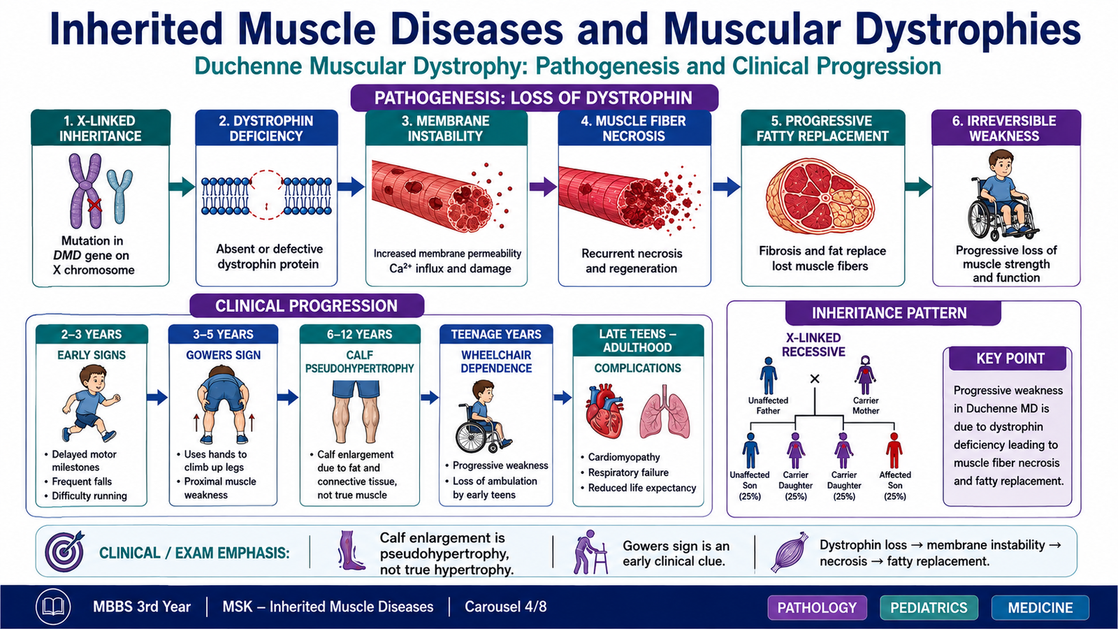
Duchenne muscular dystrophy is a severe X-linked recessive muscular dystrophy caused by pathogenic variants in the dystrophin gene. It mainly affects boys because they have only one X chromosome. Females are usually carriers, although some may develop mild manifestations.
Dystrophin links the internal actin cytoskeleton of the muscle fibre with proteins in the cell membrane and the surrounding extracellular matrix. When dystrophin is absent, contraction places excessive stress on the muscle-cell membrane. Membrane injury permits abnormal calcium entry, activates damaging enzymes and causes muscle-fibre necrosis.
Clinical presentation commonly includes:
- Delayed motor milestones.
- Difficulty running, jumping or climbing stairs.
- Frequent falls.
- Progressive symmetrical proximal weakness, especially around the pelvic girdle.
- Waddling gait.
- Increased lumbar curvature caused by weak hip muscles.
- Gowers sign when the child uses the hands to climb up the thighs while standing.
- Calf pseudohypertrophy caused by replacement of muscle with fat and connective tissue.
- Later respiratory-muscle weakness and cardiac involvement.
Diagnosis of Duchenne Muscular Dystrophy
The clinical pattern suggests the diagnosis, but laboratory and genetic investigations confirm it. Muscle enzymes rise because damaged muscle fibres release their contents into the circulation.
- Serum creatine kinase: markedly elevated, often before obvious weakness develops.
- Genetic testing: identifies the dystrophin-gene abnormality and is the preferred confirmatory approach.
- Muscle biopsy: may show dystrophic changes and absent dystrophin expression when genetic testing is inconclusive.
- Cardiac evaluation: detects associated cardiomyopathy.
- Respiratory assessment: monitors respiratory-muscle function as disease progresses.
Differential Diagnosis of Duchenne Muscular Dystrophy
The major alternatives are disorders that produce childhood weakness, delayed motor development or an abnormal gait.
- Becker muscular dystrophy: dystrophin is reduced or abnormal rather than completely absent, so onset is later and progression is slower.
- Spinal muscular atrophy: a neurogenic disorder with motor-neuron loss, hypotonia and denervation rather than primary dystrophic muscle injury.
- Congenital myopathies: weakness may be present from birth or early infancy, often with characteristic structural changes on muscle biopsy.
- Metabolic myopathies: weakness or exercise intolerance may relate to defects in glycogen, lipid or mitochondrial metabolism.
Other Congenital and Hereditary Myopathies
Congenital myopathies are inherited muscle disorders that usually present at birth or in early childhood with hypotonia, weakness, delayed motor development or feeding and respiratory difficulty. They are often classified by their characteristic structural appearance on muscle biopsy.
Important patterns include:
- Central-core disease: muscle fibres contain central areas with reduced oxidative enzyme activity; some forms are linked to abnormal calcium handling.
- Nemaline myopathy: rod-like structures are present within muscle fibres; severity ranges from mild weakness to severe neonatal disease.
- Centronuclear myopathy: nuclei are abnormally located in the centre of muscle fibres.
- Congenital muscular dystrophies: weakness begins early and biopsy shows dystrophic degeneration with fibrosis and fatty replacement.
Diagnosis is based on the age of onset, pattern of weakness, family history, serum muscle enzymes, electrophysiological findings, genetic testing and, when needed, muscle biopsy. The specific pattern of inheritance depends on the gene involved.
E. Clinical Approach to a Patient with Suspected Myopathy
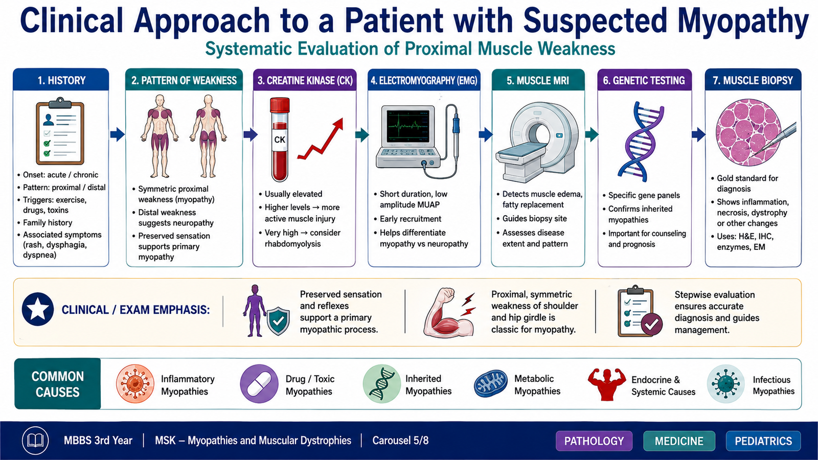
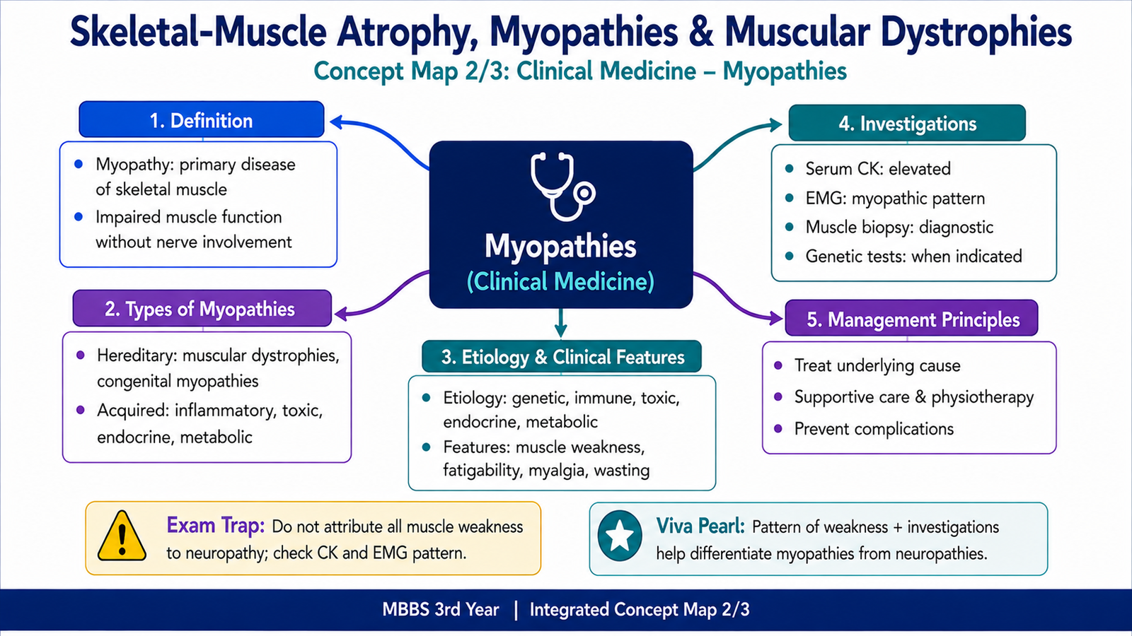
A myopathy is a primary disorder of skeletal muscle. The clinical approach aims to confirm that weakness is muscular, identify whether the disease is inherited or acquired, recognize complications and select investigations that can establish the cause.
Classification of Myopathies
Myopathies can be broadly divided into hereditary and acquired disorders. This classification guides the history, examination and choice of investigations.
- Muscular dystrophies
- Congenital structural myopathies
- Metabolic myopathies
- Mitochondrial myopathies
- Inherited channel disorders
- Inflammatory myopathies
- Toxic or drug-induced myopathies
- Endocrine and metabolic myopathies
- Infective muscle disorders
- Myopathy associated with systemic disease
History
The history should establish the onset, rate of progression and distribution of weakness. Childhood onset and a positive family history suggest an inherited disorder, whereas recent onset after medication exposure, systemic illness or inflammatory symptoms suggests an acquired cause.
Important questions include:
- When did weakness begin, and is it progressive or episodic?
- Are proximal muscles, distal muscles or both affected?
- Is weakness symmetrical?
- Is there muscle pain, tenderness, stiffness or exercise intolerance?
- Are swallowing or breathing muscles affected?
- Is there a skin rash or systemic inflammatory disease?
- Has the patient used medicines or substances associated with muscle injury?
- Is there a family history of weakness, gait abnormality or early loss of mobility?
Examination
Primary myopathies commonly cause symmetrical proximal weakness. Sensation is usually preserved because sensory nerves are not the main site of disease. Reflexes may remain present until weakness becomes advanced.
Examination should assess:
- Muscle bulk, atrophy or pseudohypertrophy.
- Distribution and severity of weakness.
- Gait and ability to rise from a chair or the floor.
- Contractures and joint range of movement.
- Skin changes suggesting dermatomyositis.
- Facial, swallowing and respiratory-muscle function.
- Cardiac findings in inherited muscular dystrophy.
- Sensory examination and reflexes to distinguish neuropathic disease.
Investigations
Investigations are selected according to the clinical pattern. No single test diagnoses every myopathy, so findings from the history, examination, laboratory tests and specialized investigations must be interpreted together.
- Serum creatine kinase: rises when muscle-cell membranes are damaged, especially in muscular dystrophy and active inflammatory muscle injury.
- Other blood tests: may assess systemic inflammation, endocrine abnormalities, electrolytes and organ complications.
- Electromyography: may support a myopathic pattern and help distinguish muscle disease from neurogenic weakness.
- Nerve-conduction studies: are useful when peripheral neuropathy is a possible alternative.
- Magnetic resonance imaging of muscle: can demonstrate edema, fatty replacement and the distribution of affected muscles.
- Genetic testing: confirms many hereditary myopathies and muscular dystrophies.
- Muscle biopsy: shows inflammation, fibre necrosis, dystrophic change, abnormal inclusions or characteristic structural abnormalities.
- Cardiac and respiratory assessment: identifies complications in disorders that affect these systems.
F. Disability, Impairment, Handicap and Measures of Health
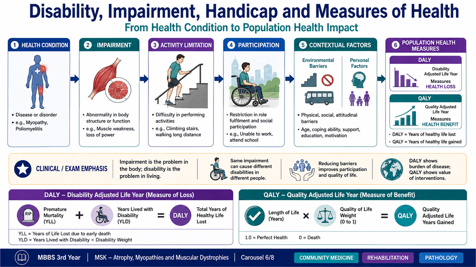
Muscle diseases and poliomyelitis may produce long-term limitations in movement, education, employment and social participation. Community medicine therefore considers not only the disease itself but also its effect on the person’s daily life and on the wider community.
Definition and Types of Disability
Disability is a limitation in functioning that arises from interaction between a person’s health condition and the physical, social or environmental barriers around that person. It may affect movement, communication, learning, self-care, work or participation in society.
Common types include:
- Physical disability: limitation of movement, mobility or physical activity.
- Sensory disability: impairment of vision, hearing or another sensory function.
- Intellectual or developmental disability: limitations in intellectual functioning and adaptive behaviour.
- Mental or psychosocial disability: restriction associated with mental-health conditions.
- Multiple disability: coexistence of more than one significant limitation.
Impairment, Disability and Handicap
These terms describe different levels of consequence. Although modern disability frameworks emphasize functioning and participation, the traditional distinctions remain important for understanding examination questions.
| Concept | Meaning | Poliomyelitis Example |
|---|---|---|
| Impairment | Loss or abnormality of a body structure or function | Paralysis and muscle wasting in one leg |
| Disability | Difficulty performing an activity in the expected manner | Difficulty walking without support |
| Handicap | Social disadvantage resulting from impairment or disability | Unable to attend school because the building is inaccessible |
The environment can increase or reduce disability. For example, a person with lower-limb weakness may participate effectively when transport, buildings, educational facilities and workplaces are accessible.
Disability-Adjusted Life Years
A disability-adjusted life year is a population measure of health loss. It combines years lost because of premature death with years lived in less than full health because of disease or disability.
A higher number of DALYs means a greater burden of disease. DALYs help public-health planners compare the effect of diseases that cause death with those that mainly produce long-term disability.
Quality-Adjusted Life Years
A quality-adjusted life year combines the length of life with the quality of health experienced during that time. One year lived in full health represents a greater health benefit than one year lived with severe limitation.
QALYs are useful when comparing the health benefit of different interventions. DALYs measure health loss, whereas QALYs measure health gained or lived in a particular state of health.
G. Poliomyelitis: Epidemiology, Determinants, Prevention and Control
Poliomyelitis is an acute viral infection that may damage motor neurons and cause flaccid paralysis. Most infected individuals do not develop paralysis, but infected persons can still spread the virus. The severe consequences of paralytic disease and the availability of effective vaccines make prevention and control major public-health priorities.
Transmission and Distribution
Poliovirus mainly spreads by the fecal–oral route. The virus enters through the mouth, multiplies in the throat and intestinal tract, and is excreted in feces. Transmission is therefore favoured by poor sanitation, unsafe water, inadequate hand hygiene and close contact.
Children who have not developed adequate immunity are especially susceptible. Areas with low vaccination coverage and poor sanitation allow continuing transmission. Population movement can carry infection from an affected area to a previously controlled area.
Important Determinants
- Inadequate routine immunization coverage.
- Missed children during vaccination activities.
- Poor sanitation and contaminated water.
- Overcrowding and close household contact.
- Population movement from areas where transmission continues.
- Weak disease surveillance and delayed response to suspected cases.
- Misinformation, refusal or difficulty accessing vaccination services.
Prevention
Immunization is the main method of prevention. Vaccination develops immunity before exposure and reduces the number of susceptible children in the community. High population immunity also interrupts transmission.
Preventive measures include:
- Complete routine poliovirus vaccination according to the approved immunization schedule.
- Additional vaccination activities where needed to close immunity gaps.
- Identification and vaccination of children who were previously missed.
- Safe water, sanitation and hand hygiene.
- Community education to address concerns and improve vaccine acceptance.
- Protection of vaccination workers and reliable access to high-risk communities.
Control Measures
Control requires rapid detection of possible transmission, laboratory confirmation and a prompt public-health response. Surveillance is essential because a single paralytic case may indicate that many silent infections are present in the community.
- Surveillance for acute flaccid paralysis: detects children with sudden flaccid weakness who require investigation.
- Appropriate specimen collection and laboratory testing: help determine whether poliovirus is responsible.
- Environmental surveillance: may detect poliovirus circulation through testing of sewage or wastewater.
- Rapid outbreak response: increases immunity around the affected population.
- Monitoring vaccination quality: identifies missed areas and groups.
- Cross-border and interregional coordination: reduces spread associated with population movement.

H. Rehabilitation after Poliomyelitis and Chronic Muscle Weakness
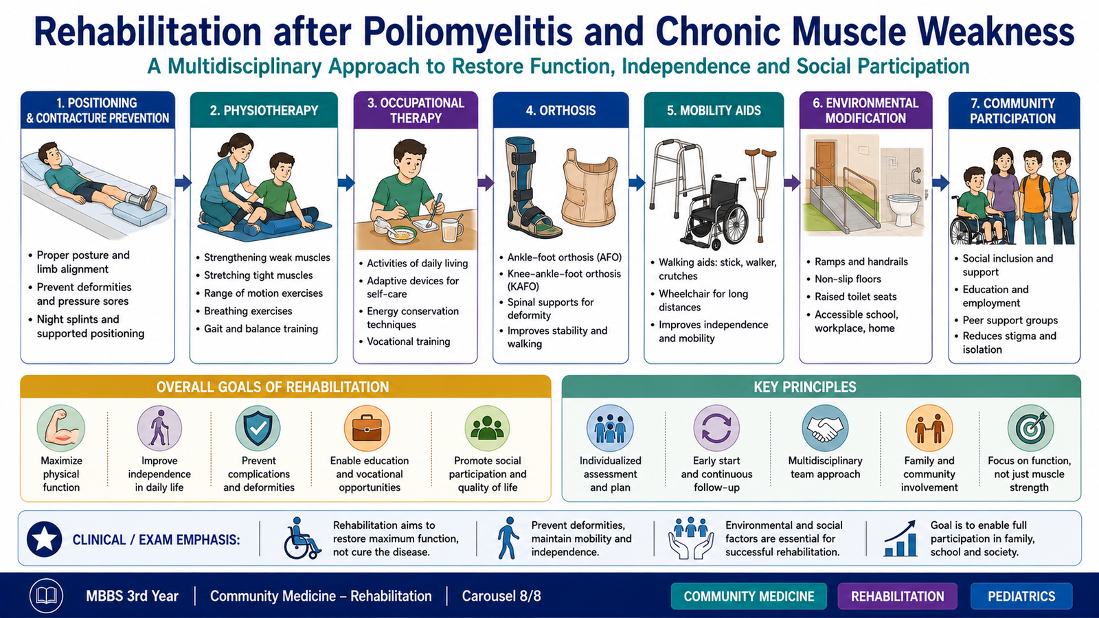
Rehabilitation is a coordinated process that helps a person achieve the greatest possible independence, function and participation after disease or injury. In poliomyelitis, the main goals are to preserve remaining muscle power, prevent deformity, improve mobility and support participation in education, work and community life.
Early Rehabilitation
During the acute stage, affected muscles are weak and vulnerable to fatigue. Care therefore focuses on correct positioning, protection of joints, prevention of pressure injury and maintenance of movement without excessive strain.
- Support the affected limbs in a functional position.
- Use gentle passive movement to reduce stiffness and contracture.
- Prevent pressure sores through regular repositioning and skin care.
- Support breathing and swallowing when related muscles are affected.
- Avoid unnecessarily exhausting severely weakened muscles.
Functional Recovery
After the acute phase, rehabilitation becomes more active. The rehabilitation plan should match the pattern of residual weakness and the person’s daily needs. Strengthening is directed toward muscles that can respond, while compensatory methods are taught for muscles with permanent paralysis.
- Physiotherapy: improves strength, flexibility, posture, balance and endurance.
- Occupational therapy: develops independence in self-care, school, work and household activities.
- Orthoses and splints: stabilize weak joints, improve alignment and reduce deformity.
- Walking aids: canes, crutches or walkers improve safe mobility.
- Wheelchair support: may be required when walking is unsafe or excessively tiring.
- Corrective orthopedic assessment: may be needed for significant contractures, deformity or joint instability.
Social, Educational and Vocational Rehabilitation
Successful rehabilitation is not limited to muscle strength. A child or adult may remain disadvantaged if buildings, transport, schools or workplaces are inaccessible. Rehabilitation must therefore reduce environmental barriers and support social participation.
- Accessible education and learning support.
- Home and workplace modifications.
- Vocational training matched to the person’s abilities.
- Psychological support and reduction of social stigma.
- Family education and caregiver support.
- Community-based rehabilitation where facility access is limited.
Prevention of Secondary Complications
Long-standing weakness changes posture and the distribution of forces across joints. Without follow-up, this may produce contractures, deformity, pain, reduced mobility and further loss of independence.
- Regular review of muscle strength and joint movement.
- Stretching and appropriate exercise.
- Monitoring of orthoses and mobility aids as the child grows.
- Prevention and treatment of pressure injury.
- Respiratory assessment when breathing muscles are weak.
- Energy conservation when activity produces excessive fatigue.
Integrated Mechanism Flow
⭐ AIM High-Yield Review
- Muscle atrophy is mainly a reduction in muscle-fibre size caused by loss of activity, nutrition, blood supply or nerve stimulation.
- Neurogenic atrophy produces small angular fibres arranged in groups.
- Myopathic injury produces scattered fibre-size variation, necrosis, regeneration and internal nuclei.
- ⭐ Perifascicular atrophy is the hallmark pathological feature of dermatomyositis.
- Polymyositis shows endomysial cytotoxic T-cell inflammation with direct muscle-fibre invasion.
- Inclusion-body myositis commonly affects older adults and shows rimmed vacuoles.
- Toxic myopathy may progress to rhabdomyolysis and myoglobin-related kidney injury.
- Muscular dystrophies cause repeated fibre necrosis followed by fatty and fibrous replacement.
- ⭐ Duchenne muscular dystrophy is an X-linked dystrophin disorder with early progressive proximal weakness.
- Gowers sign results from weakness of pelvic-girdle and proximal lower-limb muscles.
- Calf pseudohypertrophy in Duchenne muscular dystrophy is caused by fat and connective tissue, not stronger muscle.
- Markedly raised creatine kinase and confirmatory genetic testing support the diagnosis of Duchenne muscular dystrophy.
- Impairment concerns body structure or function, disability concerns activity, and handicap concerns social disadvantage.
- ⭐ DALYs measure health loss, while QALYs combine survival with quality of health.
- Poliomyelitis control depends on high vaccination coverage, surveillance of acute flaccid paralysis, rapid response and rehabilitation of affected patients.
🎥 Skeletal-Muscle Atrophy, Myopathies and Muscular Dystrophies
Watch this lecture after reading the AIM Learning Material to reinforce the pathological patterns, major myopathies and muscular dystrophies.
📌 Learning Focus
While watching, focus on:
- Neurogenic versus myopathic patterns of muscle injury
- Muscle-fibre atrophy, necrosis and regeneration
- Inflammatory and acquired myopathies
- Muscular dystrophies and dystrophin abnormalities
- Duchenne muscular dystrophy and its clinical clues
Complete the Community Medicine portion separately from the AIM Learning Material, including poliomyelitis epidemiology, prevention and control, disability concepts, DALY, QALY and rehabilitation.