
📖 Step 2 — Learning Material
🔹 1️⃣ Introduction
The nervous system is the master control and communication system of the body. It receives information from the external environment and internal organs, processes that information, and produces appropriate motor, autonomic, endocrine, emotional, and cognitive responses. Anatomically, it is located throughout the body but is centrally organized into the brain and spinal cord, with nerves extending to every region of the body.
This topic is important because all higher functions such as sensation, movement, reflexes, memory, consciousness, behavior, and autonomic regulation depend on the organization of neurons, glia, brain, spinal cord, and peripheral nerves. Clinically, understanding this foundation helps explain paralysis, sensory loss, cranial nerve lesions, autonomic dysfunction, developmental brain anomalies, and disorders of myelination.
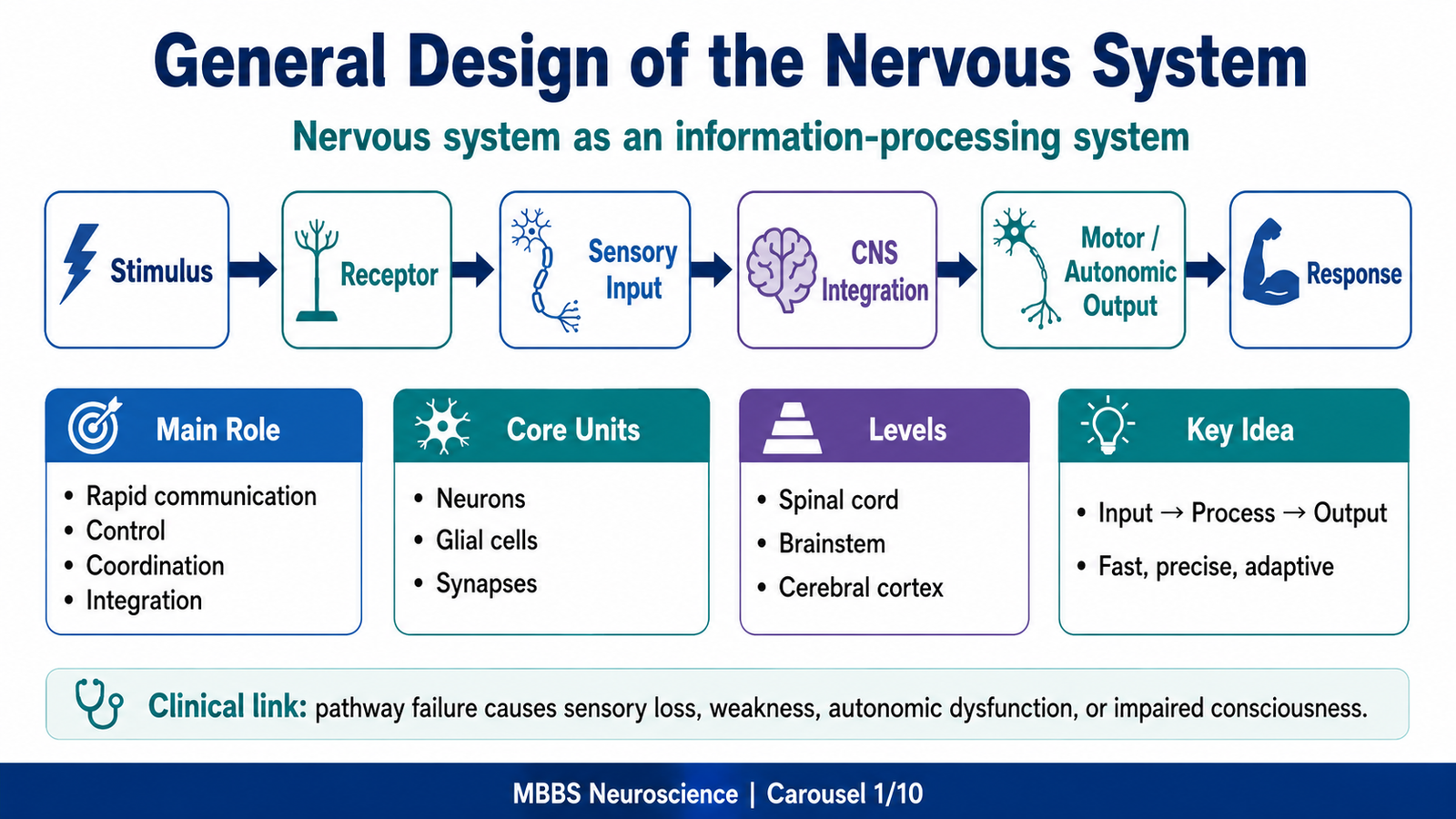
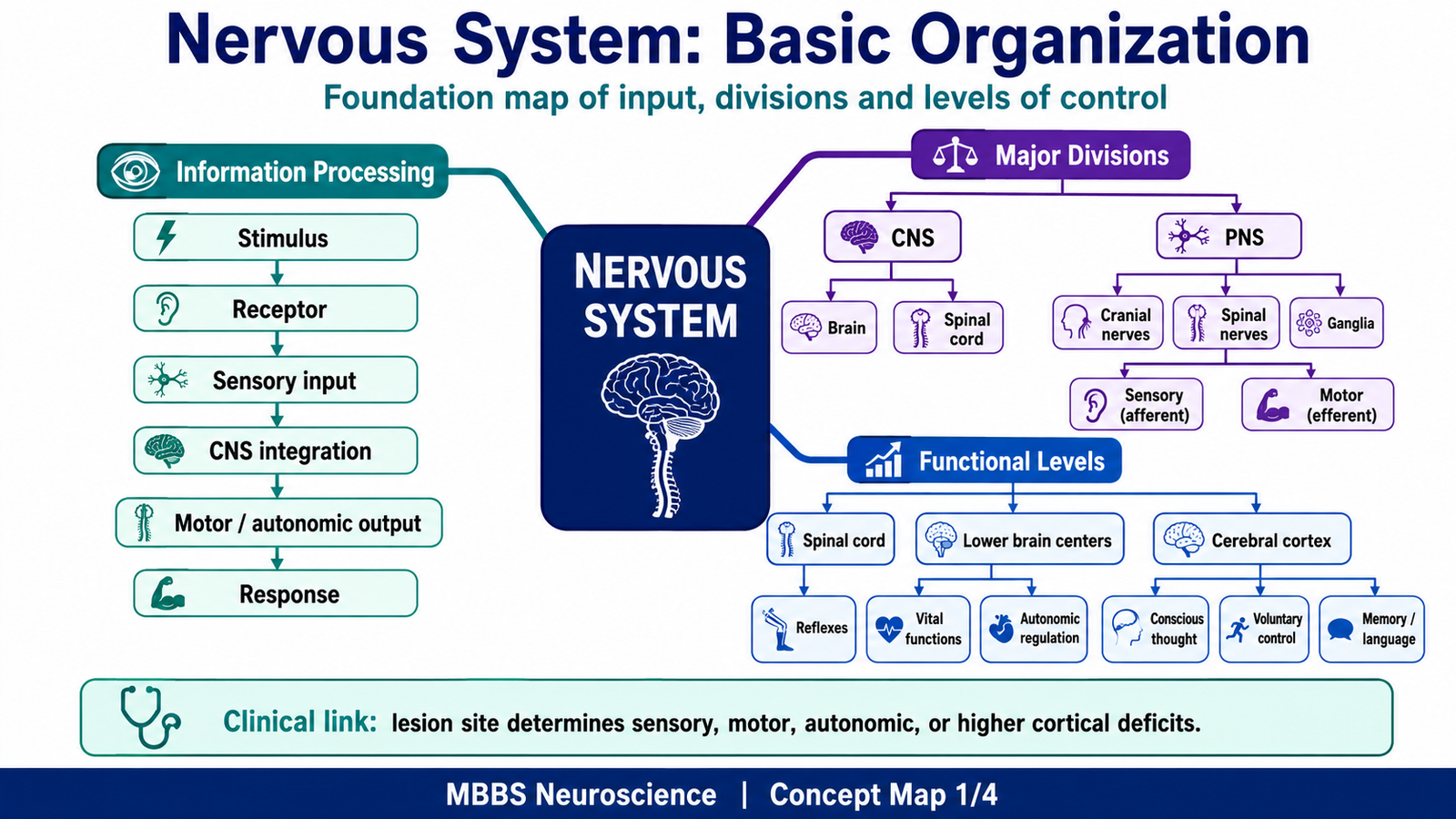
The nervous system should be studied as an information-processing system: input → processing → output → response.
🔹 2️⃣ Foundation Concepts
Key Definitions
- Nervous system: Body system responsible for rapid communication, coordination, control, and integration.
- Neuron: Structural and functional unit of the nervous system.
- Glial cells: Supportive cells that maintain, protect, nourish, insulate, and repair nervous tissue.
- Central Nervous System: Brain and spinal cord.
- Peripheral Nervous System: Cranial nerves, spinal nerves, ganglia, and peripheral nerve endings.
- Afferent pathway: Carries sensory information toward the CNS.
- Efferent pathway: Carries motor/autonomic commands away from the CNS.
- Somatic nervous system: Controls skeletal muscles and conscious sensation.
- Autonomic nervous system: Controls smooth muscle, cardiac muscle, and glands.
- Sympathetic system: Prepares body for stress, activity, and emergency responses.
- Parasympathetic system: Supports rest, digestion, conservation of energy, and maintenance functions.
Essential Terminology
- Cell body / soma: Metabolic center of neuron.
- Dendrites: Receive incoming signals.
- Axon: Conducts impulses away from cell body.
- Synapse: Functional junction between neurons or between neuron and effector.
- Myelin: Insulating sheath that increases speed of nerve impulse conduction.
- Ganglion: Collection of neuronal cell bodies outside CNS.
- Nucleus: Collection of neuronal cell bodies inside CNS.
- Tract: Bundle of nerve fibers inside CNS.
- Nerve: Bundle of nerve fibers outside CNS.
- Grey matter: Mainly neuronal cell bodies, dendrites, synapses, and unmyelinated fibers.
- White matter: Mainly myelinated axons.
Basic Overview
- The nervous system is organized into CNS and PNS.
- Functionally, it is divided into sensory, integrative, motor, somatic, and autonomic components.
- The neuron receives, processes, and transmits information.
- Glial cells do not usually conduct impulses like neurons, but they are essential for survival and function of neurons.
- The brain develops from the neural tube through primary and secondary brain vesicles.
- Developmental errors may produce major congenital anomalies of the brain and spinal cord.
🔹 3️⃣ Core Learning — Curriculum Coverage
MAJOR CONCEPT 1 — General Design of the Nervous System as an Information-Processing System
🧠 CORE
- Nervous system works by receiving sensory input, processing it, and producing output.
- It controls voluntary movement, reflexes, autonomic functions, behavior, memory, and consciousness.
- CNS acts as the main processing center.
- PNS connects CNS with receptors, muscles, glands, and organs.
- Information flow occurs through neurons and synapses.
- Glial cells maintain the environment required for neuronal function.
- The system is highly organized into levels: spinal cord, lower brain centers, and cerebral cortex.
🔬 CONCEPT EXPLAINED
The nervous system exists because the body needs a fast method of communication and control. Chemical messengers such as hormones are useful for slower and prolonged responses, but moment-to-moment control of movement, sensation, reflexes, and organ function requires rapid electrical signaling.
The basic functional plan is:
Stimulus → receptor → sensory neuron → CNS processing → motor/autonomic output → response
For example, when the hand touches a hot object, pain and temperature receptors detect the stimulus. Sensory neurons carry the information to the spinal cord. The CNS rapidly processes the signal and sends motor output to muscles, causing withdrawal. At the same time, the information may ascend to higher brain centers where pain becomes consciously perceived.
This shows that the nervous system is not merely a collection of nerves. It is an organized information-processing system designed to detect change, interpret meaning, store experience, and generate purposeful responses.
⚠️ CLINICAL IMPORTANCE
- Damage to sensory input pathways causes numbness or loss of sensation.
- Damage to motor output pathways causes weakness or paralysis.
- Damage to integrative centers may cause loss of coordination, consciousness, speech, memory, or behavior.
- Damage to autonomic pathways may disturb blood pressure, sweating, heart rate, bladder control, or bowel function.
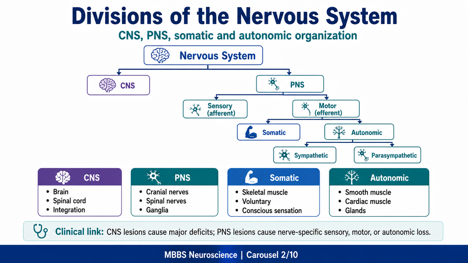
MAJOR CONCEPT 2 — Divisions of the Nervous System: CNS, PNS, Somatic and Autonomic Systems
🧠 CORE
- CNS: Brain and spinal cord; main site of processing and integration.
- PNS: Cranial nerves, spinal nerves, ganglia, and peripheral receptors.
- Somatic nervous system: Voluntary movement and conscious sensation.
- Autonomic nervous system: Involuntary control of viscera.
- Afferent division: Carries sensory input to CNS.
- Efferent division: Carries commands from CNS to muscles and glands.
- CNS and PNS are anatomically separate but functionally continuous.
🔬 CONCEPT EXPLAINED
The nervous system is divided anatomically into CNS and PNS because one part acts as the central command and processing system, while the other acts as the communication network between the CNS and the rest of the body.
The CNS includes the brain and spinal cord. It analyzes sensory information, stores memory, plans movement, regulates emotion, and controls autonomic and endocrine functions. The PNS connects this central system with the body through nerves and ganglia.
Functionally, the PNS has two major output systems. The somatic nervous system supplies skeletal muscle and allows voluntary movement. It also carries conscious sensory information such as touch, pain, temperature, and proprioception. The autonomic nervous system supplies smooth muscle, cardiac muscle, and glands. It controls involuntary functions such as heart rate, gut motility, pupil size, sweating, and blood vessel diameter.
This division is important because the same CNS may produce different types of output depending on the target organ. Skeletal muscle requires somatic motor control, while heart, gut, vessels, and glands require autonomic control.
⚠️ CLINICAL IMPORTANCE
- CNS lesions often produce complex deficits such as paralysis, altered consciousness, speech problems, or sensory level.
- PNS lesions usually produce deficits in the distribution of a specific nerve or root.
- Somatic motor damage causes weakness and loss of voluntary movement.
- Autonomic damage causes abnormal sweating, blood pressure instability, urinary retention, constipation, or sexual dysfunction.
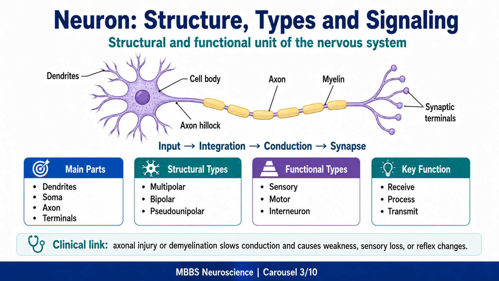
MAJOR CONCEPT 3 — Neuron: Structural and Functional Unit of the Nervous System
🧠 CORE
- Neuron is the structural and functional unit of the nervous system.
- It is specialized for excitability, conductivity, and communication.
- Main parts are cell body, dendrites, axon, and synaptic terminals.
- Dendrites receive information.
- Cell body maintains the neuron and integrates signals.
- Axon conducts impulses away from the cell body.
- Synapse transfers information to another neuron, muscle, or gland.
- Neurons are classified structurally and functionally.
🔬 CONCEPT EXPLAINED
A neuron is designed to receive, process, and transmit information. Its structure directly supports its function.
The cell body contains the nucleus and major organelles. It is the metabolic center of the neuron because it synthesizes proteins, neurotransmitters, enzymes, and structural components required for neuronal survival. If the cell body is severely damaged, the neuron may die.
The dendrites are branching processes that increase surface area for receiving incoming signals. Their branched design allows one neuron to receive information from many other neurons. This is important because nervous system function depends on integration, not isolated signaling.
The axon is a long process that carries impulses away from the cell body. Its length allows communication across long distances, such as from the spinal cord to muscles of the limb. Axons may be myelinated or unmyelinated. Myelin increases conduction speed by allowing impulses to jump between nodes of Ranvier.
The synaptic terminal communicates with the next cell. At chemical synapses, electrical activity in the presynaptic neuron causes neurotransmitter release, which then affects the postsynaptic cell.
Therefore, the neuron follows a logical direction of information flow:
Dendrite → cell body → axon hillock → axon → synaptic terminal
Structural Classification of Neurons
- Multipolar neurons: One axon and many dendrites; most common; typical motor neurons and interneurons.
- Bipolar neurons: One axon and one dendrite; found in special senses such as retina and olfactory pathway.
- Pseudounipolar neurons: Single process divides into peripheral and central branches; common in sensory ganglia.
- Anaxonic neurons: No clearly identifiable axon; involved mainly in local integration.
Functional Classification of Neurons
- Sensory neurons: Carry information from receptors to CNS.
- Motor neurons: Carry commands from CNS to muscles or glands.
- Interneurons: Connect neurons within CNS and perform integration.
⚠️ CLINICAL IMPORTANCE
- Damage to sensory neurons causes numbness, paresthesia, or loss of pain and temperature.
- Damage to motor neurons causes weakness, paralysis, wasting, or abnormal reflexes.
- Damage to interneuronal circuits may disturb coordination, reflexes, or higher processing.
- Axonal injury may interrupt communication even if the neuron cell body survives.
MAJOR CONCEPT 4 — Functional Components of a Neuron and Neural Signaling
🧠 CORE
- Neurons have input, integrative, conductive, and output zones.
- Dendrites and cell body form the receptive region.
- Axon hillock is the trigger zone for action potential generation.
- Axon is the conducting region.
- Synaptic terminals form the output region.
- Neural signaling depends on membrane excitability and ion movement.
- Synapses allow one neuron to influence another cell.
🔬 CONCEPT EXPLAINED
A neuron works because its membrane can generate and conduct electrical signals. This ability depends on ion gradients, especially sodium, potassium, calcium, and chloride. These gradients are maintained by membrane channels and energy-dependent pumps.
The input zone receives excitatory and inhibitory signals through dendrites and the cell body. These signals may bring the membrane potential closer to or farther from threshold.
The trigger zone is usually the axon hillock. If the total excitatory effect is strong enough to reach threshold, an action potential is generated.
The conducting zone is the axon. The action potential travels along the axon without losing strength. In myelinated fibers, conduction is faster because depolarization occurs mainly at nodes of Ranvier.
The output zone is the synaptic terminal. When the action potential reaches the terminal, calcium enters the presynaptic ending. Calcium triggers neurotransmitter release into the synaptic cleft. The neurotransmitter binds receptors on the postsynaptic cell and changes its activity.
This mechanism allows the nervous system to convert information into electrical signals, transmit it rapidly, and then convert it into chemical communication at synapses.
⚠️ CLINICAL IMPORTANCE
- Failure of ion gradients due to ATP depletion can impair neuronal excitability.
- Demyelination slows conduction and may cause weakness, sensory symptoms, or visual problems.
- Synaptic dysfunction may alter movement, consciousness, mood, or autonomic function.
- Calcium-dependent neurotransmitter release is essential for neuromuscular and neuronal communication.
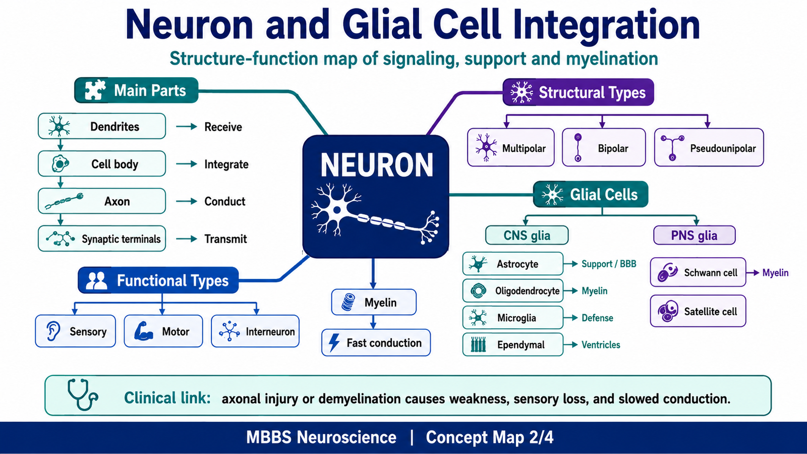
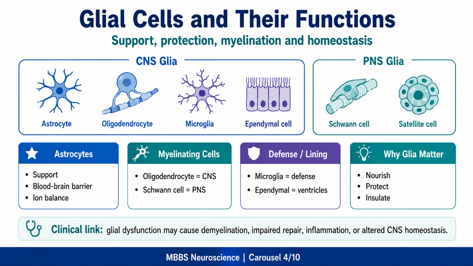
MAJOR CONCEPT 5 — Glial Cells and Their Functions
🧠 CORE
- Glial cells support neurons structurally, metabolically, and functionally.
- CNS glia include astrocytes, oligodendrocytes, microglia, and ependymal cells.
- PNS glia include Schwann cells and satellite cells.
- Glia regulate extracellular environment, myelination, immune defense, and CSF-related functions.
- They do not mainly conduct impulses like neurons.
- They are essential for normal neuronal survival and signaling.
- Glial dysfunction may produce demyelination, inflammation, or impaired CNS homeostasis.
🔬 CONCEPT EXPLAINED
Neurons are highly specialized for signaling, but they cannot function alone. They require a stable chemical environment, insulation, nutrients, protection, and structural support. Glial cells provide this essential support.
Astrocytes are star-shaped CNS glial cells. They help maintain the blood-brain barrier, regulate extracellular potassium, remove excess neurotransmitters, support neuronal metabolism, and contribute to repair after injury. They are important because neuronal function requires a stable extracellular environment.
Oligodendrocytes form myelin in the CNS. One oligodendrocyte can myelinate parts of several axons. Myelin increases conduction speed and improves efficiency.
Schwann cells form myelin in the PNS. Unlike oligodendrocytes, one Schwann cell usually myelinates one segment of one axon. Schwann cells also assist peripheral nerve regeneration.
Microglia are immune defense cells of the CNS. They remove debris and respond to injury or infection.
Ependymal cells line the ventricles and central canal. They are related to CSF circulation and ventricular lining.
Satellite cells surround neuronal cell bodies in peripheral ganglia and regulate their microenvironment.
⚠️ CLINICAL IMPORTANCE
- Loss of myelin causes slowed nerve conduction.
- CNS demyelination affects motor, sensory, visual, and coordination pathways.
- PNS demyelination may produce weakness and reduced reflexes.
- Microglial activation is important in CNS injury and inflammation.
- Ependymal or ventricular developmental problems may contribute to CSF circulation issues.
MAJOR CONCEPT 6 — General Features of Brain: Forebrain, Midbrain and Hindbrain
🧠 CORE
- Brain is the enlarged cranial part of the CNS.
- It is divided into forebrain, midbrain, and hindbrain.
- Forebrain includes cerebral hemispheres and diencephalon.
- Midbrain connects forebrain with hindbrain and contains important motor and sensory pathways.
- Hindbrain includes pons, medulla, and cerebellum.
- Brain controls consciousness, movement, sensation, autonomic regulation, coordination, behavior, and endocrine integration.
- Brain regions are anatomically distinct but functionally interconnected.
🔬 CONCEPT EXPLAINED
The brain is organized into regions that reflect both development and function.
The forebrain is the most expanded part in humans. It includes the cerebral hemispheres and diencephalon. The cerebral hemispheres are responsible for higher functions such as voluntary movement, sensation, language, memory, personality, and conscious perception. The diencephalon includes structures such as the thalamus and hypothalamus. The thalamus is a major relay station for sensory and motor information. The hypothalamus regulates autonomic, endocrine, temperature, hunger, thirst, and emotional responses.
The midbrain lies between forebrain and hindbrain. It contains ascending and descending tracts and important centers related to eye movements, visual reflexes, auditory reflexes, and motor control.
The hindbrain includes the pons, medulla oblongata, and cerebellum. The pons acts as a bridge between different parts of the CNS and contains important cranial nerve nuclei. The medulla contains vital centers for respiration, cardiovascular control, swallowing, and protective reflexes. The cerebellum coordinates movement, posture, balance, and motor learning.
Thus, the brain is arranged from higher conscious control in the cerebral cortex to essential survival functions in the brainstem.
⚠️ CLINICAL IMPORTANCE
- Forebrain lesions may cause weakness, sensory loss, aphasia, personality change, or seizures.
- Midbrain lesions may affect eye movements and motor pathways.
- Hindbrain lesions may disturb breathing, swallowing, balance, coordination, or cranial nerve function.
- Brainstem lesions are clinically serious because vital centers and major tracts are concentrated there.
MAJOR CONCEPT 7 — General Features of Spinal Cord and Spinal Enlargements
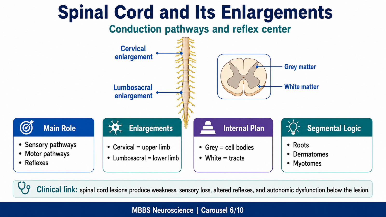
🧠 CORE
- Spinal cord is the elongated part of CNS within the vertebral canal.
- It connects brain with body through spinal nerves.
- It conducts sensory and motor information.
- It contains reflex centers.
- Cervical enlargement supplies upper limbs.
- Lumbosacral enlargement supplies lower limbs.
- Grey matter is central; white matter is peripheral.
- Segmental organization explains dermatomes, myotomes, and reflexes.
🔬 CONCEPT EXPLAINED
The spinal cord is both a conduction pathway and a reflex center. It carries sensory information upward to the brain and motor commands downward from the brain to muscles and autonomic neurons.
Anatomically, the spinal cord lies inside the vertebral canal. It is continuous above with the medulla oblongata and ends inferiorly around the upper lumbar vertebral level in adults. Spinal nerves arise segmentally from the cord.
The spinal cord has two important enlargements. The cervical enlargement is associated with nerve supply to the upper limb. The lumbosacral enlargement is associated with nerve supply to the lower limb. These enlargements exist because limb muscles require more motor neurons and sensory connections than the trunk.
In transverse section, the central grey matter contains neuronal cell bodies and synapses, while the surrounding white matter contains ascending and descending tracts. The posterior part of grey matter is mainly sensory, the anterior part is mainly motor, and the lateral horn is associated with autonomic neurons in relevant regions.
⚠️ CLINICAL IMPORTANCE
- Spinal cord injury may cause weakness, sensory loss, and autonomic dysfunction below the lesion.
- Cervical cord lesions may affect upper and lower limbs.
- Lumbosacral involvement affects lower limb, bladder, bowel, and sexual function.
- Segmental cord knowledge helps localize neurological lesions.
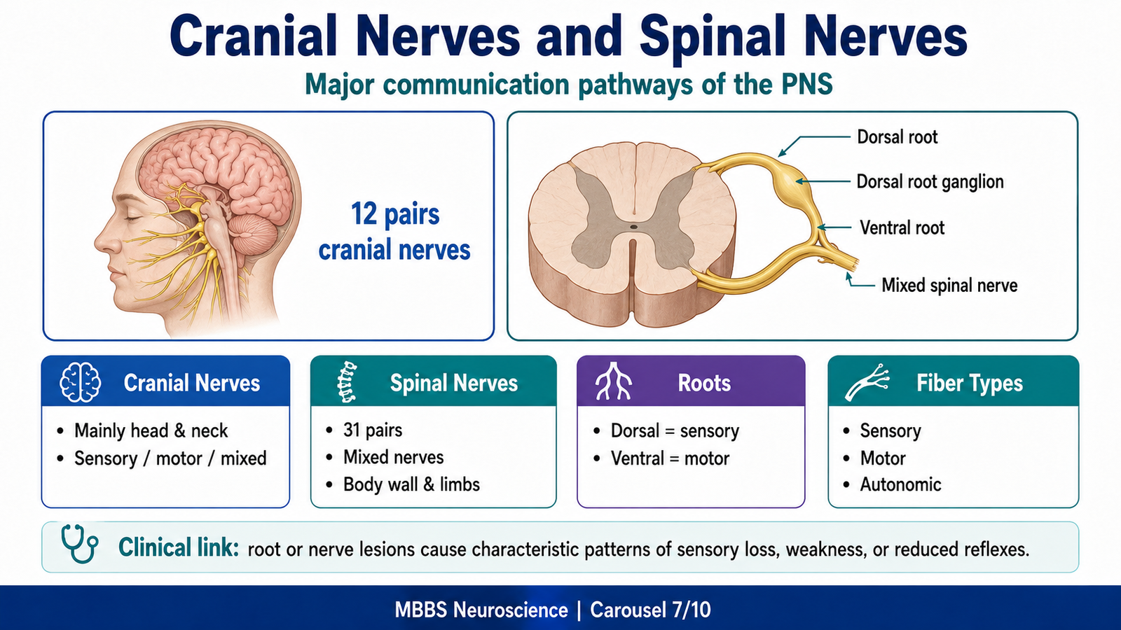
MAJOR CONCEPT 8 — General Features of Cranial Nerves and Spinal Nerves
🧠 CORE
- Cranial nerves arise mainly from the brain and brainstem.
- There are twelve pairs of cranial nerves.
- Spinal nerves arise from the spinal cord.
- Spinal nerves are formed by union of dorsal and ventral roots.
- Dorsal roots carry sensory fibers.
- Ventral roots carry motor fibers.
- Nerves may contain sensory, motor, autonomic, or mixed fibers.
- Peripheral nerves connect CNS with receptors and effectors.
🔬 CONCEPT EXPLAINED
Cranial and spinal nerves are the communication lines between the CNS and the body.
Cranial nerves mainly supply structures of the head and neck, with some extending to thoracic and abdominal viscera, especially the vagus nerve. They may be sensory, motor, or mixed. Some cranial nerves are related to special senses such as smell, vision, hearing, balance, and taste.
Spinal nerves arise in pairs from spinal cord segments. Each spinal nerve is formed by a dorsal root and a ventral root. The dorsal root carries sensory information toward the spinal cord and contains a dorsal root ganglion. The ventral root carries motor fibers away from the spinal cord. After joining, the spinal nerve becomes mixed and distributes to the body.
This arrangement is clinically important because injury at different levels produces different patterns. Dorsal root injury mainly causes sensory loss, while ventral root injury mainly causes motor weakness.
⚠️ CLINICAL IMPORTANCE
- Cranial nerve lesions produce characteristic deficits such as facial weakness, diplopia, dysphagia, hearing loss, or tongue deviation.
- Spinal nerve root compression may cause radicular pain, sensory loss, weakness, or reflex changes.
- Mixed nerve injury causes combined sensory and motor deficits.
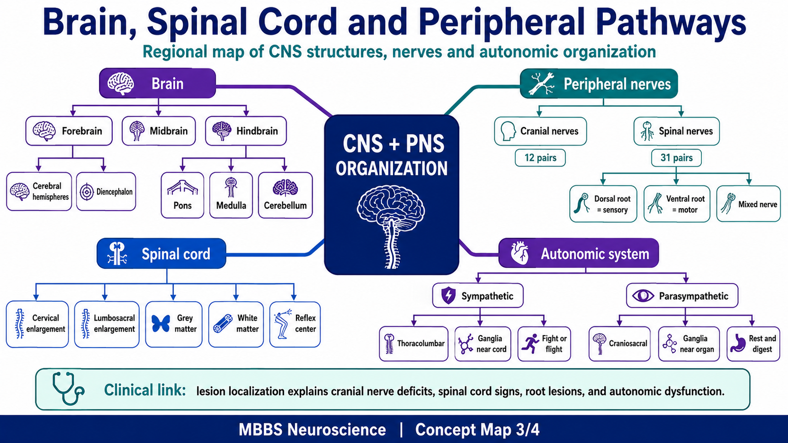
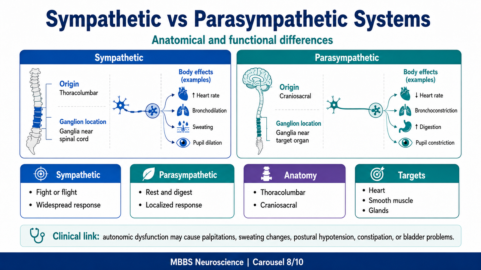
MAJOR CONCEPT 9 — Sympathetic and Parasympathetic Nervous Systems: Anatomical and Functional Differences
🧠 CORE
- Autonomic nervous system controls smooth muscle, cardiac muscle, and glands.
- Sympathetic system is thoracolumbar in origin.
- Parasympathetic system is craniosacral in origin.
- Sympathetic ganglia are usually near the spinal cord.
- Parasympathetic ganglia are usually near or within target organs.
- Sympathetic responses are widespread.
- Parasympathetic responses are more localized.
- Both systems maintain internal homeostasis.
🔬 CONCEPT EXPLAINED
The autonomic nervous system exists because many body functions must continue without conscious effort. Heart rate, blood pressure, sweating, digestion, pupil size, glandular secretion, bladder function, and gut motility must be continuously adjusted.
The sympathetic nervous system arises from thoracic and upper lumbar spinal cord segments. Its ganglia are located near the vertebral column in sympathetic chains or prevertebral ganglia. Because sympathetic fibers can diverge widely, sympathetic responses are often diffuse and coordinated. This is useful during stress, exercise, fear, blood loss, or emergency.
The parasympathetic nervous system arises from cranial nerves and sacral spinal cord segments. Its ganglia are usually near or inside the target organ. Therefore, parasympathetic effects are often more specific and localized. It promotes digestion, secretion, energy conservation, urination, defecation, and slowing of heart rate.
The anatomical difference explains the functional difference:
Sympathetic: thoracolumbar origin + ganglia near cord → widespread emergency response
Parasympathetic: craniosacral origin + ganglia near organ → localized maintenance response
⚠️ CLINICAL IMPORTANCE
- Sympathetic dysfunction may impair blood pressure control and sweating.
- Parasympathetic dysfunction may affect heart rate, gut motility, bladder emptying, and pupil response.
- Autonomic imbalance can contribute to palpitations, fainting, constipation, urinary retention, or postural hypotension.
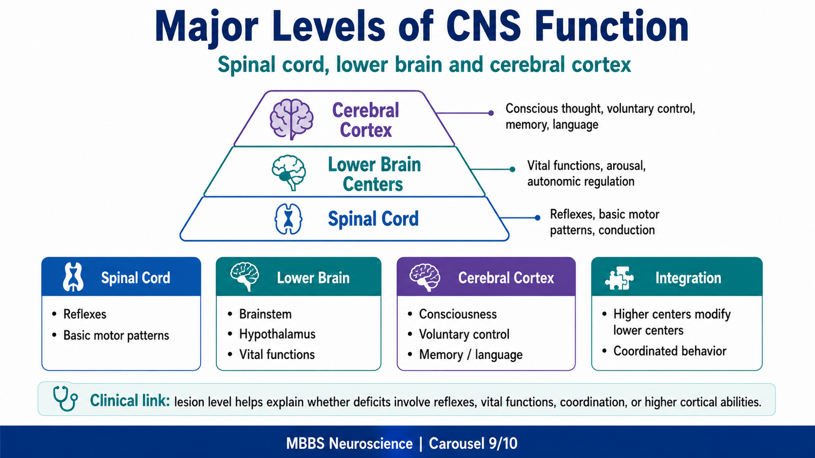
MAJOR CONCEPT 10 — Major Levels of Central Nervous System Function
🧠 CORE
- CNS function is organized into hierarchical levels.
- Spinal cord controls reflexes and basic motor patterns.
- Lower brain centers regulate vital, autonomic, and subconscious functions.
- Cerebral cortex performs conscious, voluntary, and higher intellectual functions.
- Higher centers can modify lower centers.
- Lower centers can perform essential functions without conscious control.
- Integration between levels allows coordinated behavior.
🔬 CONCEPT EXPLAINED
The CNS does not work as one uniform mass. It is organized into levels, with each level contributing specific functions.
The spinal cord level handles reflexes, basic sensory processing, and automatic motor responses. Withdrawal reflexes, stretch reflexes, and some locomotor patterns can occur through spinal circuits.
The lower brain level includes brainstem, hypothalamus, thalamus, basal ganglia, and cerebellum. These areas regulate vital functions such as respiration, cardiovascular control, posture, equilibrium, emotional responses, autonomic output, and many subconscious motor activities.
The higher brain or cortical level is responsible for conscious perception, voluntary movement, speech, reasoning, memory, planning, judgment, and learning. However, the cortex does not act alone. It depends on lower centers for posture, tone, arousal, autonomic support, and sensory relay.
This hierarchy allows efficiency. Routine functions are handled by lower centers, while higher centers are reserved for complex, conscious, and adaptive functions.
⚠️ CLINICAL IMPORTANCE
- Spinal cord lesions disturb reflexes, sensation, motor control, and autonomic function.
- Brainstem lesions may threaten life because respiratory and cardiovascular centers are present.
- Cerebellar lesions cause ataxia and loss of coordination.
- Cortical lesions may cause paralysis, aphasia, seizures, sensory neglect, or cognitive changes.
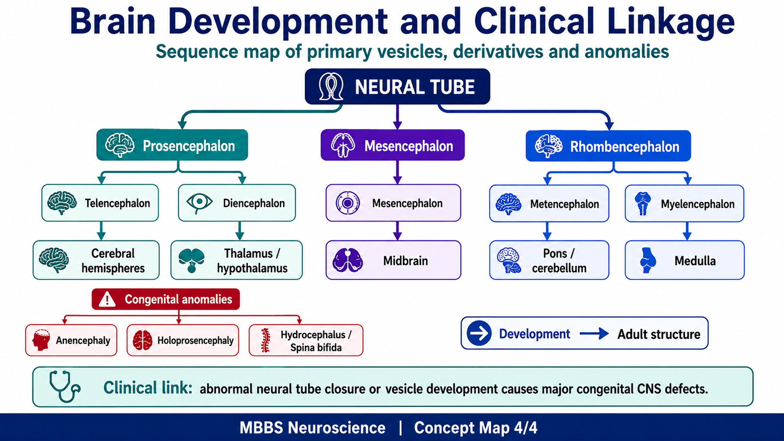
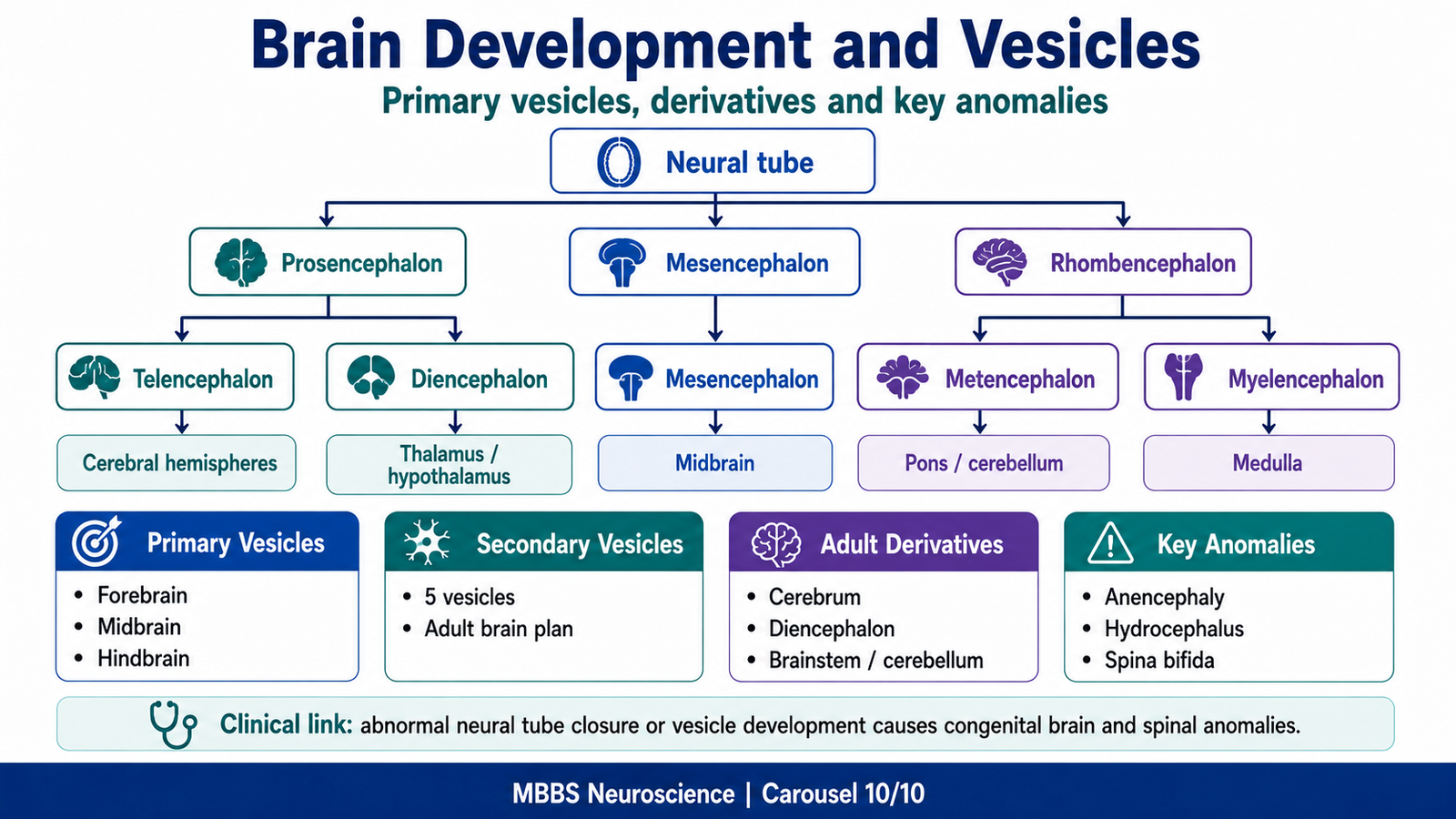
MAJOR CONCEPT 11 — Brain Development: Primary and Secondary Brain Vesicles
🧠 CORE
- Brain develops from the cranial part of the neural tube.
- Three primary brain vesicles form: prosencephalon, mesencephalon, and rhombencephalon.
- Five secondary brain vesicles form from these primary vesicles.
- Prosencephalon forms telencephalon and diencephalon.
- Mesencephalon remains as midbrain.
- Rhombencephalon forms metencephalon and myelencephalon.
- Brain vesicles give rise to adult brain regions and ventricular cavities.
- Developmental errors produce region-specific congenital anomalies.
🔬 CONCEPT EXPLAINED
The brain develops from the neural tube. The cranial end of the neural tube expands because it will form the brain. Early in development, it forms three primary brain vesicles:
- Prosencephalon — forebrain
- Mesencephalon — midbrain
- Rhombencephalon — hindbrain
These primary vesicles later form five secondary brain vesicles:
- Prosencephalon → Telencephalon + Diencephalon
- Mesencephalon → Mesencephalon
- Rhombencephalon → Metencephalon + Myelencephalon
Each vesicle forms specific adult structures. This developmental plan is important because adult brain anatomy becomes easier to understand when traced back to embryological origin.
Derivatives of Brain Vesicles
Telencephalon
- Cerebral hemispheres
- Cerebral cortex
- Basal nuclei
- Hippocampal formation
- Olfactory structures
- Lateral ventricles
Diencephalon
- Thalamus
- Hypothalamus
- Epithalamus
- Subthalamus
- Retina and optic nerve development are associated with diencephalic outgrowth
- Third ventricle
Mesencephalon
- Midbrain
- Cerebral aqueduct
Metencephalon
- Pons
- Cerebellum
- Upper part of fourth ventricle
Myelencephalon
- Medulla oblongata
- Lower part of fourth ventricle
Development → Adult Structure Link
The adult brain is not random in organization. The forebrain develops into higher centers, the midbrain remains a connecting and reflex region, and the hindbrain develops into vital centers and coordination structures. Therefore, embryology helps explain adult anatomy and clinical localization.
⚠️ CLINICAL IMPORTANCE
- Defects of forebrain development may affect cerebral hemispheres, cognition, smell, vision-related pathways, and hypothalamic functions.
- Midbrain developmental problems may affect eye movement pathways and connection between forebrain and hindbrain.
- Hindbrain developmental defects may affect breathing, swallowing, posture, balance, and cerebellar coordination.
MAJOR CONCEPT 12 — Development of Prosencephalon, Mesencephalon and Rhombencephalon with Congenital Anomalies
🧠 CORE
- Prosencephalon gives rise to telencephalon and diencephalon.
- Mesencephalon remains relatively undivided and forms midbrain.
- Rhombencephalon forms metencephalon and myelencephalon.
- Folding and expansion shape the developing brain.
- Ventricular cavities develop along with brain vesicles.
- Congenital anomalies depend on the region affected.
- Early developmental defects usually cause severe malformations.
- Clinical depth should focus on mechanism and functional consequence.
🔬 CONCEPT EXPLAINED
The prosencephalon undergoes major expansion to form the cerebral hemispheres and diencephalon. Because the cerebral cortex is responsible for higher function, defects in forebrain development may cause severe neurological impairment. Failure of proper division of the forebrain may result in holoprosencephaly, where cerebral hemispheres do not separate normally.
The mesencephalon forms the midbrain. It is less expanded than the forebrain but remains important because it contains pathways and nuclei related to eye movements, visual reflexes, auditory reflexes, and motor control.
The rhombencephalon forms the hindbrain. Its derivatives include pons, cerebellum, and medulla. Developmental problems in this region may affect coordination, balance, respiration, swallowing, and brainstem-related cranial nerve functions.
Congenital anomalies of the nervous system commonly occur when neural tube closure, brain vesicle formation, ventricular development, or regional differentiation is abnormal.
Important Congenital Anomalies — Undergraduate Plan
Anencephaly
- Failure of cranial neural tube closure.
- Major parts of brain and skull vault fail to develop.
- Severe and incompatible with life.
Spina bifida
- Failure of vertebral arch closure, often with neural tube involvement.
- May affect spinal cord and nerve roots.
- Functional consequence may include lower limb weakness, sensory loss, and bladder/bowel dysfunction.
Hydrocephalus
- Excess CSF accumulation within ventricles.
- May occur due to obstruction of CSF flow, overproduction, or impaired absorption.
- Causes ventricular dilatation and pressure effects on developing brain.
Holoprosencephaly
- Failure of normal division of prosencephalon into cerebral hemispheres.
- Can affect forebrain, facial development, and higher neurological function.
Arnold–Chiari malformation
- Hindbrain-related malformation with downward displacement of cerebellar/brainstem structures.
- May disturb CSF flow and brainstem/cerebellar function.
⚠️ CLINICAL IMPORTANCE
- Developmental brain anomalies are tested because they link embryology with adult functional loss.
- Early neural tube defects affect major brain and spinal structures.
- Vesicle-specific defects help localize which adult structures may be abnormal.
- Clinical learning should focus on cause, affected structure, and functional consequence rather than detailed management.
⚙️ 4️⃣ Functional Flow
Structure → Function → Outcome
Neuron
- Structure: Dendrites, cell body, axon, synaptic terminal
- Function: Receive, integrate, conduct, and transmit information
- Outcome: Rapid communication and coordinated response
Myelin
- Structure: Insulating sheath around axons
- Function: Increases conduction speed
- Outcome: Fast motor, sensory, and reflex activity
CNS
- Structure: Brain and spinal cord
- Function: Processing, integration, memory, motor planning, reflex control
- Outcome: Coordinated body control and higher function
PNS
- Structure: Cranial nerves, spinal nerves, ganglia
- Function: Connects CNS with body
- Outcome: Sensation, movement, autonomic regulation
Spinal Cord Enlargements
- Structure: Cervical and lumbosacral enlargements
- Function: Extra neurons for limb innervation
- Outcome: Fine control and sensory supply of upper and lower limbs
Brain Vesicles
- Structure: Primary and secondary embryonic vesicles
- Function: Form adult brain regions
- Outcome: Explains adult brain organization and congenital anomalies
🩺 5️⃣ Clinical Correlation
1. Demyelination
Myelin normally increases the speed and efficiency of impulse conduction. If myelin is damaged, nerve impulses slow down or fail to conduct properly. This can cause weakness, sensory symptoms, visual problems, poor coordination, or reduced reflexes depending on the pathway involved.
2. Peripheral Nerve Injury
Peripheral nerve damage interrupts communication between CNS and body. If sensory fibers are affected, numbness or pain may occur. If motor fibers are affected, weakness or paralysis occurs. If autonomic fibers are affected, sweating, blood flow, or visceral function may be disturbed.
3. Spinal Cord Lesion
A spinal cord lesion affects ascending sensory tracts, descending motor tracts, reflex pathways, and autonomic pathways. The level of lesion determines the pattern of deficit. Higher lesions produce more extensive functional loss.
4. Cranial Nerve Lesion
Cranial nerve lesions produce localized deficits related to the function of that nerve, such as facial weakness, visual disturbance, hearing loss, abnormal eye movements, dysphagia, or tongue weakness.
5. Hydrocephalus
Hydrocephalus occurs when CSF accumulates in ventricles. The ventricles enlarge and pressure may damage developing brain tissue. It is important because it connects embryology, ventricles, CSF flow, and neurological function.
6. Neural Tube Defects
Failure of neural tube closure may produce severe congenital anomalies such as anencephaly or spina bifida. These defects demonstrate the importance of early nervous system development.
📌 6️⃣ Summary Points
- The nervous system works as an information-processing system: input → integration → output.
- CNS consists of brain and spinal cord; PNS consists of cranial nerves, spinal nerves, ganglia, and peripheral receptors.
- The neuron is the structural and functional unit of the nervous system.
- Dendrites receive signals, cell body maintains and integrates, axon conducts, and synapse transmits.
- Neurons are structurally classified as multipolar, bipolar, pseudounipolar, and anaxonic.
- Neurons are functionally classified as sensory, motor, and interneurons.
- Glial cells support neurons through myelination, nutrition, protection, homeostasis, and repair.
- Brain is broadly organized into forebrain, midbrain, and hindbrain.
- Spinal cord enlargements correspond to upper limb and lower limb innervation.
- Sympathetic system is thoracolumbar, while parasympathetic system is craniosacral.
- Brain develops from primary and secondary brain vesicles.
- Congenital anomalies become understandable when linked to neural tube closure and brain vesicle development.