
📖 Step 2 — Learning Material
🔹 1️⃣ Introduction
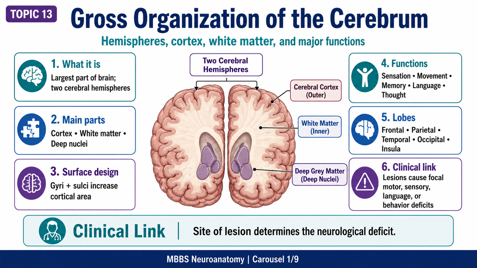
The cerebrum is the largest and most highly developed part of the brain, responsible for higher mental functions such as consciousness, memory, language, voluntary movement, sensation, reasoning, emotion, and personality. It occupies the supratentorial cranial cavity and forms the major bulk of the forebrain.
The cerebral hemispheres are divided externally into lobes by important sulci and gyri, while internally they contain grey matter nuclei and white matter fiber systems that connect different cortical and subcortical regions.
The diencephalon lies deep within the cerebral hemispheres around the third ventricle and includes clinically important structures such as the thalamus and hypothalamus. The thalamus acts mainly as a sensory relay station, while the hypothalamus regulates autonomic, endocrine, emotional, and homeostatic functions.
Histologically, the cerebral cortex shows a layered arrangement of neurons, especially pyramidal and granular cells. Recognition of cortical layers is important because different cortical areas are specialized for motor, sensory, and associative functions.
Clinically, lesions of the cerebrum and diencephalon can produce paralysis, sensory loss, aphasia, visual defects, memory disturbances, altered consciousness, endocrine imbalance, autonomic dysfunction, and behavioral changes.
🔹 2️⃣ Foundation Concepts
Key Definitions
• Cerebrum: Largest part of the brain, made of two cerebral hemispheres connected by commissural fibers, especially the corpus callosum.
• Cerebral hemisphere: One half of the cerebrum; each hemisphere has surfaces, lobes, sulci, gyri, cortex, white matter, and deep grey matter.
• Cerebral cortex: Outer layer of grey matter covering the cerebral hemispheres; responsible for higher cortical functions.
• Grey matter: Region containing neuronal cell bodies, dendrites, synapses, and neuroglia.
• White matter: Region mainly containing myelinated nerve fibers connecting different parts of the nervous system.
• Sulcus: A groove on the brain surface.
• Gyrus: A raised fold of cerebral cortex between sulci.
• Lobe: A major anatomical region of the cerebral hemisphere separated by prominent sulci.
• Association fibers: White matter fibers connecting cortical areas within the same hemisphere.
• Commissural fibers: Fibers connecting corresponding or related areas of the two cerebral hemispheres.
• Projection fibers: Fibers connecting cerebral cortex with lower parts of CNS such as thalamus, brainstem, and spinal cord.
• Corpus callosum: Largest commissural fiber bundle connecting the two cerebral hemispheres.
• Thalamus: Major diencephalic grey matter mass that relays sensory and motor information to the cerebral cortex.
• Hypothalamus: Diencephalic structure forming the floor and lower lateral wall of the third ventricle; regulates autonomic, endocrine, temperature, hunger, thirst, emotion, and circadian functions.
🔹 3️⃣ Core Learning — Curriculum Coverage
MAJOR CONCEPT 1: Gross Organization of the Cerebrum
🧠 CORE
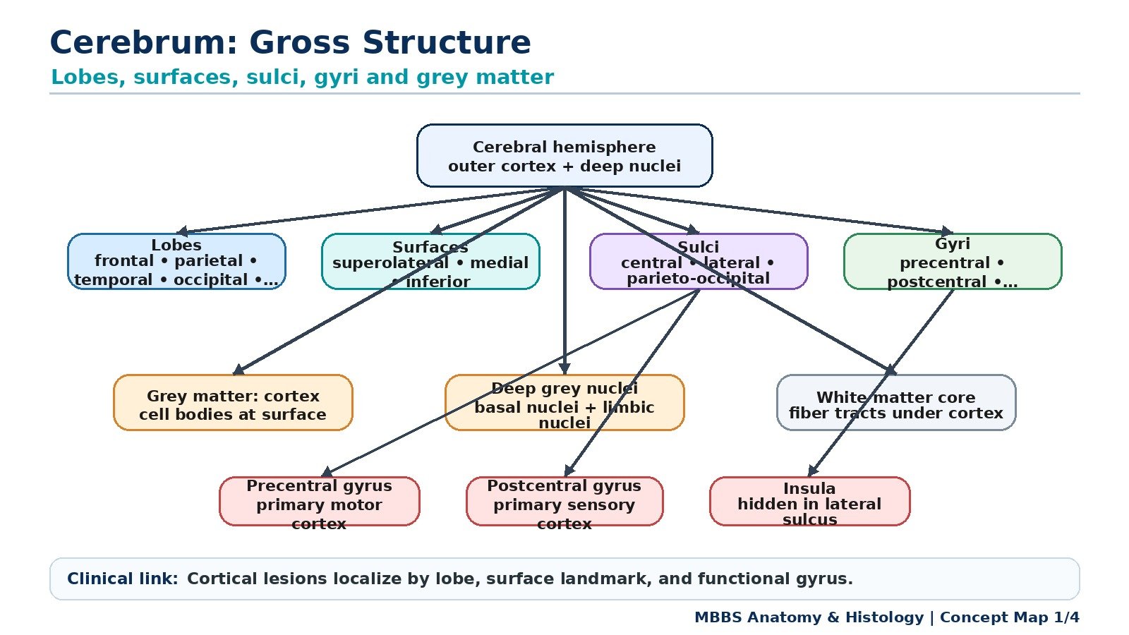
• The cerebrum consists of two cerebral hemispheres separated by the longitudinal fissure.
• Each hemisphere has an outer grey matter cortex, inner white matter, and deep grey matter nuclei.
• The two hemispheres are connected mainly by the corpus callosum.
• The cerebral surface is folded into gyri and sulci to increase cortical surface area.
• Major lobes include frontal, parietal, temporal, occipital, and insula.
• Cerebral hemispheres control contralateral body functions due to crossing of many motor and sensory pathways.
• The cerebrum performs sensory perception, voluntary movement, language, memory, personality, and conscious thought.
🔬 CONCEPT EXPLAINED
The cerebrum is designed to maximize processing capacity within the limited space of the cranial cavity. Instead of being smooth, its surface is folded into gyri and sulci. This folding increases the surface area of the cerebral cortex, allowing more neurons to be packed into the skull. Therefore, the anatomical design directly supports higher intellectual and sensory-motor functions.
Each cerebral hemisphere contains an outer layer of grey matter called the cerebral cortex. This cortex contains neuronal cell bodies and synaptic connections, making it the main site of information processing. Beneath the cortex lies white matter, which contains myelinated fibers that transmit information rapidly between cortical areas, between the two hemispheres, and between the cortex and lower CNS.
The cerebrum does not work as one uniform mass. Different cortical regions are specialized. The frontal lobe is strongly related to voluntary motor activity, planning, judgment, personality, and speech production. The parietal lobe processes general somatic sensations such as touch, pain, pressure, proprioception, and spatial awareness. The temporal lobe is important for hearing, memory, emotion, and language comprehension. The occipital lobe is the primary visual processing area. The insula is hidden deep in the lateral sulcus and is related to visceral sensation, taste, autonomic integration, and emotional awareness.
This organization allows the brain to localize functions while still integrating them through white matter connections. For example, when a student hears a question, the temporal cortex processes sound, association cortex interprets meaning, frontal cortex plans the answer, and motor cortex controls speech muscles. This shows how structure and function are integrated.
⚠️ CLINICAL IMPORTANCE
Damage to the cerebrum produces deficits according to the area involved. A frontal lobe lesion may cause weakness, personality change, poor judgment, or expressive aphasia. A parietal lesion may cause sensory loss or neglect. A temporal lesion may cause memory disturbance, hearing-related deficits, or receptive aphasia. An occipital lesion may cause visual field defects. Therefore, clinical signs help localize cerebral lesions.
MAJOR CONCEPT 2: Lobes, Surfaces, Sulci and Gyri of the Cerebral Hemisphere
🧠 CORE
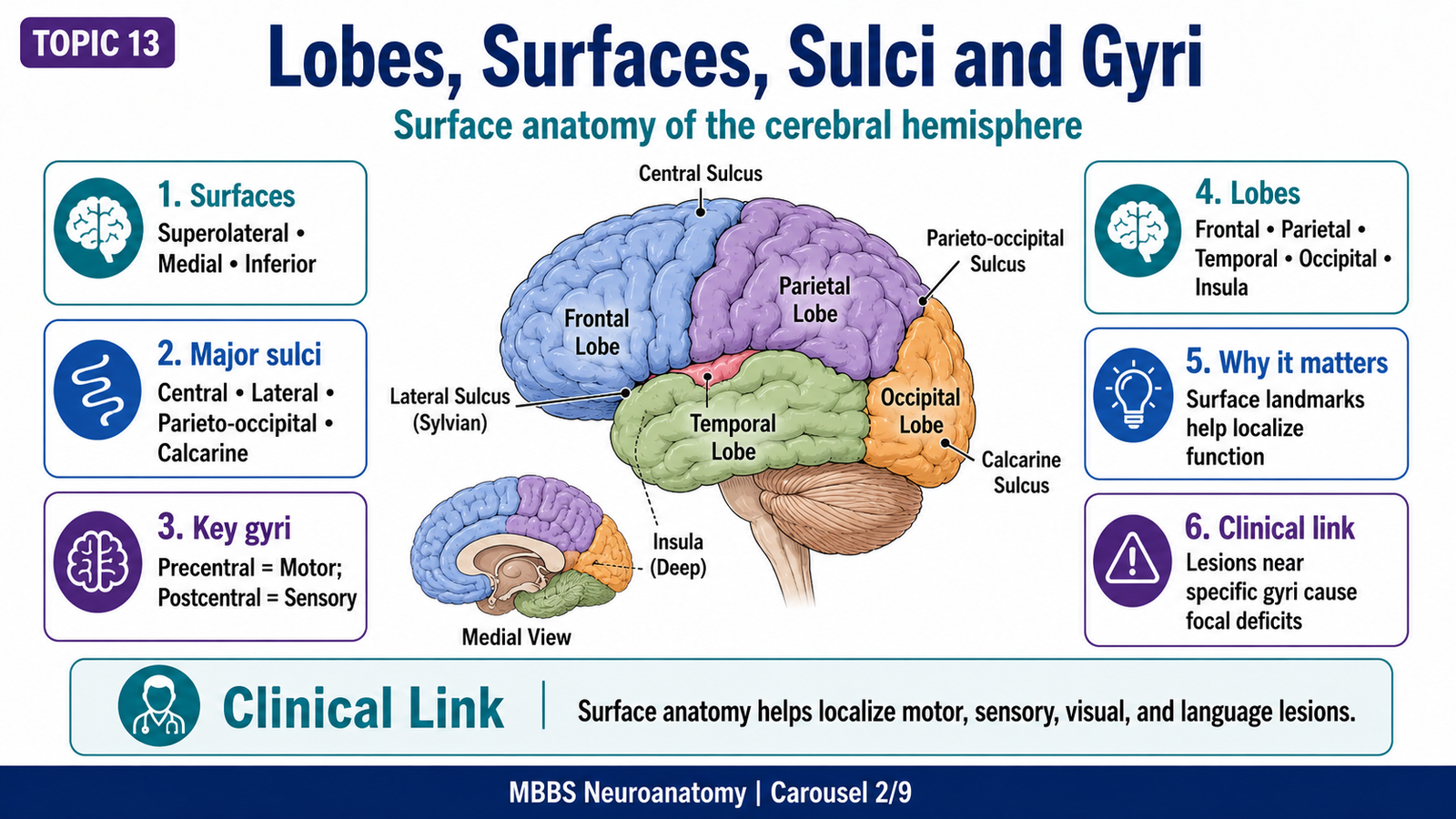
• Each cerebral hemisphere has superolateral, medial, and inferior surfaces.
• Important sulci include central sulcus, lateral sulcus, parieto-occipital sulcus, and calcarine sulcus.
• The central sulcus separates frontal and parietal lobes.
• The lateral sulcus separates temporal lobe from frontal and parietal lobes.
• The parieto-occipital sulcus helps separate parietal and occipital lobes.
• Gyri around major sulci contain important functional cortical areas.
• Surface anatomy is essential for localizing cortical functions.
🔬 CONCEPT EXPLAINED
To understand cerebral function, the student must first understand the map of the cerebral surface. The cerebral hemisphere has three major surfaces. The superolateral surface is the large convex surface seen from the side. The medial surface faces the opposite hemisphere across the longitudinal fissure. The inferior surface rests partly on the anterior and middle cranial fossae and tentorium cerebelli.
The central sulcus is one of the most important sulci. It separates the frontal lobe anteriorly from the parietal lobe posteriorly. The gyrus immediately anterior to it is the precentral gyrus, which contains the primary motor cortex. The gyrus immediately posterior to it is the postcentral gyrus, which contains the primary somatosensory cortex. This arrangement is clinically important because lesions anterior and posterior to the central sulcus produce different deficits: motor weakness anteriorly and sensory loss posteriorly.
The lateral sulcus is another major landmark. It separates the temporal lobe below from the frontal and parietal lobes above. Deep inside the lateral sulcus lies the insula. The temporal lobe contains the auditory cortex, memory-related structures, and language comprehension areas in the dominant hemisphere.
On the medial surface, the parieto-occipital sulcus and calcarine sulcus are important. The calcarine sulcus is especially important because the primary visual cortex is located along its margins. Therefore, lesions around the calcarine sulcus can produce visual field defects.
The lobes are not just anatomical divisions; they represent functional specialization. The frontal lobe is placed anteriorly, supporting planning, executive control, and motor output. The parietal lobe lies posterior to the central sulcus, receiving body sensations. The occipital lobe lies posteriorly, receiving visual input. The temporal lobe lies laterally and inferiorly, suited for auditory, memory, and language functions.
⚠️ CLINICAL IMPORTANCE
Knowledge of sulci and gyri helps localize cortical lesions in stroke, trauma, tumors, and epilepsy. For example, damage to the precentral gyrus causes contralateral upper motor neuron weakness, while damage to the postcentral gyrus causes contralateral sensory loss. Occipital lobe lesions may cause visual field defects, and dominant temporal or frontal lesions may produce aphasia.
MAJOR CONCEPT 3: Distribution of Grey Matter in the Cerebral Hemispheres
🧠 CORE
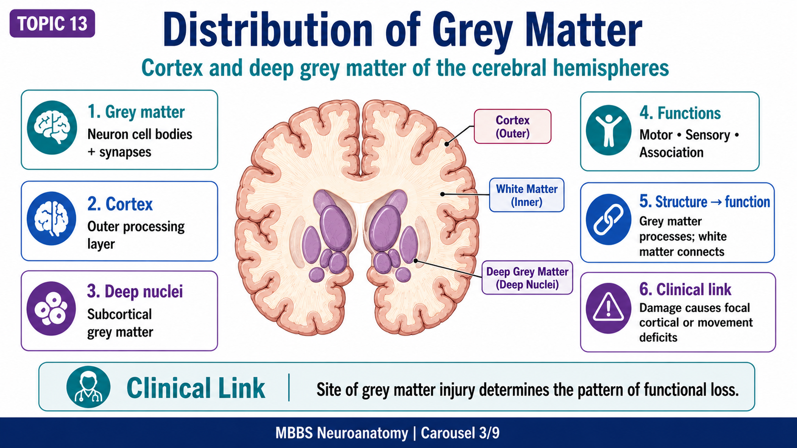
• Grey matter contains neuronal cell bodies, dendrites, synapses, and neuroglia.
• In the cerebral hemispheres, grey matter is present in the cerebral cortex and deep nuclei.
• Cerebral cortex forms the outer functional layer of the hemispheres.
• Deep grey matter includes basal nuclei and related subcortical nuclei.
• Grey matter is responsible for processing, integration, planning, and response generation.
• The cortex is organized into functional sensory, motor, and association areas.
• Grey matter damage produces functional loss depending on the involved region.
🔬 CONCEPT EXPLAINED
Grey matter is the processing tissue of the nervous system. In the cerebral hemispheres, it is mainly distributed in two locations: superficially as the cerebral cortex and deeply as subcortical grey matter nuclei.
The cerebral cortex forms a continuous sheet over the surface of the hemisphere. It receives information, analyzes it, stores memories, generates conscious perception, and initiates voluntary action. Because different areas of cortex have different inputs and outputs, cortical function is regionally specialized.
Deep grey matter includes the basal nuclei, which are functionally important in motor control. Although the basal nuclei are not the main focus of this topic, students should remember that not all cerebral grey matter is cortical. Some grey matter lies deep within the hemisphere and participates in movement regulation and behavioral circuits.
The structure-function relationship is clear: grey matter contains synapses and neuronal cell bodies, so it is suited for processing information. White matter contains myelinated axons, so it is suited for transmitting information. Therefore, proper brain function requires both grey matter processing and white matter communication.
⚠️ CLINICAL IMPORTANCE
Damage to cortical grey matter causes loss of the function represented in that cortical region. Motor cortex damage causes contralateral weakness. Sensory cortex damage causes sensory loss. Association cortex damage may cause neglect, apraxia, agnosia, language disturbance, or impaired judgment. Deep grey matter lesions may disturb movement control.
MAJOR CONCEPT 4: White Matter Fibers of the Cerebrum
🧠 CORE
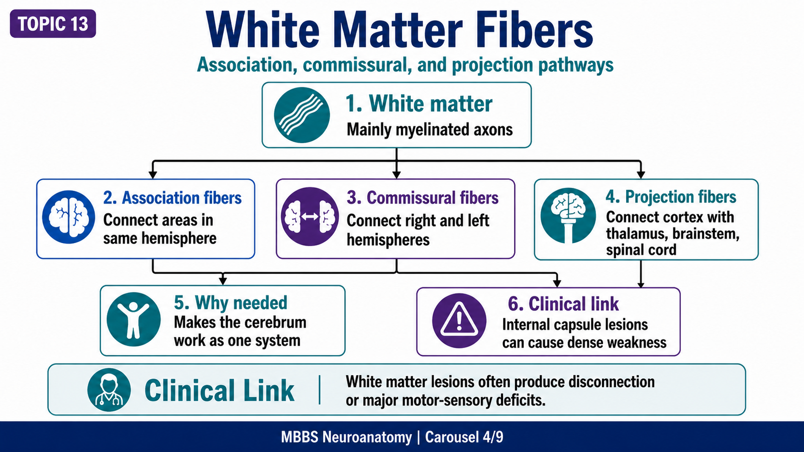
• Cerebral white matter consists mainly of myelinated axons.
• It connects different cortical and subcortical areas.
• Three major types are association, commissural, and projection fibers.
• Association fibers connect areas within the same hemisphere.
• Commissural fibers connect the two hemispheres.
• Projection fibers connect cortex with thalamus, brainstem, and spinal cord.
• White matter allows the cerebrum to work as an integrated system.
🔬 CONCEPT EXPLAINED
The cerebral cortex cannot function properly if each cortical area works in isolation. White matter fibers solve this problem by acting as communication pathways. They allow information to move rapidly from one brain area to another.
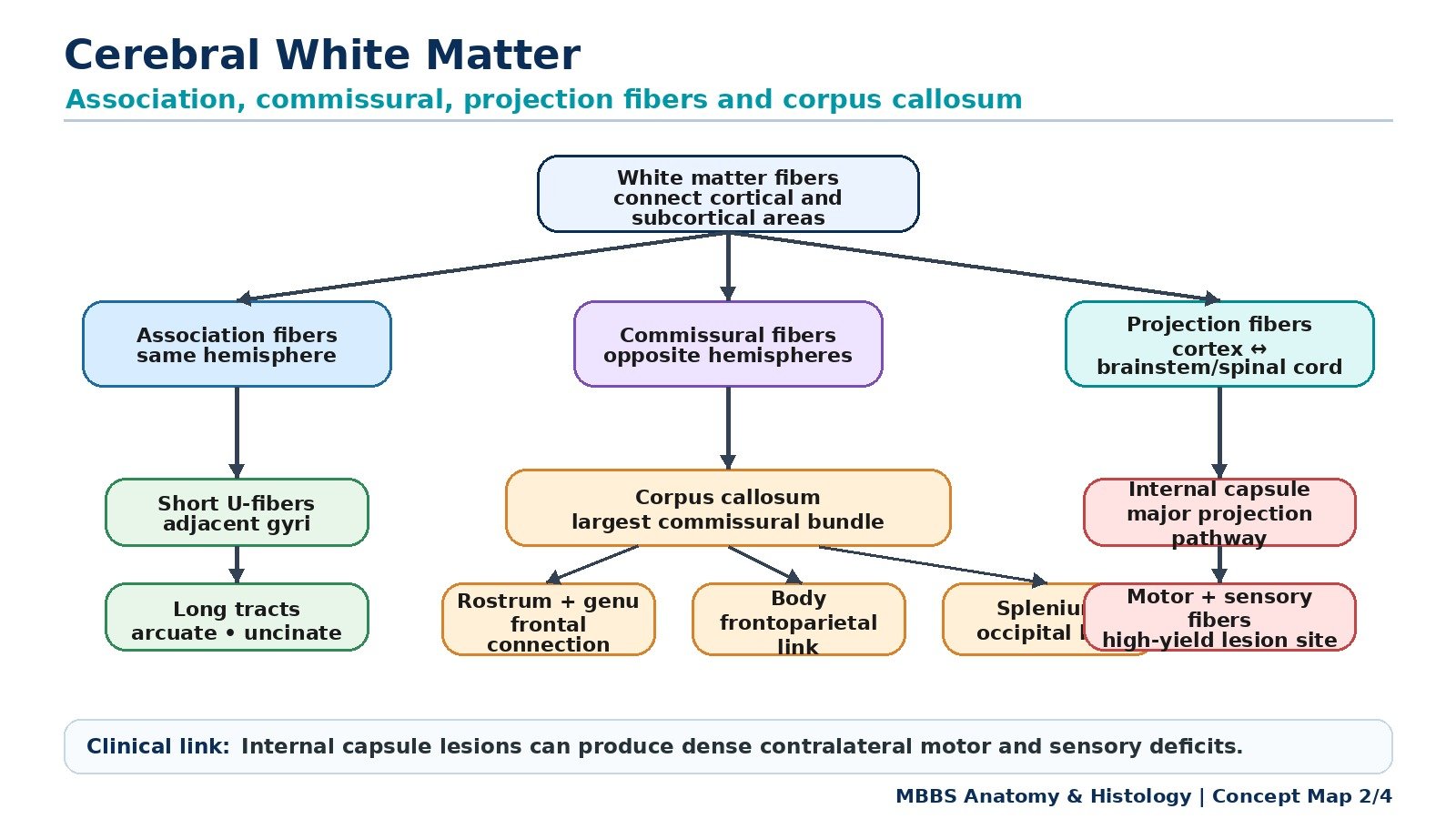
Association fibers connect cortical areas within the same cerebral hemisphere. Short association fibers connect neighboring gyri, while long association fibers connect distant lobes. For example, language requires association between auditory comprehension areas, visual areas, and speech production areas. If association fibers are damaged, cortical areas may remain intact but fail to communicate properly.
Commissural fibers connect the two cerebral hemispheres. This is necessary because many functions require bilateral coordination. The largest commissural bundle is the corpus callosum. Other commissural fibers include the anterior commissure and posterior commissure. Commissural connections allow information from one hemisphere to influence the other.
Projection fibers connect the cerebral cortex with lower CNS structures. They include ascending fibers carrying sensory information to cortex and descending fibers carrying motor commands from cortex to brainstem and spinal cord. Projection fibers pass through important regions such as the internal capsule. Because many fibers are compactly arranged there, a small lesion can cause major motor or sensory deficits.
Therefore, white matter is not just filling material. It is the communication system of the cerebrum. Grey matter processes information, but white matter distributes it.
⚠️ CLINICAL IMPORTANCE
White matter lesions can disconnect brain regions. Damage to projection fibers in the internal capsule may cause dense contralateral weakness or sensory loss. Damage to association fibers may cause higher cortical disconnection syndromes. Damage to commissural fibers may impair interhemispheric communication.
MAJOR CONCEPT 5: Corpus Callosum
🧠 CORE
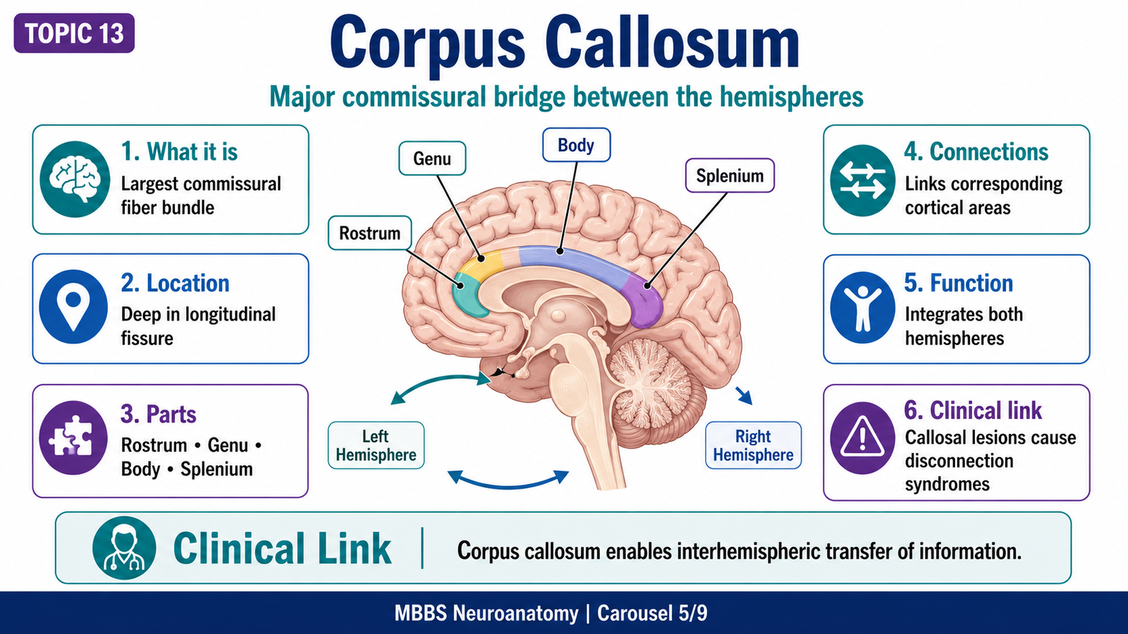
• Corpus callosum is the largest commissural fiber bundle of the brain.
• It connects corresponding and related cortical areas of the two cerebral hemispheres.
• It lies at the bottom of the longitudinal fissure and forms the roof of the lateral ventricles.
• Main parts are rostrum, genu, body, and splenium.
• Genu fibers connect frontal regions.
• Body fibers connect large areas of frontal, parietal, and temporal cortices.
• Splenium fibers connect occipital and posterior cortical regions.
• It allows coordination between right and left cerebral hemispheres.
🔬 CONCEPT EXPLAINED
The two cerebral hemispheres are anatomically separate, but functionally they must cooperate. The corpus callosum provides the main structural pathway for this cooperation. It contains millions of commissural fibers that cross the midline and connect cortical areas of both hemispheres.
The corpus callosum has four main parts. The rostrum is the thin anterior-inferior part. The genu is the curved anterior part. The body forms the long central portion. The splenium is the thick posterior end. These parts are not merely anatomical names; they reflect different fiber distributions. The genu mainly connects frontal lobes, which are involved in planning and executive functions. The body connects broad cortical regions, while the splenium connects posterior areas, especially visual association regions.
Functionally, the corpus callosum allows information processed in one hemisphere to be shared with the other. This is important because many functions show hemispheric dominance. For example, language is usually dominant in the left hemisphere, while spatial awareness is more strongly represented in the right hemisphere. Callosal fibers allow integration of these specialized functions.
⚠️ CLINICAL IMPORTANCE
Lesions of the corpus callosum may cause disconnection between hemispheres. In severe callosal damage, one hemisphere may not access information processed by the other. Clinically, callosal lesions can occur due to trauma, tumors, demyelination, infarction, or congenital agenesis. Students should remember the corpus callosum as the major interhemispheric bridge.
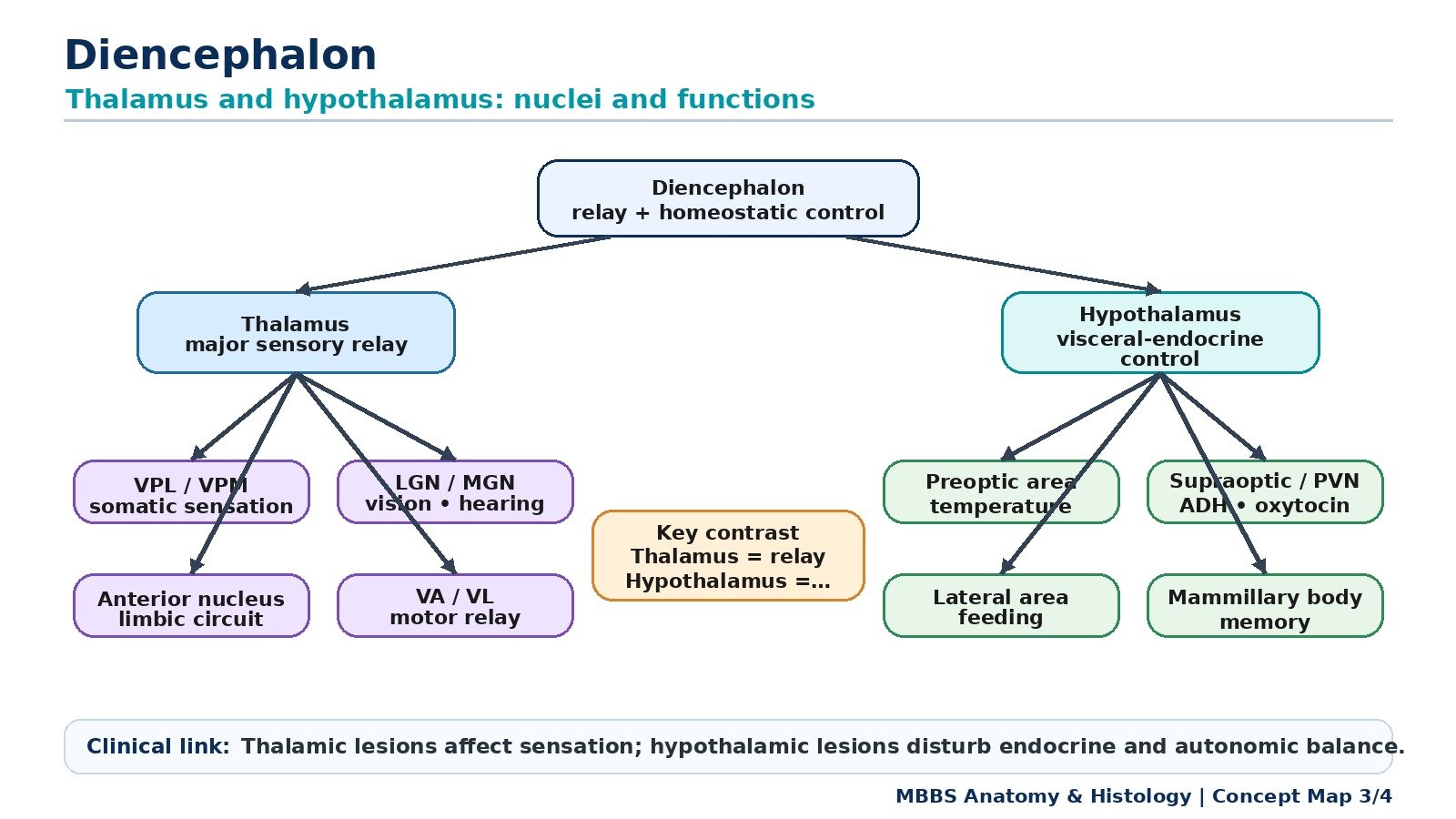
MAJOR CONCEPT 6: Diencephalon — General Organization
🧠 CORE
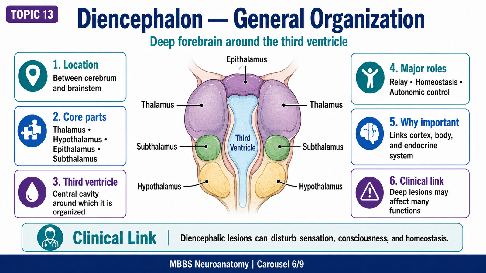
• The diencephalon is part of the forebrain located deep between the cerebral hemispheres.
• It surrounds the third ventricle.
• Major components include thalamus, hypothalamus, epithalamus, and subthalamus.
• Thalamus is mainly a relay and integration center.
• Hypothalamus is mainly a homeostatic, autonomic, and endocrine control center.
• Diencephalon links sensory, motor, emotional, autonomic, and endocrine functions.
• It is clinically important in consciousness, sensation, endocrine control, temperature regulation, and behavior.
🔬 CONCEPT EXPLAINED
The diencephalon lies between the cerebral hemispheres and the brainstem. It is strategically located because information traveling to and from the cerebral cortex often passes through or is regulated by diencephalic structures.
The thalamus forms the major mass of the diencephalon. It is sometimes called the gateway to the cerebral cortex because most sensory information, except smell, passes through thalamic nuclei before reaching the cortex. However, the thalamus is not a passive relay. It filters, modifies, and integrates information before forwarding it.
The hypothalamus lies below the thalamus and forms the floor and lower lateral wall of the third ventricle. Its position allows it to connect the nervous system with the endocrine system through the pituitary gland. It also controls autonomic responses and homeostatic drives such as hunger, thirst, temperature regulation, sleep-wake rhythm, and emotional expression.
Therefore, the diencephalon acts as a deep control hub. The cortex allows conscious perception and voluntary behavior, while the diencephalon helps regulate the internal state of the body and controls the flow of information reaching the cortex.
⚠️ CLINICAL IMPORTANCE
Diencephalic lesions can produce sensory disturbances, altered consciousness, endocrine abnormalities, autonomic dysfunction, appetite changes, temperature dysregulation, emotional disturbance, and sleep disorders. Because the diencephalon is deep, small lesions may affect multiple important pathways.
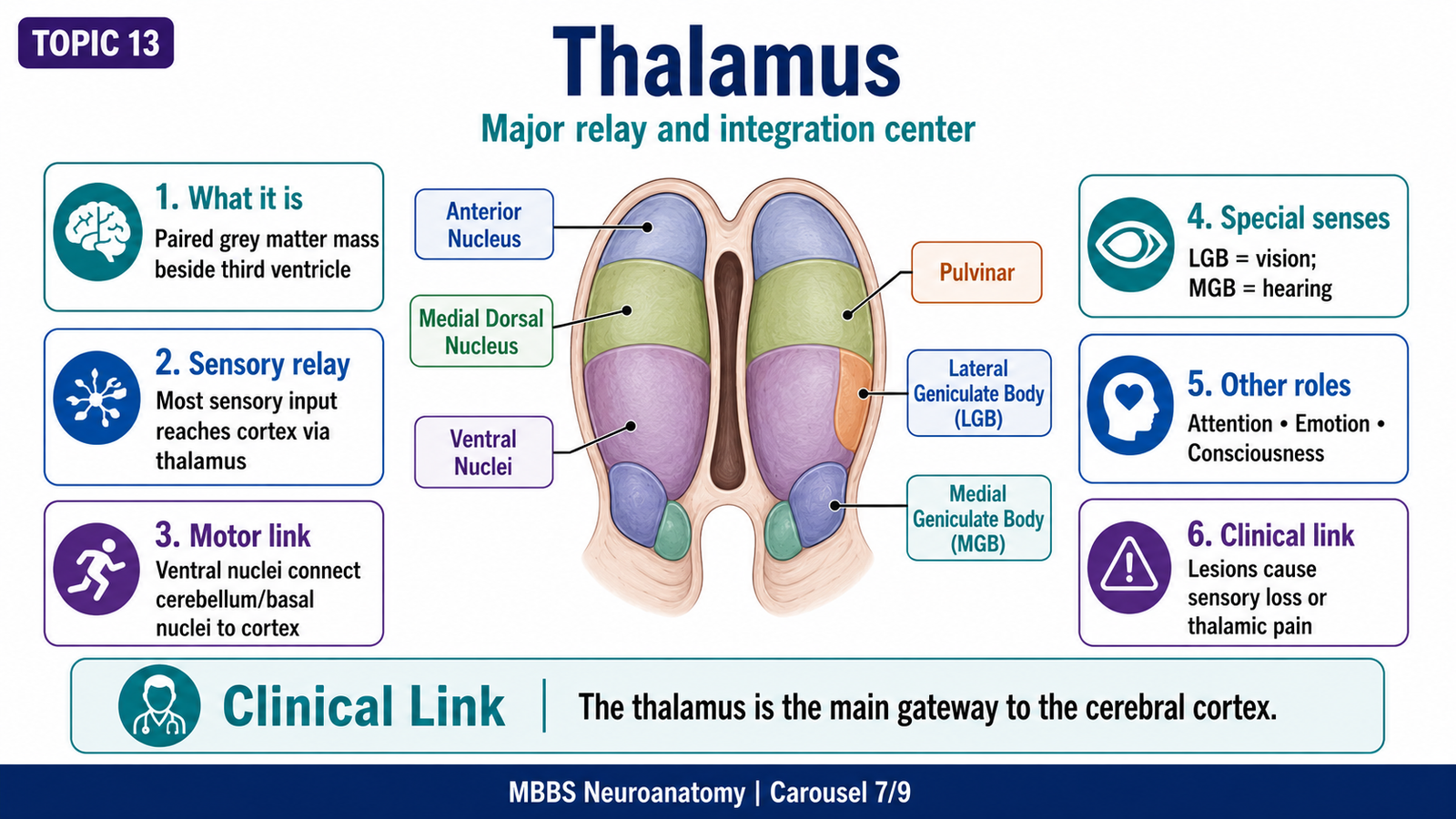
MAJOR CONCEPT 7: Thalamus — Structure and Important Nuclei
🧠 CORE
• Thalamus is a paired oval mass of grey matter on each side of the third ventricle.
• It forms much of the lateral wall of the third ventricle.
• It receives sensory, motor, limbic, and cortical inputs.
• It relays information to specific cortical areas.
• Major nuclei include anterior, medial, ventral, lateral, pulvinar, lateral geniculate, and medial geniculate nuclei.
• Ventral posterior nuclei relay somatic sensation.
• Lateral geniculate body relays vision.
• Medial geniculate body relays hearing.
• Thalamus is important in sensation, motor coordination, emotion, attention, and consciousness.
🔬 CONCEPT EXPLAINED
The thalamus is best understood as a selective relay and integration station. Almost all sensory information reaching the cerebral cortex passes through the thalamus. This includes touch, pain, temperature, proprioception, vision, and hearing. The major exception is olfaction, which reaches cortical areas without first relaying through the thalamus.
Structurally, each thalamus is an oval grey matter mass located deep in the brain. The two thalami lie on either side of the third ventricle. The internal organization of the thalamus is based on nuclei, and each nucleus has specific connections and functions.
The ventral posterior nucleus is important for general somatic sensation. It receives sensory information from the body and face and sends it to the primary somatosensory cortex. Therefore, it is essential for conscious perception of touch, pain, temperature, and proprioception.
The lateral geniculate nucleus is the visual relay nucleus. It receives input from the optic tract and sends fibers to the visual cortex. The medial geniculate nucleus is the auditory relay nucleus. It receives auditory pathway input and projects to the auditory cortex.
The ventral anterior and ventral lateral nuclei are involved in motor control. They receive information from basal nuclei and cerebellum and project to motor areas of the cerebral cortex. This allows planning and coordination of voluntary movement.
The anterior nucleus is related to limbic functions and memory circuits. The medial dorsal nucleus is connected with the prefrontal cortex and is involved in emotion, behavior, and cognition. The pulvinar participates in visual association and attention.
Thus, the thalamus is not only a sensory relay station. It integrates sensory, motor, emotional, and cognitive information before it reaches cortical awareness.
⚠️ CLINICAL IMPORTANCE
Thalamic lesions may cause contralateral sensory loss, abnormal pain syndromes, movement problems, memory disturbance, attention deficits, or altered consciousness. A lesion involving visual relay nuclei can affect visual pathways, while lesions involving auditory relay nuclei may affect hearing-related processing.
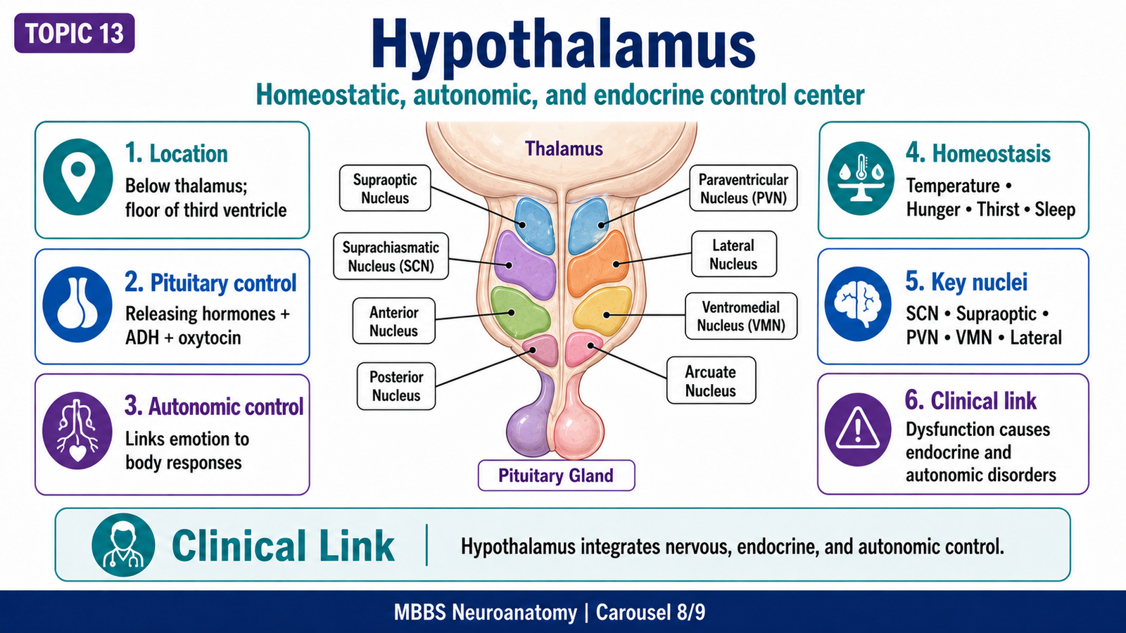
MAJOR CONCEPT 8: Hypothalamus — Structure and Important Nuclei
🧠 CORE
• Hypothalamus lies below the thalamus and forms the floor and lower lateral wall of the third ventricle.
• It is a major regulator of homeostasis.
• It controls autonomic nervous system activity.
• It regulates pituitary endocrine function.
• It participates in temperature control, hunger, thirst, sleep, emotion, and circadian rhythm.
• Important nuclei include supraoptic, paraventricular, arcuate, ventromedial, lateral, anterior, posterior, and suprachiasmatic nuclei.
• It links nervous, endocrine, autonomic, and emotional responses.
🔬 CONCEPT EXPLAINED
The hypothalamus is small in size but extremely important functionally. It maintains internal stability of the body by monitoring internal conditions and producing appropriate autonomic, endocrine, and behavioral responses.
The hypothalamus controls the pituitary gland in two major ways. First, neurons in the supraoptic and paraventricular nuclei produce hormones such as antidiuretic hormone and oxytocin, which travel to the posterior pituitary for release. Second, hypothalamic releasing and inhibiting hormones regulate the anterior pituitary through the hypophyseal portal system. This allows the hypothalamus to influence growth, thyroid function, adrenal function, reproduction, and lactation.
The hypothalamus also controls autonomic output. In general, anterior hypothalamic regions are more associated with parasympathetic and heat-loss responses, while posterior hypothalamic regions are more associated with sympathetic and heat-conservation responses. This is important for temperature regulation, cardiovascular responses, sweating, shivering, and stress reactions.
Different nuclei have different functions. The suprachiasmatic nucleus regulates circadian rhythm using light-related input. The lateral hypothalamic area is associated with hunger and feeding behavior. The ventromedial nucleus is associated with satiety. The arcuate nucleus is important in endocrine regulation and appetite-related signaling. The anterior hypothalamus helps heat loss, while the posterior hypothalamus helps heat conservation.
The hypothalamus also connects with limbic structures. This explains why emotions produce bodily responses such as tachycardia, sweating, dry mouth, altered appetite, or changes in sleep.
⚠️ CLINICAL IMPORTANCE
Hypothalamic dysfunction can cause endocrine imbalance, diabetes insipidus, obesity or reduced feeding, temperature dysregulation, sleep disturbance, autonomic instability, emotional changes, and reproductive problems. For exams, the key idea is that hypothalamus links body homeostasis with endocrine and autonomic control.
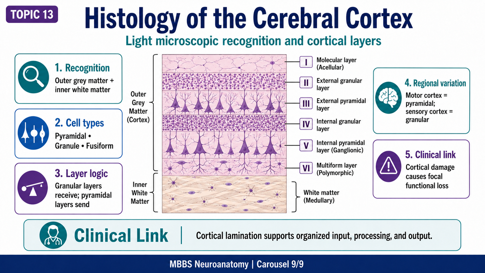
MAJOR CONCEPT 9: Histology of the Cerebral Cortex
🧠 CORE
• Cerebral cortex is grey matter covering the cerebral hemispheres.
• It is composed of neurons, neuroglia, nerve fibers, and blood vessels.
• Neurons are arranged in layers, especially in the neocortex.
• The typical neocortex has six layers.
• Main neuron types include pyramidal cells, stellate/granule cells, fusiform cells, and horizontal cells.
• Motor cortex has prominent pyramidal cells.
• Sensory cortex has prominent granular layers.
• Histological recognition depends on layered grey matter and neuronal arrangement.
🔬 CONCEPT EXPLAINED
The cerebral cortex is the microscopic basis of higher brain function. On light microscopy, it appears as a cellular grey matter layer at the surface of the cerebrum. Beneath it lies paler white matter containing mainly myelinated axons. This contrast between cellular cortex and fiber-rich white matter is an important recognition feature.
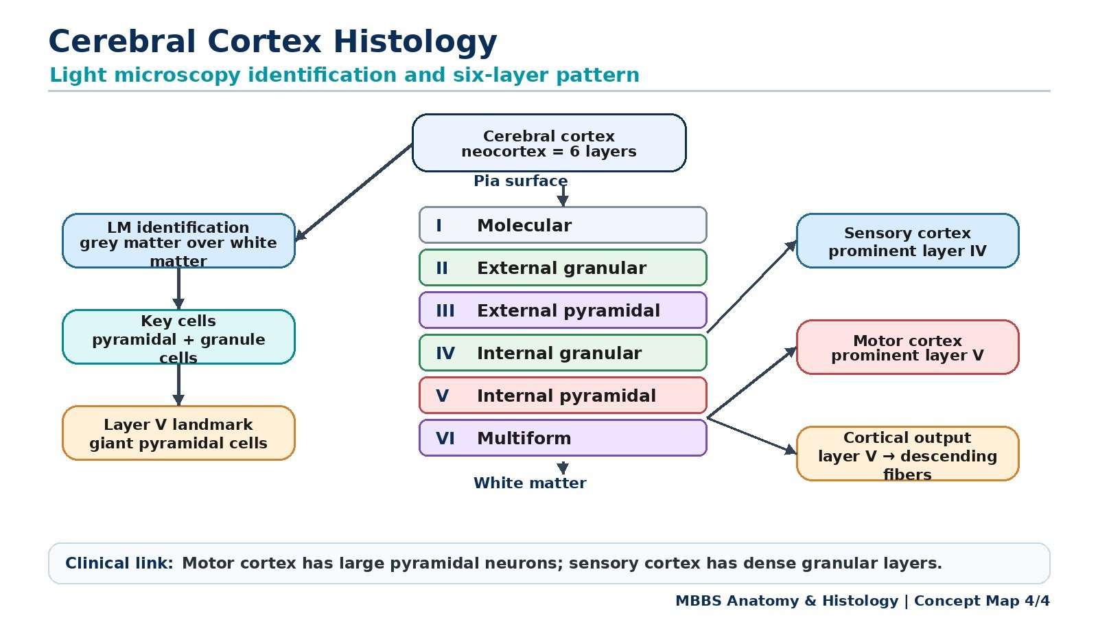
The cerebral cortex is arranged in layers because different neurons receive, process, and send information in organized directions. The typical neocortex has six layers from superficial to deep:
Layer I — Molecular layer:
This is the most superficial layer. It contains relatively few neuronal cell bodies and many nerve fibers and synapses. Its pale appearance and low cellularity help identify it microscopically.
Layer II — External granular layer:
This layer contains small granule cells and small pyramidal cells. It participates in intracortical connections.
Layer III — External pyramidal layer:
This layer contains pyramidal neurons of increasing size. It gives rise mainly to association and commissural fibers connecting cortical areas.
Layer IV — Internal granular layer:
This layer contains many granule cells. It is especially well developed in sensory cortex because sensory input from the thalamus terminates here.
Layer V — Internal pyramidal layer:
This layer contains large pyramidal neurons. In the primary motor cortex, it contains very large pyramidal cells called Betz cells. These neurons give rise to major descending motor pathways.
Layer VI — Multiform layer:
This deepest cortical layer contains different cell shapes, including fusiform cells. It sends fibers mainly to the thalamus.
The structure-function relationship is very important. Sensory cortex receives large amounts of input; therefore, its granular layers are prominent. Motor cortex sends strong output to lower motor centers; therefore, its pyramidal layer is prominent. This explains why histological differences reflect function.
Microscopic Appearance and Recognition Features
On light microscopy, cerebral cortex is recognized by:
• Outer grey matter and inner white matter.
• Layered arrangement of neurons.
• Superficial molecular layer with fewer cells.
• Deeper layers containing pyramidal, granule, and fusiform cells.
• Pyramidal neurons with triangular cell bodies and apical dendrites directed toward the surface.
• Granule cells appearing as small, densely arranged neurons.
• White matter appearing paler due to myelinated fibers and fewer neuronal cell bodies.
Cell Arrangement
Cells are arranged in laminae rather than in glands, follicles, cords, or tubules. This layered arrangement is the most important exam recognition feature. Pyramidal cells are especially important because they are the main output neurons of the cortex. Granule cells are important input-processing neurons, especially in sensory areas.
Tissue Layers
The six layers of neocortex should be remembered as:
- Molecular layer
- External granular layer
- External pyramidal layer
- Internal granular layer
- Internal pyramidal layer
- Multiform layer
A simple functional memory is:
• Granular layers receive input.
• Pyramidal layers send output.
• Molecular layer supports synaptic interaction.
• Multiform layer communicates with thalamus.
⚠️ CLINICAL IMPORTANCE
Damage to the cerebral cortex produces functional deficits depending on the involved cortical area. Motor cortex damage causes contralateral weakness. Sensory cortex damage causes contralateral sensory loss. Association cortex damage produces higher cortical dysfunction. Histologically, cortical disease may affect neurons, myelin, synapses, or blood supply, leading to disturbed brain function.
⚙️ 4️⃣ Functional Flow
The cerebrum and diencephalon function as an integrated system.
The cerebral cortex provides conscious perception, voluntary action, language, memory, judgment, and personality. Its folded surface increases processing area, while its layered histological structure supports organized input and output.
The white matter connects cortical areas with each other, connects the two hemispheres, and connects the cortex with lower CNS centers. This allows isolated cortical functions to become coordinated behavior.
The corpus callosum allows right and left hemispheres to communicate. This is essential because some functions are lateralized, such as language and spatial processing.
The thalamus controls the flow of sensory, motor, limbic, and cognitive information to the cortex. It ensures that information reaching consciousness is organized and directed to the correct cortical area.
The hypothalamus regulates the internal state of the body. It adjusts autonomic, endocrine, emotional, sleep, appetite, thirst, and temperature responses. This means that higher brain activity is linked with body homeostasis.
Histologically, the cerebral cortex demonstrates how microscopic structure supports function. Granular layers receive input, pyramidal layers send output, and different cortical areas show different layer dominance according to their function.
Therefore:
Structure → Function → Outcome
• Gyri and sulci increase surface area → more cortical neurons → higher intellectual capacity.
• Grey matter contains cell bodies and synapses → information processing → perception and decision-making.
• White matter contains myelinated fibers → rapid communication → integrated brain function.
• Corpus callosum connects hemispheres → bilateral coordination → unified cognition.
• Thalamus relays and filters information → organized cortical awareness → meaningful sensation and motor planning.
• Hypothalamus controls autonomic and endocrine systems → homeostasis → survival and adaptation.
• Cortical layers organize input and output → efficient processing → specialized cortical function.
🩺 5️⃣ Clinical Correlation
1. Cerebral Cortex Lesions
A lesion of the cerebral cortex produces symptoms according to the affected functional area. Damage to the precentral gyrus causes contralateral weakness because this region contains the primary motor cortex. Damage to the postcentral gyrus causes contralateral sensory loss because it contains the primary somatosensory cortex. Damage to the occipital cortex causes visual field defects because it processes visual input.
2. Aphasia
In the dominant hemisphere, frontal and temporal language areas are clinically important. Damage to frontal language areas may impair speech production, while damage to temporal language areas may impair comprehension. This shows that language depends not only on one area but also on association pathways connecting different cortical regions.
3. Internal Capsule and Projection Fiber Lesions
Projection fibers are compactly arranged in the internal capsule. A small stroke in this region can interrupt many descending motor fibers at once. As a result, the patient may develop dense contralateral weakness. This is an important example of how anatomical compactness increases clinical severity.
4. Corpus Callosum Lesions
Damage to the corpus callosum can disconnect the two hemispheres. This may impair transfer of information from one hemisphere to the other. The essential exam concept is that corpus callosum is the major commissural pathway for interhemispheric communication.
5. Thalamic Lesions
A thalamic lesion may produce contralateral sensory loss because thalamic nuclei relay sensory information to the cortex. Some patients may develop abnormal pain perception due to disturbed sensory processing. Thalamic involvement may also affect consciousness, attention, memory, or movement depending on the nuclei involved.
6. Hypothalamic Dysfunction
Hypothalamic lesions can disturb temperature regulation, thirst, hunger, endocrine function, sleep rhythm, and autonomic responses. For example, damage affecting ADH-related pathways may cause diabetes insipidus, while damage to appetite-regulating nuclei may cause abnormal feeding behavior.
7. Histological Cortical Damage
Microscopic damage to cortical neurons, synapses, or myelinated fibers can impair higher functions. Loss of pyramidal neurons in motor cortex affects motor output, while damage in sensory cortex affects sensory perception. Histology therefore helps explain why different cortical regions produce different clinical deficits.
📌 6️⃣ Summary Points
- The cerebrum consists of two hemispheres with outer cortex, inner white matter, and deep grey matter.
- The central sulcus separates the frontal and parietal lobes; precentral gyrus is motor, postcentral gyrus is sensory.
- The lateral sulcus separates temporal lobe from frontal and parietal lobes; the insula lies deep within it.
- Grey matter processes information; white matter transmits information.
- Association fibers connect areas within the same hemisphere.
- Commissural fibers connect the two hemispheres; the corpus callosum is the largest commissural bundle.
- Projection fibers connect cerebral cortex with thalamus, brainstem, and spinal cord.
- Corpus callosum has four main parts: rostrum, genu, body, and splenium.
- Thalamus is the major relay and integration station for sensory, motor, limbic, and cortical information.
- Hypothalamus regulates autonomic, endocrine, emotional, temperature, hunger, thirst, and sleep functions.
- The cerebral cortex has six layers; granular layers mainly receive input, pyramidal layers mainly send output.
- On light microscopy, cerebral cortex is recognized by outer grey matter, inner white matter, and layered neuronal arrangement.