
📖 Step 2 — Learning Material
🔹 1️⃣ Introduction
The nervous system receives continuous information from the external environment and from inside the body. This information is detected by sensory receptors, which convert physical or chemical stimuli into electrical signals that the nervous system can understand. Receptors are present in the skin, muscles, joints, viscera, special sense organs, blood vessels, and internal tissues.
This topic is important because sensation is not simply “feeling”; it is a carefully coded physiological process in which the nervous system identifies the type, site, strength, and duration of a stimulus. Pain, touch, pressure, temperature, proprioception, vision, hearing, smell, and taste all depend on receptor function and neural coding.
Clinically, damage to receptors, sensory nerves, spinal pathways, or cortical sensory areas may cause numbness, tingling, loss of proprioception, abnormal pain, referred pain, or sensory mislocalization. Therefore, understanding receptors and coding forms the foundation for neurological examination and sensory pathway interpretation.
🔹 2️⃣ Foundation Concepts
Key Definitions
- Sensory receptor: A specialized nerve ending or cell that detects a specific form of stimulus and converts it into an electrical signal.
- Stimulus: Any physical or chemical change capable of activating a receptor.
- Sensory transduction: Conversion of stimulus energy into a change in receptor membrane potential.
- Receptor potential: A graded local potential produced in a receptor when it is stimulated.
- Generator potential: A graded potential produced directly in the sensory nerve ending; if large enough, it generates action potentials.
- Modality: The type of sensation, such as touch, pain, temperature, pressure, vibration, vision, hearing, smell, or taste.
- Labeled line principle: Each sensory pathway is dedicated to one sensory modality; stimulation of that pathway is interpreted as that specific sensation.
- Doctrine of specific nerve energies: The brain interprets activity in a particular sensory nerve pathway as a specific type of sensation, regardless of how that pathway is stimulated.
- Law of projection: Sensation is perceived as coming from the receptor’s peripheral location, even if the pathway is stimulated at another point.
- Threshold: Minimum stimulus strength required to generate a receptor potential sufficient to produce action potentials.
- Neuronal pool: A group of interconnected neurons in the CNS that process and relay sensory signals.
Essential Terminology
- Exteroceptors: Receptors detecting stimuli from outside the body.
- Interoceptors / visceroceptors: Receptors detecting stimuli from internal organs.
- Proprioceptors: Receptors detecting body position, movement, muscle length, and tension.
- Mechanoreceptors: Detect mechanical deformation.
- Thermoreceptors: Detect temperature change.
- Nociceptors: Detect tissue-damaging stimuli.
- Chemoreceptors: Detect chemical changes.
- Photoreceptors: Detect light.
- Frequency coding: Stronger receptor stimulation produces a higher frequency of action potentials.
- Spatial coding: Stimulus location is identified by which receptor field and pathway are activated.
- Divergence: One input spreads to many neurons.
- Convergence: Many inputs combine onto fewer neurons.
- Prolongation: Neural circuits continue firing briefly after the original stimulus has stopped.
🔹 3️⃣ Core Learning — Curriculum Coverage
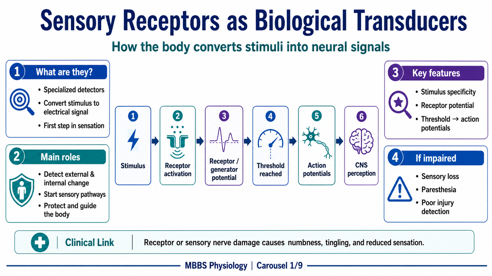
Sensory Receptors as Biological Transducers
🧠 CORE
- Sensory receptors are the first link between the body and nervous system.
- Their main function is to detect specific stimuli and convert them into electrical signals.
- Each receptor is specialized for a particular type of energy, such as mechanical, thermal, chemical, or light energy.
- The first electrical response is usually a graded receptor potential.
- If the receptor potential reaches threshold, action potentials are generated in the sensory neuron.
- Receptors allow the CNS to monitor both external environment and internal body state.
- Failure of receptors or sensory nerves produces sensory loss, abnormal sensation, or poor motor coordination.
🔬 CONCEPT EXPLAINED
To understand sensation, the first idea is that the brain does not directly receive touch, heat, pain, or pressure. Instead, it receives electrical impulses traveling in sensory nerves. The job of a sensory receptor is to convert a real-world stimulus into this electrical language. This process is called sensory transduction.
The initiating event is a stimulus, such as pressure on skin, stretching of a muscle, increased temperature, tissue injury, or a chemical change in blood. The purpose of the receptor is to detect that specific change early enough for the body to respond. For example, touch allows recognition of objects, pain protects from injury, proprioception guides movement, and chemoreception helps regulate respiration.
When a stimulus acts on a receptor, it changes the permeability of the receptor membrane. Usually, ion channels open or close. This produces a local change in membrane potential called a receptor potential. If this graded potential is small, no action potential may occur. If it becomes large enough to reach threshold at the nearby sensory nerve membrane, action potentials are produced and travel toward the CNS.
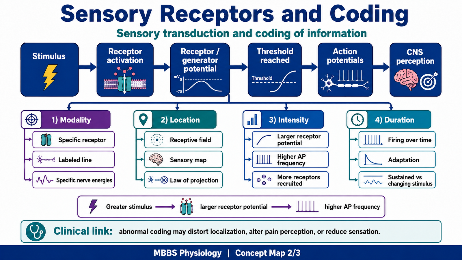
The important cause → effect relationship is:
Stronger stimulus → larger receptor potential → higher chance of reaching threshold → increased frequency of action potentials → stronger sensation perceived by CNS.
This system exists because graded receptor potentials alone cannot travel long distances. They are local signals. Action potentials, however, can travel from the receptor to the spinal cord, brainstem, thalamus, and cerebral cortex without losing strength. Therefore, the body first converts the stimulus into a graded local signal and then into action potentials for long-distance transmission.
Control and regulation occur at different levels. At the receptor level, threshold determines whether a stimulus is strong enough to be detected. At the sensory neuron level, action potential frequency carries information about stimulus intensity. At the CNS level, neuronal pools, inhibition, convergence, divergence, and cortical interpretation refine the message.
The physiological advantage is that the nervous system can detect weak stimuli, respond strongly to dangerous stimuli, and avoid unnecessary responses to insignificant stimuli. If this mechanism is impaired, the person may fail to detect injury, may experience numbness, or may feel abnormal sensations such as tingling, burning, or exaggerated pain.
⚠️ CLINICAL IMPORTANCE
Damage to sensory receptors or peripheral nerves reduces sensory input. In diabetic neuropathy, for example, damage to peripheral sensory fibers may impair pain, temperature, or touch sensation. The clinical consequence is that injury may occur without the patient noticing it, especially in the feet.
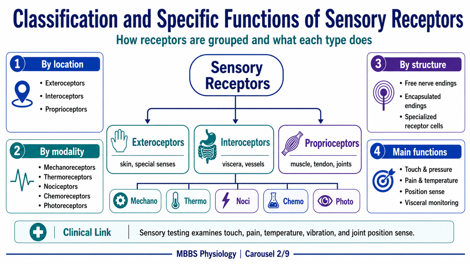
Classification and Specific Functions of Sensory Receptors
🧠 CORE
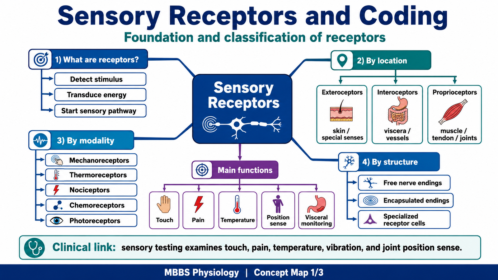
- Receptors can be classified by location, stimulus modality, and structural organization.
- Location-based classes include exteroceptors, interoceptors, and proprioceptors.
- Modality-based classes include mechanoreceptors, thermoreceptors, nociceptors, chemoreceptors, and photoreceptors.
- Different receptors have different specific functions.
- Receptor structure is closely related to the type of stimulus detected.
- Histological organization helps explain receptor function, especially in skin, muscle, tendon, and special sensory tissues.
- Clinical sensory testing depends on understanding receptor type and function.
🔬 CONCEPT EXPLAINED
After understanding what receptors do, the next step is to classify them. Classification is important because different receptors are designed for different jobs. A receptor in the skin detecting light touch is not built like a receptor in the carotid body detecting oxygen tension, and neither behaves like a muscle spindle detecting muscle stretch.
Classification According to Location in the Body
Exteroceptors are located near the body surface and detect external stimuli. They include receptors for touch, pressure, pain, temperature, and special senses such as vision and hearing. Their purpose is to help the body interact safely with the external environment.
Interoceptors, also called visceroceptors, are located in internal organs and blood vessels. They detect stretch, pressure, chemical changes, and internal pain. For example, baroreceptors detect blood pressure changes, while chemoreceptors detect changes in oxygen, carbon dioxide, and hydrogen ion concentration.
Proprioceptors are found in muscles, tendons, and joints. They detect muscle length, muscle tension, joint position, and movement. Their purpose is to allow posture, balance, coordinated movement, and awareness of body position without looking.
Classification According to Stimulus Modality
Mechanoreceptors respond to mechanical deformation. Examples include touch receptors in skin, pressure receptors, vibration receptors, muscle spindles, Golgi tendon organs, and baroreceptors. When mechanical force deforms the receptor membrane, mechanically gated ion channels open, causing a receptor potential.
Thermoreceptors respond to temperature change. Warm and cold receptors help maintain body protection and temperature awareness. Extreme temperatures may also activate nociceptors because they can damage tissues.
Nociceptors respond to potentially damaging stimuli. These may be mechanical, thermal, or chemical. Their main purpose is protection. Pain is unpleasant because it forces the body to withdraw, rest, or protect injured tissue.
Chemoreceptors respond to chemical changes. They are important in taste, smell, blood gas regulation, pH regulation, and respiratory control.
Photoreceptors respond to light. They are present in the retina and convert light energy into neural signals for vision.
Histological and Structural Recognition Features
From a histology point of view, sensory receptors may appear as free nerve endings, encapsulated nerve endings, or specialized receptor cells.
Free nerve endings are simple, unencapsulated terminal branches of sensory neurons. They are widely distributed in skin, connective tissue, and many internal tissues. They mainly detect pain, temperature, crude touch, and itch. Their simple microscopic structure suits their function because tissue injury or temperature change can directly affect the exposed nerve endings.
Encapsulated receptors have nerve endings surrounded by connective tissue capsules or specialized cells. Examples include Meissner corpuscles, Pacinian corpuscles, muscle spindles, and Golgi tendon organs. The capsule modifies the mechanical stimulus before it reaches the nerve ending. This structure → function relationship allows the receptor to detect specific types of deformation such as light touch, vibration, stretch, or tension.
Merkel discs are associated with specialized epithelial cells in the skin and are important for fine touch and pressure. Their arrangement near the epidermis helps detect sustained contact and texture.
Meissner corpuscles are found in dermal papillae of hairless skin, such as fingertips. They are oval, encapsulated structures arranged close to the epidermis, making them suitable for light touch and low-frequency vibration.
Pacinian corpuscles are large onion-like encapsulated receptors located deeper in dermis, subcutaneous tissue, and around joints. Their concentric lamellae allow rapid adaptation, making them excellent for detecting vibration and deep pressure.
Muscle spindles are stretch receptors located within skeletal muscle. They contain specialized intrafusal muscle fibers arranged parallel to ordinary muscle fibers. This arrangement allows them to detect muscle length and rate of change of length.
Golgi tendon organs are located in tendons near the muscle-tendon junction. They are arranged in series with skeletal muscle fibers and detect muscle tension. This helps protect muscles and tendons from excessive force.
Specific Functions of Important Receptors
Each receptor type performs a specific physiological function because its structure is matched to the stimulus it detects. Touch receptors help identify object shape and surface. Pressure receptors detect deeper mechanical deformation. Vibration receptors detect rapidly changing mechanical stimuli. Muscle spindles detect stretch and support reflex control of posture. Golgi tendon organs detect tension and protect against excessive contraction. Nociceptors detect tissue injury. Chemoreceptors monitor chemical composition of body fluids. Photoreceptors provide visual information.
Therefore, receptor classification is not only descriptive; it explains how the nervous system divides the body’s sensory world into meaningful categories.
⚠️ CLINICAL IMPORTANCE
Loss of proprioceptors or their pathways causes poor coordination and unsteady gait, especially when the eyes are closed. Loss of nociceptor function may prevent pain perception and increase risk of injury. Damage to mechanoreceptors or large sensory fibers may reduce vibration and fine touch sensation.
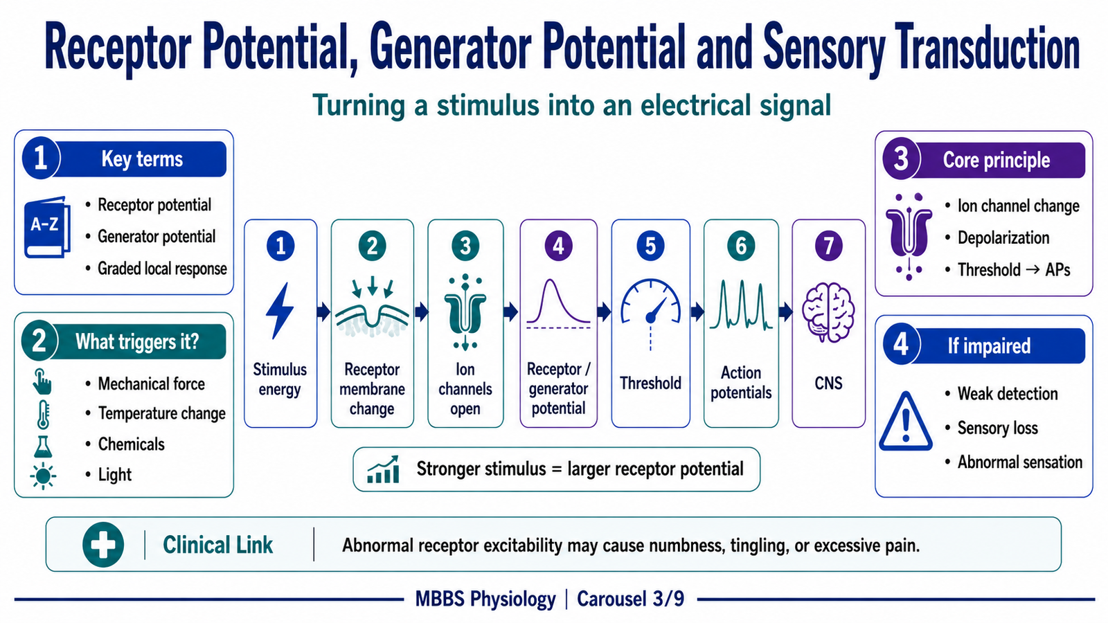
Receptor Potential, Generator Potential and Sensory Transduction
🧠 CORE
- Sensory transduction converts stimulus energy into electrical activity.
- The first electrical signal is usually a graded receptor potential.
- Receptor potentials vary in size according to stimulus strength.
- If threshold is reached, action potentials are generated.
- Action potentials carry sensory information to the CNS.
- Receptor potential amplitude influences action potential frequency.
- Transduction is the key step that changes physical or chemical information into neural language.
🔬 CONCEPT EXPLAINED
The central mechanism in receptor physiology is sensory transduction. A stimulus alone has no meaning to the nervous system until it is converted into electrical activity. The initiating event may be pressure, stretch, heat, cold, tissue damage, light, or chemical change. The purpose of transduction is to allow the nervous system to interpret these different forms of energy using a common signal: changes in membrane potential and action potentials.
When a stimulus affects a receptor, it changes ion movement across the receptor membrane. In many receptors, sodium or other cations enter the cell or nerve ending, producing depolarization. This depolarization is the receptor potential. It is graded, meaning its size depends on stimulus strength. A weak stimulus produces a small receptor potential, while a strong stimulus produces a larger receptor potential.
If the receptor itself is part of the sensory neuron, the receptor potential is often called a generator potential. When this generator potential reaches threshold at the trigger zone of the sensory fiber, action potentials begin. The receptor potential itself does not travel far, but the action potentials travel long distances to the CNS.
The sequence is:
Stimulus → receptor membrane change → ion channel opening/closing → receptor potential → threshold reached → action potentials → CNS interpretation.
The cause → effect relationship is very important. A stronger stimulus opens more channels or changes membrane conductance more strongly. This produces a larger receptor potential. A larger receptor potential reaches threshold more easily and keeps the sensory fiber above threshold for longer. As a result, action potentials are generated at a higher frequency.
Control occurs through receptor threshold, receptor adaptation, local membrane excitability, and central processing. Some receptors require only slight deformation to respond, while others need strong or damaging stimuli. This allows the body to separate harmless stimuli from potentially dangerous ones.
The physiological advantage is sensitivity with selectivity. The receptor can respond according to stimulus strength rather than giving an all-or-none local response. But once threshold is crossed, action potentials ensure reliable long-distance transmission. If this mechanism fails, a stimulus may not be detected, may be detected weakly, or may be misinterpreted as abnormal sensation.
⚠️ CLINICAL IMPORTANCE
Abnormal receptor excitability can produce paresthesia, such as tingling or pins-and-needles sensations. Reduced receptor or nerve excitability can cause numbness. Excessive activation of nociceptors during tissue injury produces pain, which has protective value but may become harmful if persistent.
Doctrine of Specific Nerve Energies, Modality and Labeled Line Principle
🧠 CORE
- The CNS identifies sensation partly by which sensory pathway is activated.
- Each sensory modality has dedicated receptors and pathways.
- The labeled line principle explains how modality is preserved from receptor to cortex.
- The doctrine of specific nerve energies states that stimulation of a specific sensory pathway produces its specific sensation.
- The brain interprets signals according to the pathway, not simply according to the original stimulus.
- This explains why pressure on the eye may be perceived as light.
- Failure of pathway integrity causes modality-specific sensory loss.
🔬 CONCEPT EXPLAINED
Once receptors convert stimuli into action potentials, the next question is: how does the brain know whether the signal represents touch, pain, heat, vibration, or light? The answer is that the nervous system uses labeled pathways.
A sensory receptor is usually most sensitive to one form of energy. For example, photoreceptors respond to light, thermoreceptors respond to temperature, and mechanoreceptors respond to deformation. These receptors connect to specific sensory fibers, spinal cord pathways, thalamic relays, and cortical areas. This dedicated pathway is called a labeled line.
The labeled line principle means that activity in a particular line is interpreted as a particular modality. If a pain pathway is activated, the brain perceives pain. If a visual pathway is activated, the brain perceives light. The CNS does not need to inspect the original stimulus directly; it interprets the signal according to the activated pathway.
This leads to the doctrine of specific nerve energies. The doctrine states that the type of sensation experienced depends on which nerve pathway is stimulated, not necessarily on the exact form of stimulation. For example, mechanical pressure on the eyeball may stimulate the retina and optic pathway. Even though the stimulus is pressure, the perception may be flashes of light because the visual pathway has been activated.
The purpose of this organization is accuracy and speed. If each modality has its own pathway, the brain can rapidly interpret signals without confusion. The cause → effect relationship is:
Specific receptor activated → specific sensory fiber activated → specific CNS pathway activated → specific cortical area activated → specific modality perceived.
Control and regulation occur through receptor specificity, synaptic organization, and cortical mapping. Sensory pathways maintain separation between different modalities, although integration can occur at higher levels.
The physiological advantage is that the nervous system can preserve the identity of a stimulus from the periphery to consciousness. If this system fails because of pathway damage, a patient may lose one modality while others remain intact. For example, fine touch and proprioception may be impaired while crude touch or pain may be relatively preserved, depending on the damaged pathway.
⚠️ CLINICAL IMPORTANCE
The labeled line principle is important in neurological examination. Testing pain, temperature, vibration, proprioception, and light touch separately helps identify which sensory pathway may be affected. It also explains referred and phantom sensations, where the CNS interprets activity according to pathway organization.

Law of Projection and Localization of Sensation
🧠 CORE
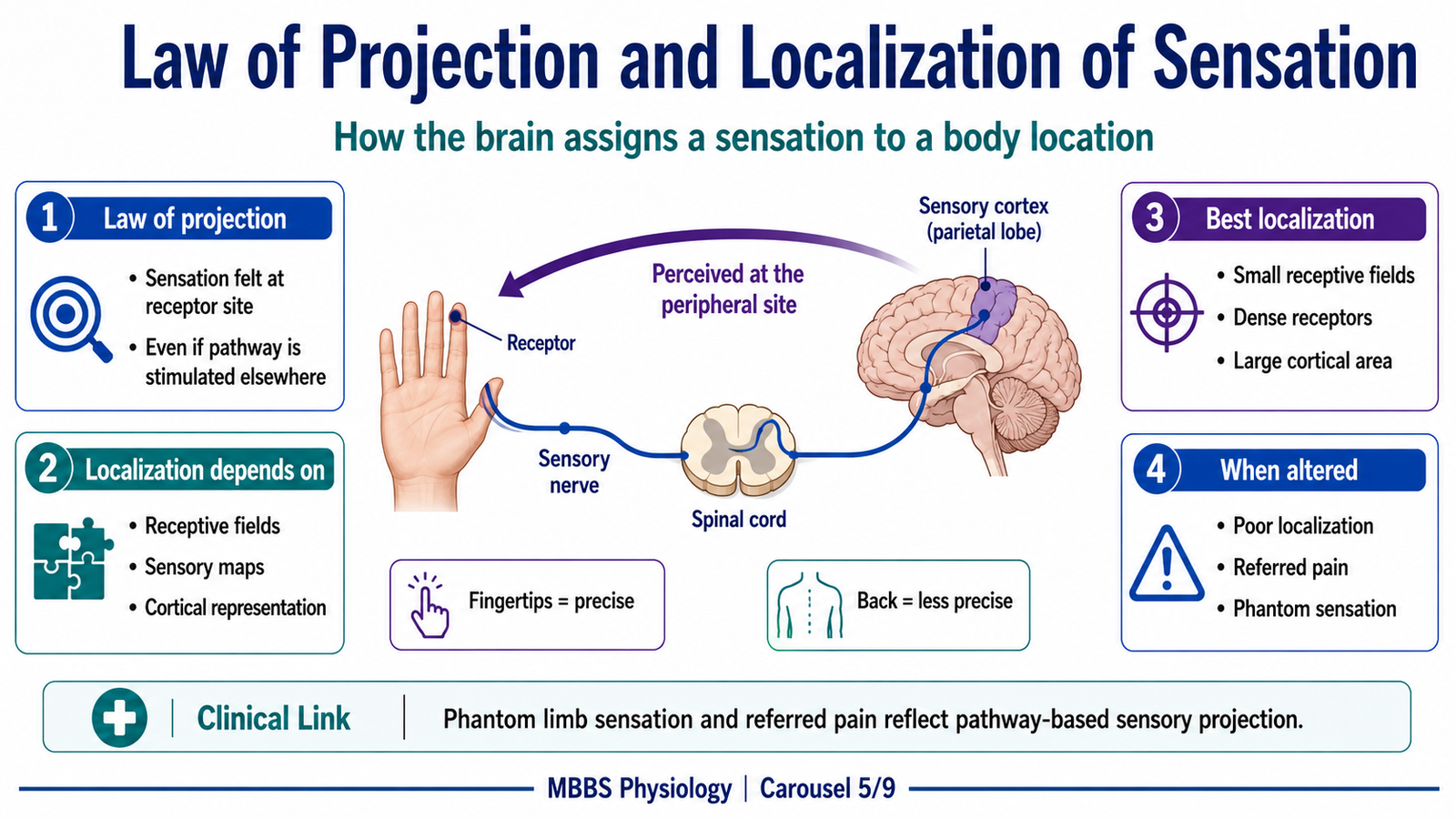
- The law of projection explains how the brain assigns sensation to a body location.
- Sensation is perceived at the peripheral receptor site.
- This occurs even if the sensory pathway is stimulated at a more central point.
- Localization depends on receptor fields and cortical sensory maps.
- Smaller receptive fields allow more precise localization.
- Sensory projection is essential for protective and skilled responses.
- Disturbance of projection can cause mislocalized pain or phantom sensations.
🔬 CONCEPT EXPLAINED
After identifying the type of sensation, the nervous system must identify where it came from. This is explained by the law of projection. According to this law, when a sensory pathway is activated, the brain projects the sensation back to the peripheral area normally served by that pathway.
For example, if a sensory nerve from the hand is stimulated, the brain interprets the sensation as coming from the hand, even if the actual stimulation occurs along the nerve trunk. This happens because sensory pathways are organized according to body maps. Each receptor field is connected through specific neurons to specific regions of the somatosensory cortex.
The initiating event may be stimulation of a receptor, peripheral nerve, spinal pathway, thalamic relay, or cortical area. The purpose of projection is to allow the body to localize stimuli accurately and respond appropriately. If the foot touches a sharp object, the person must know that the stimulus is in the foot, not simply that pain exists somewhere.
The sequence is:
Peripheral receptor field → sensory nerve → spinal cord/brainstem relay → thalamus → sensory cortex → perception projected to body region.
The cause → effect relationship is:
Activation of a pathway serving a body region → cortical interpretation of that pathway → sensation localized to that body region.
Control depends on the organization of receptive fields and cortical representation. Areas such as fingertips and lips have many receptors with small receptive fields and large cortical representation, allowing fine localization. Areas like the back have larger receptive fields and less precise localization.
The physiological advantage is accurate protective and functional behavior. The body can withdraw the exact part exposed to danger and can perform fine sensory discrimination during tasks such as writing, palpation, and object recognition.
⚠️ CLINICAL IMPORTANCE
Phantom limb sensation after amputation illustrates projection. The limb is absent, but activity in the remaining sensory pathway may still be perceived as coming from the missing limb. Referred pain also involves misinterpretation of sensory input due to convergence of visceral and somatic afferents.
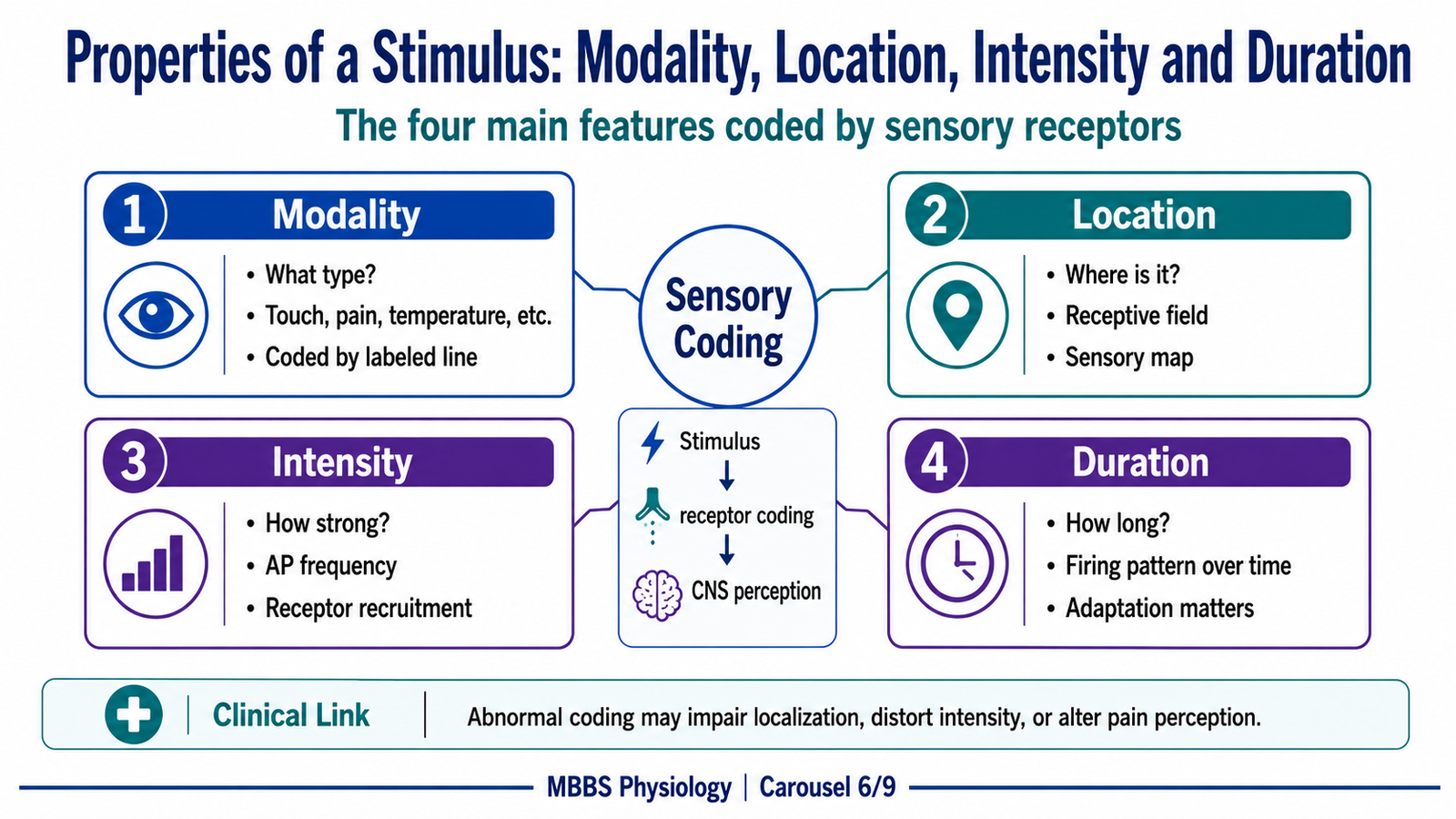
Properties of Stimulus: Modality, Location, Intensity and Duration
🧠 CORE
- Every sensation must be coded for modality, location, intensity, and duration.
- Modality is coded by labeled lines.
- Location is coded by receptive fields and sensory maps.
- Intensity is mainly coded by action potential frequency and recruitment of receptors.
- Duration is coded by the pattern of receptor firing over time.
- Receptor adaptation helps distinguish changing from constant stimuli.
- These properties allow the CNS to build a meaningful sensory perception.
🔬 CONCEPT EXPLAINED
The CNS does not simply ask whether a receptor is active or inactive. It extracts several important properties from each stimulus: what type it is, where it is, how strong it is, and how long it lasts.
Modality
Modality means the type of sensation. It is determined by receptor specificity and labeled lines. A thermoreceptor pathway produces temperature sensation, while a mechanoreceptor pathway produces touch or pressure. This prevents confusion between sensory types.
Location
Location is determined by which receptor fields are activated. Each receptor monitors a specific area called a receptive field. When that receptor and pathway are activated, the CNS localizes the sensation to that field. Smaller receptive fields allow better localization because each receptor represents a smaller area.
Intensity
Intensity means stimulus strength. Since action potentials are all-or-none, a single action potential cannot be “small” or “large.” Therefore, the nervous system codes intensity mainly by frequency of action potentials and by number of receptors recruited.
A weak stimulus produces a small receptor potential. If threshold is reached, action potentials occur at low frequency. A stronger stimulus produces a larger receptor potential, keeping the sensory fiber above threshold for longer and producing a higher frequency of action potentials. Very strong stimuli may also activate neighboring receptors, increasing the number of active sensory fibers.
Thus:
Greater stimulus intensity → larger receptor potential → higher action potential frequency → recruitment of more receptors → stronger perceived sensation.
Duration
Duration means how long the stimulus continues. Some receptors continue firing as long as the stimulus remains. These are useful for monitoring sustained information, such as posture and muscle stretch. Other receptors respond strongly at the beginning or during change but reduce firing during constant stimulation. This is called adaptation and is useful for detecting change, movement, or vibration.
The purpose of duration coding is to help the CNS decide whether a stimulus is constant, changing, or newly appearing. A constant harmless stimulus, such as clothes touching the skin, becomes less noticeable because adaptation reduces unnecessary sensory load. But important sustained stimuli, such as muscle stretch or pain from tissue injury, may continue to be signaled.
Control and regulation depend on receptor type, stimulus strength, adaptation rate, and central filtering. The physiological advantage is efficiency: the nervous system pays more attention to important changes while still monitoring essential continuous information.
⚠️ CLINICAL IMPORTANCE
Loss of intensity coding may cause reduced ability to judge stimulus strength. Abnormal pain pathways may exaggerate intensity, causing hyperalgesia. Poor localization may occur in large receptive fields or pathway lesions. Loss of duration coding may impair vibration sense or proprioception.
Frequency of Action Potentials and Threshold Level of Receptor Potential
🧠 CORE
- Receptor potentials are graded, but action potentials are all-or-none.
- Threshold determines when a receptor potential produces action potentials.
- Stronger receptor potentials produce higher action potential frequency.
- Frequency coding is a major method for coding stimulus intensity.
- Receptor potential amplitude depends on stimulus strength.
- Action potential frequency is interpreted by the CNS as stronger or weaker sensation.
- Failure of threshold or frequency coding alters sensory perception.
🔬 CONCEPT EXPLAINED
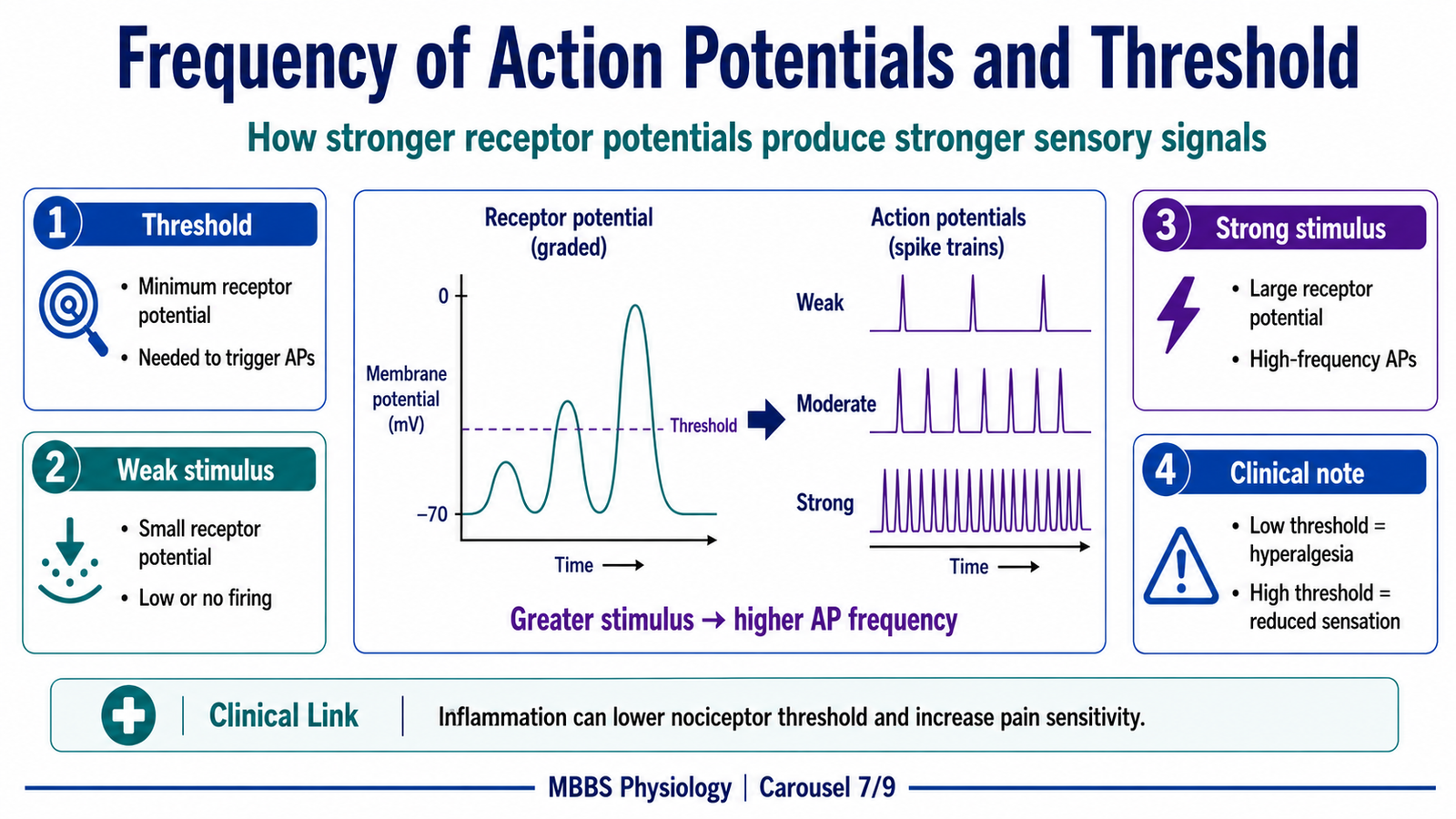
A very important physiological problem is how the nervous system represents different stimulus strengths when action potentials are all-or-none. The answer is frequency coding.
The initiating event is a stimulus acting on a receptor. The purpose of frequency coding is to convert different stimulus intensities into different rates of nerve impulses. A weak stimulus causes slight receptor depolarization. If this depolarization does not reach threshold, no action potentials occur and the stimulus is not consciously detected. If threshold is reached, action potentials begin.
As stimulus strength increases, receptor potential amplitude increases. This causes the sensory neuron to fire more frequently. Therefore, the CNS interprets a higher firing frequency as a stronger stimulus.
The sequence is:
Weak stimulus → small receptor potential → may not reach threshold → no sensation or weak sensation.
Moderate stimulus → threshold reached → low-frequency action potentials → mild sensation.
Strong stimulus → large receptor potential → high-frequency action potentials → strong sensation.
The cause → effect relationship is direct and examinable:
Stimulus strength controls receptor potential size, and receptor potential size controls action potential frequency.
Control occurs through threshold level, receptor sensitivity, stimulus duration, and adaptation. Some receptors are highly sensitive and reach threshold easily. Others, such as nociceptors, usually require stronger or damaging stimuli, which prevents harmless stimuli from being interpreted as pain.
The physiological advantage is that the CNS can grade sensation without changing the size of action potentials. This preserves the reliability of nerve conduction while still allowing the perception of weak, moderate, and strong stimuli.
⚠️ CLINICAL IMPORTANCE
In neuropathy, sensory fibers may fail to fire normally even when receptors are stimulated, causing reduced sensation. In inflammatory states, nociceptor threshold may decrease, so normally mild stimuli may become painful. This is one mechanism behind tenderness and hyperalgesia.
Relaying of Sensory Signals Through Neuronal Pools
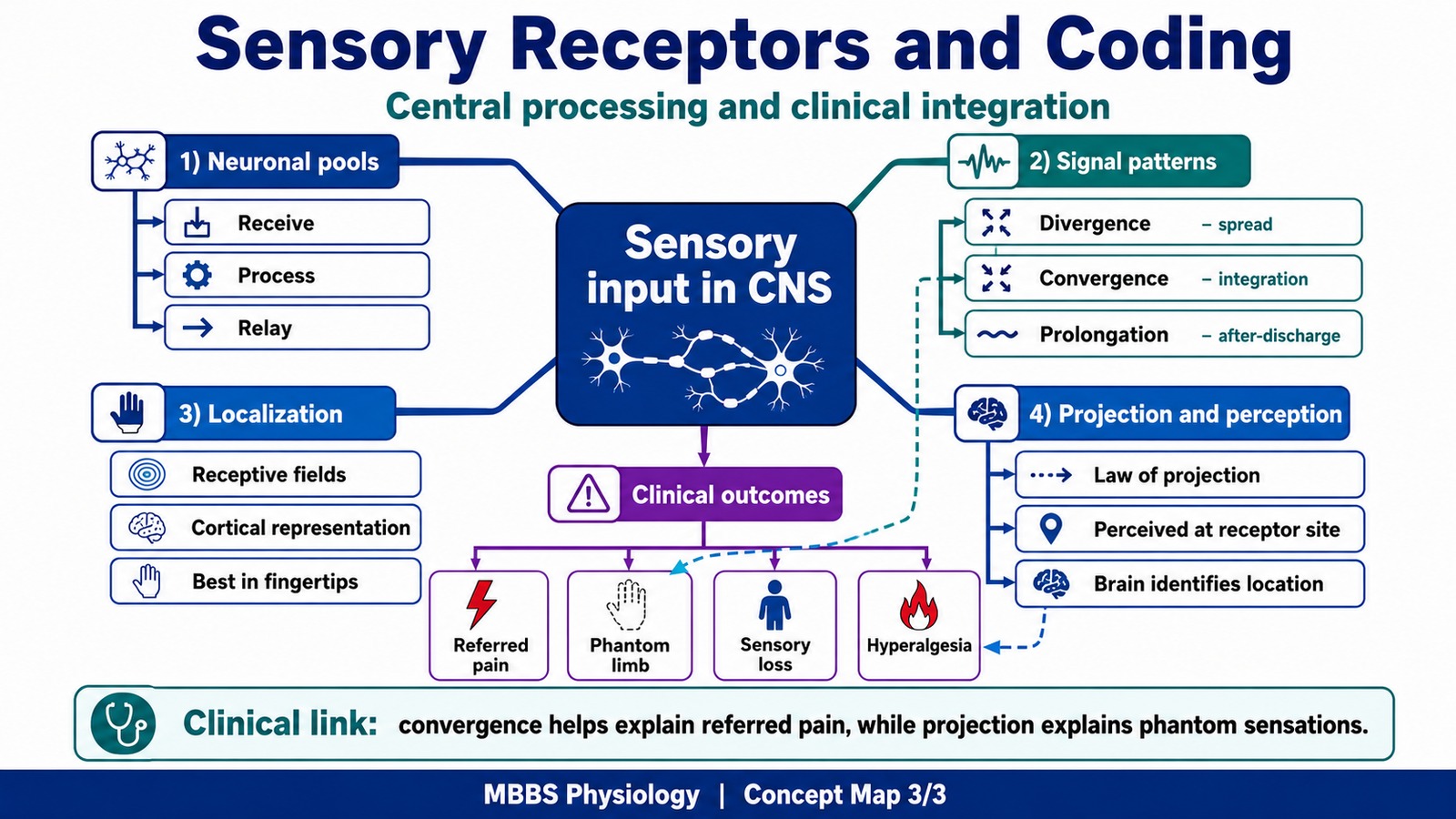
🧠 CORE
- Sensory information is processed by groups of neurons called neuronal pools.
- A neuronal pool receives input, processes it, and sends output to other neurons.
- Sensory signals may be amplified, distributed, focused, or prolonged.
- Neuronal pools allow integration of multiple sensory inputs.
- They are important in reflexes, perception, and coordinated responses.
- Divergence, convergence, and prolongation are major patterns of signal processing.
- Abnormal processing may contribute to exaggerated pain or sensory confusion.
🔬 CONCEPT EXPLAINED
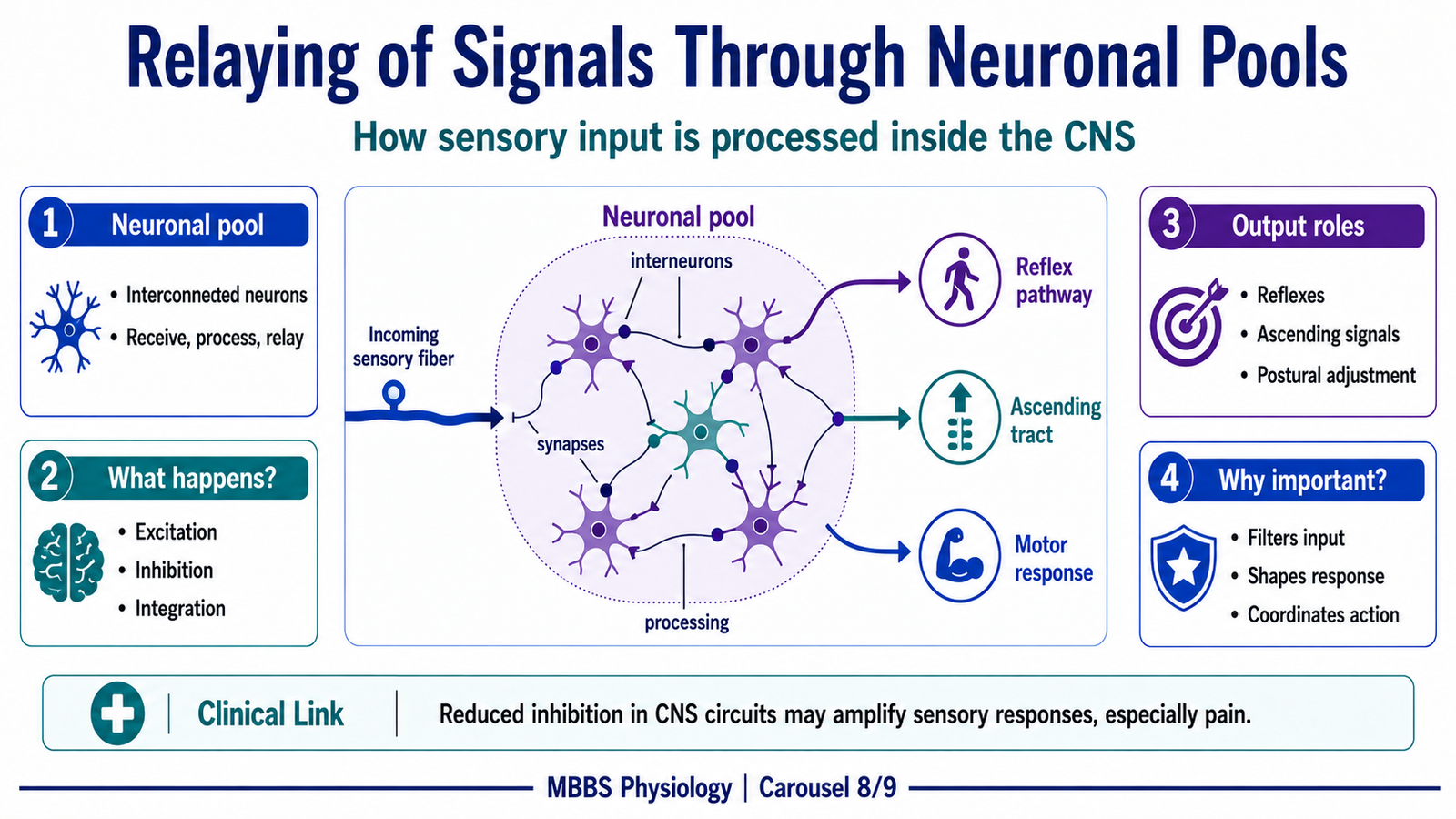
After action potentials enter the CNS, the signal is not passed like a simple wire message. Instead, it enters neuronal pools, where groups of neurons process sensory information. This processing determines how widely the signal spreads, how strongly it influences other neurons, and how long its effect continues.
The initiating event is arrival of sensory impulses from receptors into the spinal cord, brainstem, thalamus, or cortex. The purpose of neuronal pools is to transform raw sensory input into useful information for reflexes, perception, posture, movement, and protective responses.
A neuronal pool may contain excitatory neurons, inhibitory neurons, interneurons, and output neurons. When sensory input enters the pool, some neurons may be strongly excited, some weakly excited, and some inhibited. This creates organized processing rather than random spread.
The sequence is:
Sensory input → synaptic transmission in neuronal pool → excitation/inhibition of interneurons → processed output → relay to reflex pathways, ascending tracts, or higher centers.
Cause → effect relationships are important. Strong sensory input activates more neurons in the pool. Inhibitory interneurons may sharpen the signal by reducing activity in neighboring pathways. Excitatory connections may amplify or distribute the signal. This allows the CNS to respond appropriately to the importance of the stimulus.
Control and regulation occur through synaptic strength, inhibitory circuits, facilitation, fatigue, descending modulation, and receptor input frequency. The physiological advantage is flexibility. A simple stimulus can trigger a spinal reflex, inform the brain, adjust posture, and prepare motor responses at the same time.
⚠️ CLINICAL IMPORTANCE
Abnormal neuronal pool processing can increase pain sensitivity or produce persistent pain after injury. Reduced inhibition in sensory circuits may allow excessive spread of impulses, contributing to exaggerated responses.
Divergence, Convergence and Prolongation of Signals in the CNS
🧠 CORE
- Divergence spreads one input to many neurons.
- Convergence combines many inputs onto one or fewer neurons.
- Prolongation allows a signal to continue after the original stimulus stops.
- These mechanisms help the CNS process sensory information efficiently.
- Divergence is useful for reflex spread and parallel processing.
- Convergence is useful for integration but may reduce localization accuracy.
- Prolongation helps maintain responses when continued activity is needed.
🔬 CONCEPT EXPLAINED
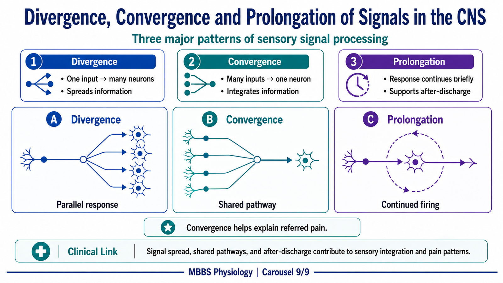
Once sensory signals enter neuronal pools, three important processing patterns become especially important: divergence, convergence, and prolongation.
Divergence
Divergence means that one incoming sensory fiber stimulates many neurons. The purpose is to distribute information widely. For example, a painful stimulus may activate withdrawal reflex pathways, ascending pain pathways, autonomic responses, and emotional responses.
The initiating event is a sensory input entering the CNS. The signal branches through interneurons and activates multiple output pathways. The cause → effect relationship is:
One input → multiple neurons activated → wider CNS response.
The physiological advantage is that important stimuli can produce coordinated responses. If the hand touches a hot object, divergence helps activate withdrawal muscles, inhibit opposing muscles, alert consciousness, and trigger autonomic changes.
If divergence is excessive or poorly controlled, sensory signals may spread too widely, contributing to poorly localized pain or exaggerated responses.
Convergence
Convergence means that many input fibers synapse onto one neuron or a smaller group of neurons. The purpose is integration. The CNS can combine information from multiple receptors and body regions.
The sequence is:
Multiple sensory inputs → common interneuron or projection neuron → integrated output.
The advantage is that the CNS can compare and combine signals. However, convergence may reduce precision. If visceral and somatic afferents converge onto the same spinal neurons, the brain may misinterpret visceral pain as coming from the body surface. This contributes to referred pain.
Prolongation
Prolongation means that the output of a neuronal pool continues for a short time after the original input stops. This may occur because of reverberating circuits or after-discharge in interneuronal networks. The purpose is to maintain a response long enough to be useful.
For example, a protective reflex may need to continue briefly even after the initial stimulus changes. Prolongation helps stabilize neural responses and prevents the CNS from reacting only in isolated flashes.
Control occurs through inhibitory neurons, synaptic fatigue, and descending modulation. The physiological advantage is continuity of response. If prolongation is excessive, abnormal persistent sensations or pain may occur.
⚠️ CLINICAL IMPORTANCE
Convergence explains important exam concepts such as referred pain. Divergence explains why a painful stimulus can cause local withdrawal, emotional discomfort, and autonomic effects. Prolonged activity in pain pathways may contribute to chronic pain mechanisms at a basic undergraduate level.
⚙️ 4️⃣ Functional Flow
| Structure | Function | Outcome |
|---|---|---|
| Free nerve endings | Detect pain, temperature, crude touch, itch | Protective awareness of harmful or changing stimuli |
| Encapsulated receptors | Detect touch, pressure, vibration, stretch | Precise mechanical sensation |
| Pacinian corpuscles | Detect vibration and deep pressure | Awareness of rapidly changing mechanical stimuli |
| Meissner corpuscles | Detect light touch in hairless skin | Fine tactile discrimination |
| Merkel discs | Detect sustained pressure and texture | Recognition of shape and surface detail |
| Muscle spindles | Detect muscle length and stretch | Posture, tone, stretch reflex, coordinated movement |
| Golgi tendon organs | Detect muscle tension | Protection from excessive contraction |
| Chemoreceptors | Detect chemical composition | Regulation of respiration and internal homeostasis |
| Receptor potential | Converts stimulus into graded electrical signal | Determines whether action potentials will occur |
| Sensory nerve fiber | Conducts action potentials to CNS | Long-distance transmission of sensory information |
| Neuronal pools | Process and integrate sensory input | Reflexes, perception, amplification, modulation |
| Somatosensory cortex | Interprets modality, intensity, and location | Conscious sensory perception |
The overall integration is:
Stimulus → receptor structure → receptor potential → action potential frequency → CNS pathway → neuronal pool processing → cortical interpretation → sensation and response.
This shows that sensation depends on both peripheral receptor design and central nervous system interpretation. A receptor detects the stimulus, but the CNS gives it meaning.
🩺 5️⃣ Clinical Correlation
Peripheral Neuropathy
Peripheral neuropathy damages sensory fibers. If sensory action potentials cannot travel normally, the CNS receives weak or abnormal input. This causes numbness, tingling, burning, or loss of vibration and pain sensation. The mechanism is:
Nerve damage → impaired sensory conduction → reduced or abnormal CNS input → sensory loss or paresthesia.
Loss of Proprioception
Damage to proprioceptors or their pathways impairs awareness of joint position and movement. The patient may become unsteady, especially in the dark or when eyes are closed, because vision can no longer compensate. The mechanism is:
Proprioceptive pathway damage → poor position sense → impaired posture and coordination.
Hyperalgesia
Inflammation can lower the threshold of nociceptors. A stimulus that normally causes mild pain may produce exaggerated pain. The mechanism is:
Tissue injury → inflammatory mediators → nociceptor sensitization → lower threshold → increased pain response.
Referred Pain
Visceral and somatic sensory fibers may converge onto the same spinal neurons. The brain is more accustomed to receiving pain from somatic structures, so it may project visceral pain to a body surface area. The mechanism is:
Visceral afferent input + somatic afferent convergence → shared CNS pathway → pain perceived at somatic site.
Phantom Limb Sensation
After amputation, sensory pathways and cortical maps related to the missing limb may remain active. The brain projects activity to the original limb area. The mechanism is:
Residual pathway activity → cortical interpretation → sensation projected to absent limb.
Loss of Vibration Sense
Vibration depends strongly on rapidly adapting mechanoreceptors and large sensory fibers. Damage to these fibers may reduce vibration sense, which is commonly tested clinically using a tuning fork.
📌 6️⃣ Summary Points
- Sensory receptors convert physical or chemical stimuli into electrical signals.
- Sensory transduction produces a graded receptor potential.
- If receptor potential reaches threshold, action potentials are generated.
- Stronger stimulus produces larger receptor potential and higher action potential frequency.
- Receptor classification may be based on location, modality, or structure.
- Exteroceptors detect external stimuli, interoceptors detect internal stimuli, and proprioceptors detect body position and movement.
- Mechanoreceptors detect deformation; thermoreceptors detect temperature; nociceptors detect tissue injury; chemoreceptors detect chemicals; photoreceptors detect light.
- The labeled line principle explains how the CNS identifies sensory modality.
- The doctrine of specific nerve energies means stimulation of a specific pathway produces its specific sensation.
- The law of projection explains why sensation is perceived at the peripheral receptor location.
- Divergence spreads sensory signals, convergence integrates them, and prolongation maintains them.
- Referred pain is an important clinical example of convergence in sensory pathways.