
📖 Step 2 — Learning Material
🔹 1️⃣ Introduction
Synaptic transmission is the process by which one neuron communicates with another neuron, muscle cell, or gland cell. It is the basic mechanism through which the nervous system receives information, processes it, stores it, and produces responses. Synapses are present throughout the CNS and PNS, especially in the brain, spinal cord, autonomic ganglia, neuromuscular junctions, and sensory pathways. In the CNS, synaptic activity determines whether a neuron remains silent or generates an action potential. This topic is clinically important because many neurological and psychiatric disorders result from abnormal neurotransmitter release, receptor function, excitation–inhibition imbalance, or impaired brain metabolism. Understanding synaptic transmission also explains the basis of drugs acting on the nervous system, seizures, Parkinsonism, depression, tetanus, botulism, hypoxia, and hypoglycemia. Therefore, synaptic transmission connects physiology, biochemistry, neural function, and clinical medicine into one integrated concept.
🔹 2️⃣ Foundation Concepts
Key Definitions
- Synapse: A functional junction between two neurons or between a neuron and an effector cell where signals are transmitted.
- Presynaptic neuron: The neuron that sends the signal.
- Postsynaptic neuron: The neuron that receives the signal.
- Synaptic cleft: The narrow space between presynaptic and postsynaptic membranes.
- Neurotransmitter: A chemical messenger released from the presynaptic terminal to influence the postsynaptic cell.
- Excitatory postsynaptic potential: A graded depolarization that brings the neuron closer to threshold.
- Inhibitory postsynaptic potential: A graded hyperpolarization or stabilization that moves the neuron away from threshold.
- Threshold: The critical membrane potential at which an action potential is generated.
- Axon initial segment: The region of the axon where action potentials are usually initiated.
- Second messenger: An intracellular signaling molecule that transmits receptor activation into cellular responses.
- Neural metabolism: The biochemical processes that provide energy and molecules needed for neuronal activity.
Essential Terminology
- Ionotropic receptor: A receptor directly linked to an ion channel.
- Metabotropic receptor: A receptor linked to G-protein and second messenger systems.
- Quantal release: Release of neurotransmitter in packets from synaptic vesicles.
- Synaptic delay: The time required for neurotransmitter release, diffusion, receptor binding, and postsynaptic response.
- Summation: Addition of multiple postsynaptic potentials.
- Facilitation: Increased synaptic response due to repeated activity.
- Synaptic fatigue: Decreased synaptic transmission after prolonged activity.
- Reuptake: Removal of neurotransmitter back into presynaptic neuron or glial cell.
- Excitation–inhibition balance: The functional balance between excitatory and inhibitory inputs in the CNS.
Basic Overview
- Neurons communicate mainly through chemical synapses.
- An action potential reaching the presynaptic terminal causes calcium entry.
- Calcium triggers vesicle fusion and neurotransmitter release.
- Neurotransmitter binds receptors on the postsynaptic membrane.
- Postsynaptic response may be excitatory or inhibitory.
- The neuron integrates many inputs at the soma and axon initial segment.
- If threshold is reached, a new action potential is generated.
- Synaptic function depends strongly on energy metabolism and neurotransmitter synthesis.
🔹 3️⃣ Core Learning — Curriculum Coverage
1. Synapses: Definition, Classification and Physiological Structure
🧠 CORE
- Synapses are specialized junctions for communication between neurons or between neurons and effector cells.
- They may be chemical or electrical.
- Chemical synapses are most common in the human nervous system.
- A chemical synapse has presynaptic terminal, synaptic cleft, and postsynaptic membrane.
- Synaptic vesicles contain neurotransmitters.
- The postsynaptic membrane contains specific receptors.
- Synapses allow direction, regulation, integration, and plasticity of neuronal signals.
- CNS synapses may be axodendritic, axosomatic, axoaxonic, dendrodendritic, or neuromuscular.
🔬 CONCEPT EXPLAINED
A synapse is not simply a gap between neurons; it is a highly specialized communication site. The nervous system receives millions of signals every second, but these signals must be controlled, selected, strengthened, weakened, or blocked according to need. Synapses make this possible.
There are two main types of synapses. Electrical synapses allow direct ionic current flow through gap junctions. They are fast and useful where synchronized activity is needed. However, they are less flexible. Chemical synapses use neurotransmitters and are slightly slower, but they allow amplification, inhibition, modulation, learning, memory, and drug action. Therefore, chemical synapses dominate in the CNS.
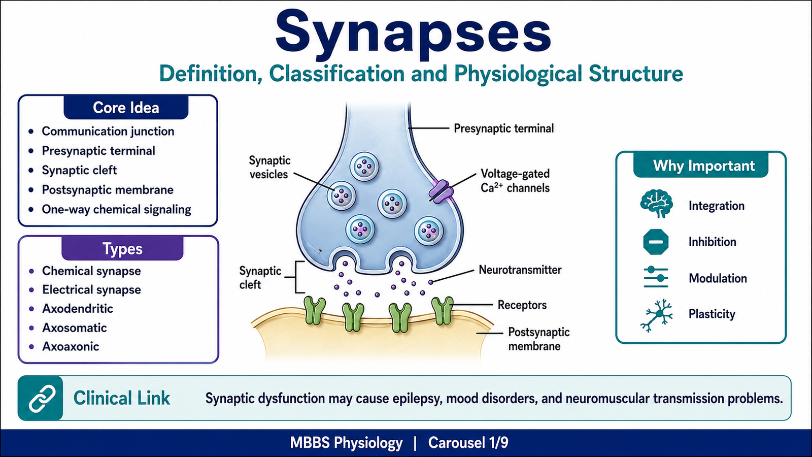
A typical chemical synapse has three important parts. The presynaptic terminal contains synaptic vesicles filled with neurotransmitter and voltage-gated calcium channels. The synaptic cleft is a narrow extracellular space through which neurotransmitter diffuses. The postsynaptic membrane contains receptors and ion channels that convert chemical signals into electrical or biochemical responses.
The structure of a synapse explains its function. The presynaptic side is designed for neurotransmitter release. The postsynaptic side is designed for detection and response. The cleft separates the two membranes so that signaling can be regulated chemically rather than by simple electrical continuity. As a result, the nervous system gains control over whether a signal excites, inhibits, or modulates the next neuron.
Synapses can be classified according to site of contact. Axodendritic synapses are common and usually influence dendritic input. Axosomatic synapses strongly affect neuronal firing because they are close to the cell body. Axoaxonic synapses regulate neurotransmitter release from another axon terminal and are important in presynaptic inhibition or facilitation. This arrangement allows the CNS to process information at multiple levels before a final response is produced.
⚠️ Clinical Importance
Damage or dysfunction of synapses can disturb neuronal communication even when neurons are structurally intact. Disorders of synaptic transmission are involved in epilepsy, Parkinson disease, depression, anxiety, myasthenic syndromes, tetanus, botulism, and many drug effects.
2. Presynaptic Events: How an Action Potential Releases Neurotransmitter
🧠 CORE
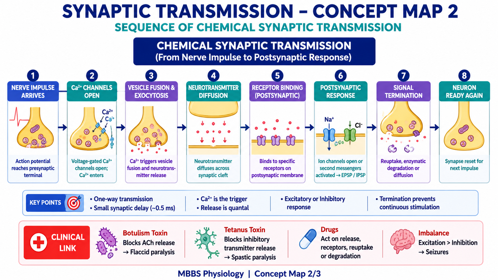
- Neurotransmitter release begins when an action potential reaches the presynaptic terminal.
- Depolarization opens voltage-gated calcium channels.
- Calcium enters the terminal down its electrochemical gradient.
- Calcium binds vesicle-associated proteins and triggers vesicle fusion.
- Neurotransmitter is released into the synaptic cleft by exocytosis.
- Release is quantal because vesicles release packets of transmitter.
- Neurotransmitter diffuses across the cleft and binds postsynaptic receptors.
- Signal termination occurs by reuptake, enzymatic degradation, or diffusion.
🔬 CONCEPT EXPLAINED
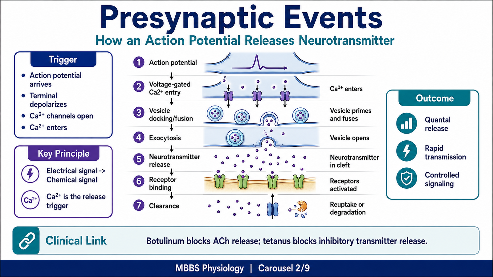
The initiating event in chemical synaptic transmission is the arrival of an action potential at the presynaptic terminal. The purpose of this mechanism is to convert an electrical signal in the presynaptic neuron into a chemical signal that can cross the synaptic cleft and influence the next cell.
When the action potential reaches the nerve terminal, it depolarizes the presynaptic membrane. This depolarization opens voltage-gated calcium channels. Calcium concentration is much higher outside the neuron than inside, so calcium rapidly enters the terminal. This calcium entry is the key trigger for neurotransmitter release.
Inside the terminal, synaptic vesicles are already docked near the presynaptic membrane. Calcium binds to calcium-sensitive vesicle proteins such as synaptotagmin and promotes interaction of vesicle fusion proteins. This leads to fusion of the vesicle membrane with the presynaptic membrane. As a result, neurotransmitter is released into the synaptic cleft by exocytosis.
This process shows a clear cause–effect sequence:
Action potential → terminal depolarization → calcium channel opening → calcium influx → vesicle fusion → neurotransmitter release → receptor activation.
The physiological advantage of this mechanism is control. The neuron does not release neurotransmitter randomly; release occurs mainly when electrical activity reaches the terminal. Calcium acts as the link between electrical activity and chemical release. More calcium entry usually produces more vesicle fusion, so the strength of transmission can be adjusted.
After release, neurotransmitter must be removed quickly; otherwise, the postsynaptic neuron would remain continuously activated. Termination occurs by reuptake into presynaptic terminals or glial cells, enzymatic breakdown, or diffusion away from the synapse. For example, acetylcholine is broken down by acetylcholinesterase, while many monoamines are removed by reuptake transporters.
If this mechanism is impaired, synaptic communication fails. Reduced calcium entry or blocked vesicle fusion decreases transmitter release and causes weakness or loss of function. Excessive transmitter release or impaired removal can cause overexcitation, abnormal movements, autonomic symptoms, or seizures.
⚠️ Clinical Importance
Botulinum toxin blocks acetylcholine release by interfering with vesicle fusion proteins, causing flaccid paralysis. Tetanus toxin blocks inhibitory transmitter release in the spinal cord, causing excessive motor neuron activity and spastic paralysis.
3. Properties of Synaptic Transmission
🧠 CORE
- Chemical synaptic transmission is usually one-way.
- It has synaptic delay because chemical steps take time.
- It allows convergence and divergence of signals.
- It can show summation, facilitation, inhibition, fatigue, and plasticity.
- Synaptic strength can increase or decrease depending on activity.
- Synapses act as decision-making points in neural circuits.
- Repeated activity may alter future responses.
- These properties allow learning, coordination, filtering, and protection from overactivity.
🔬 CONCEPT EXPLAINED
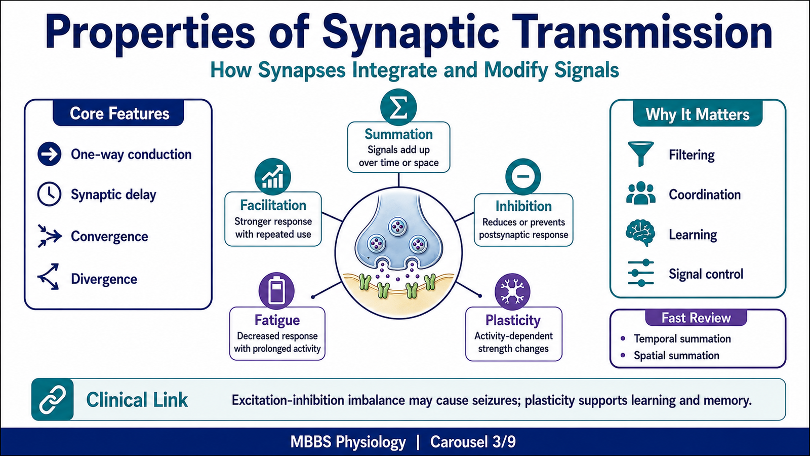
Synaptic transmission has special properties that make the nervous system more than a simple wire network. Unlike electrical conduction along an axon, synaptic transmission involves chemical release, diffusion, receptor binding, and postsynaptic response. Because of these steps, chemical synapses show a small synaptic delay. This delay is usually very short, but it is important because it shows that transmission is not purely electrical.
Chemical synapses usually conduct signals in one direction because neurotransmitter vesicles are located presynaptically and receptors are located postsynaptically. This creates organized flow of information through neural pathways.
Synapses also allow convergence and divergence. In convergence, many presynaptic neurons influence one postsynaptic neuron. This allows integration of multiple signals. In divergence, one neuron influences many postsynaptic neurons, allowing distribution of information to several pathways. These properties are essential for reflexes, sensory processing, motor control, and higher brain functions.
Another major property is summation. A single excitatory input may be too weak to produce an action potential. However, if many excitatory inputs arrive together, or if repeated inputs arrive rapidly, their effects add together. This can bring the neuron to threshold.
Synapses can also show facilitation. If presynaptic activity occurs repeatedly, residual calcium may remain inside the terminal. This calcium increases the probability of vesicle release during the next impulse, producing a stronger postsynaptic response. This is useful in short-term enhancement of synaptic transmission.
In contrast, synaptic fatigue occurs when repeated stimulation reduces transmission, often because neurotransmitter vesicles become temporarily depleted or postsynaptic receptors become less responsive. Fatigue is protective because it can limit excessive neuronal discharge.
Synaptic plasticity is the ability of synapses to change their strength. This is the physiological basis of learning and memory. Therefore, synapses are not fixed junctions; they are dynamic control points where the nervous system modifies information processing according to experience and need.
⚠️ Clinical Importance
Excessive excitation or reduced inhibition can produce seizures. Synaptic fatigue may protect circuits from continuous overactivity. Abnormal synaptic plasticity is involved in learning disorders, chronic pain, addiction, and neuropsychiatric disease.
4. Neurotransmitters: Characteristics, Classification, CNS Actions and Biosynthesis
🧠 CORE
- Neurotransmitters are chemical messengers released from neurons.
- They must be synthesized, stored or available, released, act on receptors, and be removed.
- CNS neurotransmitters may be excitatory, inhibitory, or modulatory.
- Major CNS transmitters include glutamate, GABA, glycine, acetylcholine, dopamine, noradrenaline, serotonin, histamine, peptides, nitric oxide, and endocannabinoids.
- Biosynthesis depends on amino acids, glucose metabolism, enzymes, cofactors, and vesicular storage.
- Neurotransmitter metabolism links biochemistry with neural function.
- Altered neurotransmitter levels cause functional and clinical effects.
- Many nervous system drugs act by modifying neurotransmitter synthesis, release, receptors, reuptake, or degradation.
🔬 CONCEPT EXPLAINED
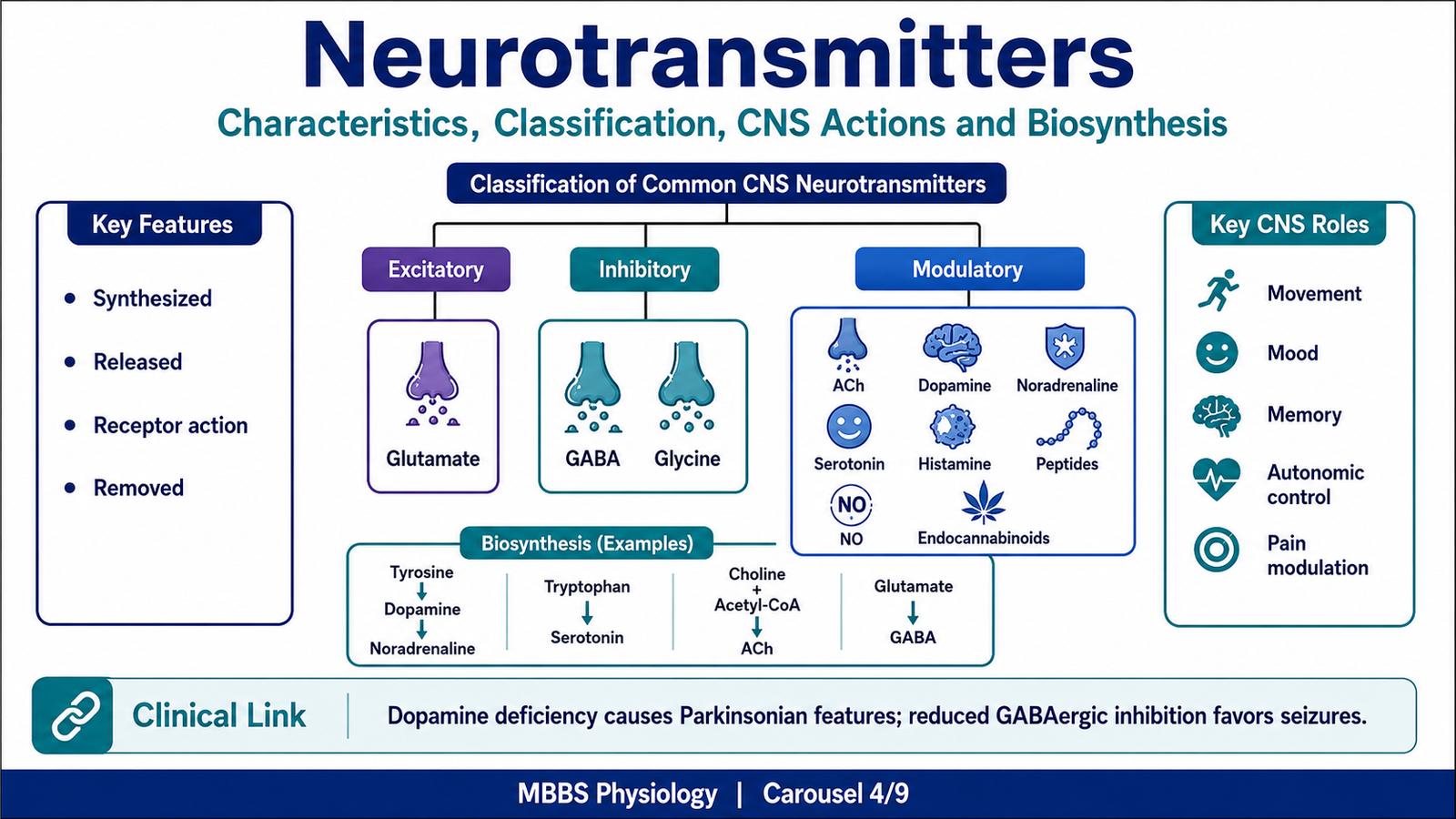
A neurotransmitter is a chemical signal used by neurons to communicate. For a substance to be considered a neurotransmitter, it should be synthesized or present in the neuron, released when the neuron is activated, produce a specific response in the postsynaptic cell, and have a mechanism for removal or inactivation. These characteristics are important because they distinguish true neurotransmitters from other chemicals present in nervous tissue.
Neurotransmitters can be classified in several ways. Functionally, they may be excitatory, inhibitory, or modulatory. Excitatory transmitters increase the chance that the postsynaptic neuron will fire. Inhibitory transmitters decrease that chance. Modulatory transmitters may not directly excite or inhibit strongly but adjust neuronal responsiveness, synaptic strength, mood, attention, sleep, reward, or autonomic function.
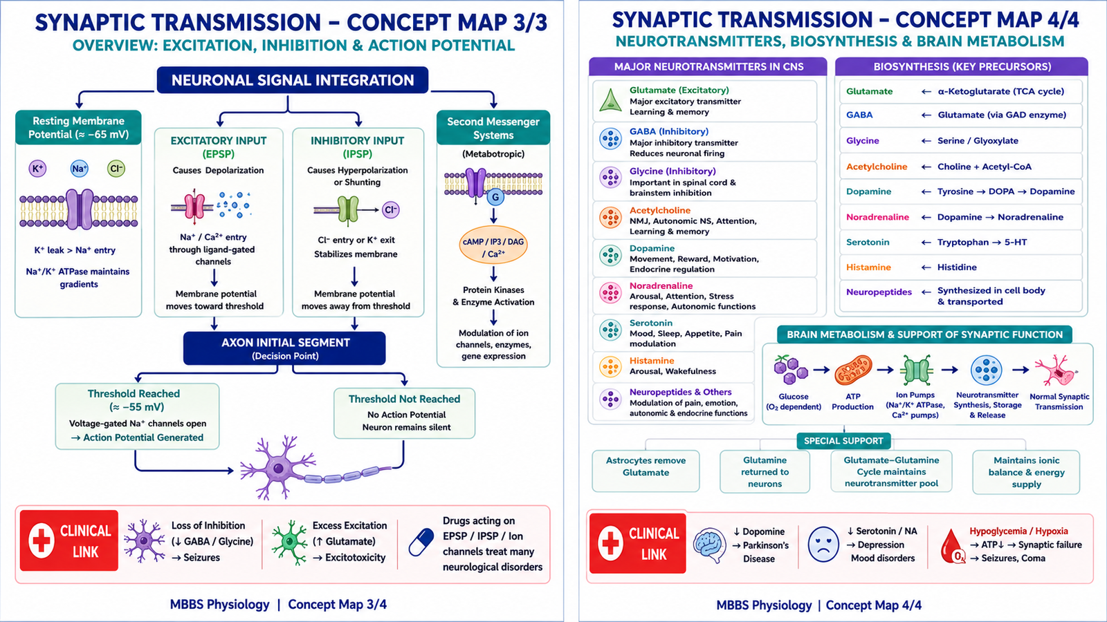
The major excitatory neurotransmitter in the CNS is glutamate. It opens cation channels through receptors such as AMPA and NMDA receptors, producing depolarization. Glutamate is essential for learning and memory, but excessive glutamate can damage neurons by excitotoxicity.
The major inhibitory neurotransmitter in the brain is GABA. GABA acts mainly through GABA-A receptors, which increase chloride conductance, and GABA-B receptors, which act through G-proteins to increase potassium conductance or reduce calcium entry. The result is inhibition of neuronal firing. In the spinal cord and brainstem, glycine is also an important inhibitory neurotransmitter.
Acetylcholine is important in the neuromuscular junction, autonomic nervous system, attention, arousal, and memory. It is synthesized from choline and acetyl-CoA by choline acetyltransferase. Acetyl-CoA links acetylcholine synthesis to energy metabolism because it comes from carbohydrate and fatty acid metabolism.
Dopamine, noradrenaline, and adrenaline are catecholamines synthesized from tyrosine. Tyrosine is converted to DOPA by tyrosine hydroxylase, then to dopamine. Dopamine can be converted to noradrenaline, and noradrenaline can be converted to adrenaline in appropriate cells. Dopamine is important in movement, reward, motivation, and endocrine regulation. Noradrenaline is important in arousal, attention, stress responses, and autonomic function.
Serotonin is synthesized from tryptophan and is involved in mood, sleep, appetite, pain modulation, and many CNS functions. Histamine is synthesized from histidine and contributes to arousal and wakefulness. Neuropeptides are synthesized in the cell body as larger precursor proteins and transported down the axon. They often act slowly and modulate pain, autonomic function, emotion, and endocrine responses.
Some transmitters are unusual. Nitric oxide is synthesized from arginine and diffuses freely across membranes. It is not stored in vesicles like classic neurotransmitters. Endocannabinoids are lipid-derived messengers produced on demand from membrane lipids and often act retrogradely, meaning they move from postsynaptic neuron back to presynaptic terminal to regulate transmitter release.
Biochemically, neurotransmitter synthesis depends on functional molecules such as amino acids, acetyl-CoA, enzymes, vitamin cofactors, vesicular transporters, and energy supply. Therefore, nervous tissue metabolism is not separate from neurotransmission; it directly supports synaptic function.
⚠️ Clinical Importance
Reduced dopamine in basal ganglia circuits is associated with Parkinsonism. Altered serotonin and noradrenaline signaling is involved in mood disorders. Reduced GABAergic inhibition or excessive glutamatergic excitation may produce seizures. Drugs used in neurology and psychiatry often act by changing neurotransmitter levels or receptor activity.
5. Postsynaptic Receptors and Mechanism of Neurotransmitter Action
🧠 CORE
- Neurotransmitters act by binding receptors on the postsynaptic membrane.
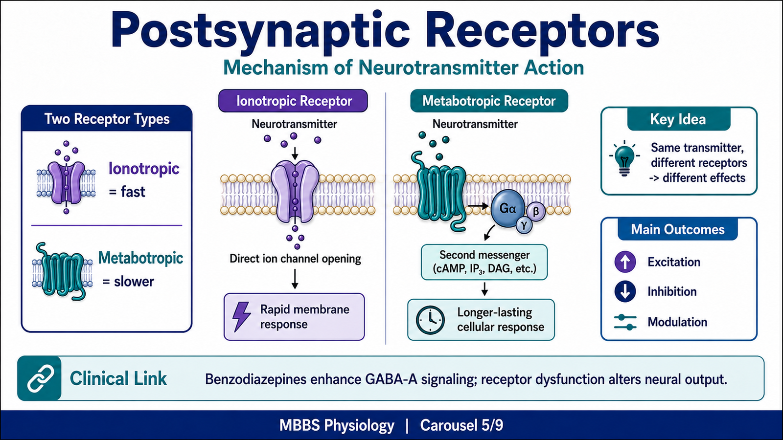
- Receptors may be ionotropic or metabotropic.
- Ionotropic receptors produce fast responses by opening ion channels.
- Metabotropic receptors produce slower, longer-lasting responses through G-proteins.
- Postsynaptic effects depend on receptor type, not only the neurotransmitter.
- The same neurotransmitter can excite or inhibit depending on receptor subtype.
- Second messenger systems amplify and prolong synaptic effects.
- Receptor function determines whether the postsynaptic neuron is excited, inhibited, or modulated.
🔬 CONCEPT EXPLAINED
After neurotransmitter is released into the synaptic cleft, it diffuses to the postsynaptic membrane and binds to specific receptors. The purpose of receptors is to convert a chemical signal into a physiological response. This response may be electrical, metabolic, or genomic depending on the receptor type.
Ionotropic receptors are ligand-gated ion channels. When neurotransmitter binds, the channel opens directly. Ions move according to their electrochemical gradients, producing rapid changes in membrane potential. For example, glutamate acting on AMPA receptors allows mainly sodium entry, producing depolarization. GABA acting on GABA-A receptors allows chloride movement, producing inhibition. These responses are fast and useful for rapid sensory, motor, and reflex activity.
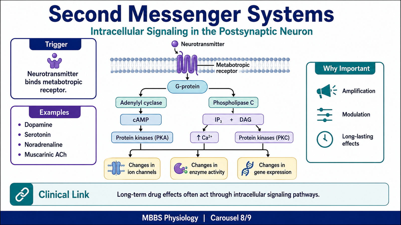
Metabotropic receptors do not open ion channels directly. Instead, they activate G-proteins and second messenger systems. These systems may open or close ion channels indirectly, alter enzyme activity, change neurotransmitter release, or modify gene expression. Responses are slower than ionotropic responses but last longer and can strongly regulate neuronal behavior.
The important physiological principle is that the effect of a neurotransmitter depends on the receptor. Acetylcholine can excite skeletal muscle through nicotinic receptors but slow the heart through muscarinic receptors. Similarly, dopamine, serotonin, and noradrenaline have multiple receptor subtypes with different effects.
Second messenger systems increase the power and flexibility of neurotransmission. A single receptor activation can influence many intracellular molecules. Common second messengers include cyclic AMP, IP3, DAG, calcium, and protein kinases. These systems can change membrane excitability, synaptic strength, enzyme activity, and long-term neuronal function.
Cause–effect reasoning is central here:
Neurotransmitter binding → receptor activation → ion channel change or second messenger activation → postsynaptic membrane response → altered neuronal excitability.
If receptor function is impaired, neurotransmitter release may be normal but the postsynaptic response becomes abnormal. This can cause weakness, abnormal movement, altered consciousness, mood disturbance, or seizures depending on the circuit involved.
⚠️ Clinical Importance
Many drugs act on postsynaptic receptors. Benzodiazepines enhance GABA-A receptor function and increase inhibition. Antipsychotic drugs often affect dopamine receptors. Some toxins, autoimmune diseases, and genetic disorders alter receptor function and produce neurological symptoms.
6. Excitatory and Inhibitory Postsynaptic Potentials
🧠 CORE
- Postsynaptic potentials are graded local changes in membrane potential.
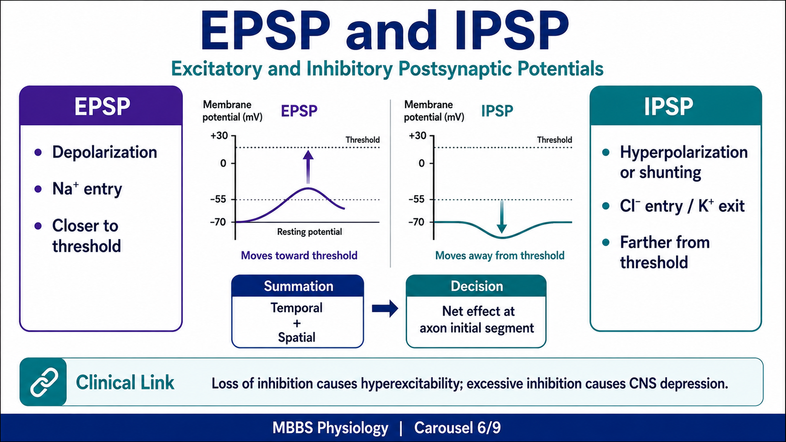
- EPSP brings the membrane potential closer to threshold.
- IPSP moves the membrane away from threshold or reduces the effect of excitation.
- EPSPs commonly result from sodium or calcium entry.
- IPSPs commonly result from chloride entry or potassium exit.
- Postsynaptic potentials are not all-or-none.
- They can summate spatially and temporally.
- Their final balance determines whether the neuron fires.
🔬 CONCEPT EXPLAINED
The postsynaptic neuron receives many excitatory and inhibitory signals at the same time. These signals do not automatically produce action potentials. Instead, they produce small graded changes in membrane potential called postsynaptic potentials.
An excitatory postsynaptic potential is a local depolarization. The initiating event is binding of an excitatory neurotransmitter, commonly glutamate, to postsynaptic receptors. The purpose of EPSP is to increase the probability that the postsynaptic neuron will generate an action potential. When excitatory channels open, sodium usually enters the cell more than potassium leaves. This makes the inside of the neuron less negative. As a result, the membrane potential moves closer to threshold.
An inhibitory postsynaptic potential reduces the chance of firing. The initiating event is binding of an inhibitory neurotransmitter such as GABA or glycine. The purpose of IPSP is to prevent excessive or inappropriate neuronal activity. Inhibition may occur by chloride entry, which makes the inside more negative, or by potassium exit, which also hyperpolarizes the membrane. In some cases, inhibition occurs by shunting, where increased membrane conductance reduces the effect of excitatory inputs even without marked hyperpolarization.
The relationship between EPSP and IPSP determines neuronal output. EPSPs push the neuron toward firing, while IPSPs pull it away from firing or stabilize it. The neuron therefore acts like an integrator. It does not respond to one signal alone; it calculates the net effect of many inputs.
There are two important types of summation. Temporal summation occurs when repeated signals from the same synapse arrive rapidly and add together. Spatial summation occurs when signals from different synapses arrive at the same time and add together. If the total depolarization reaches threshold at the axon initial segment, an action potential is generated.
The physiological advantage of this system is precision. The CNS can strengthen important signals, suppress irrelevant signals, coordinate movements, control reflexes, and prevent uncontrolled discharge.
⚠️ Clinical Importance
Loss of inhibition can cause hyperexcitability and seizures. Excess inhibition can produce sedation, reduced consciousness, or impaired motor activity. Many sedative and antiepileptic drugs work by enhancing inhibitory transmission or reducing excitatory transmission.
7. Resting Membrane Potential, Threshold and Generation of Action Potential at the Axon Initial Segment
🧠 CORE
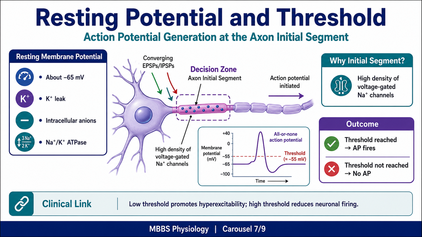
- The neuronal soma has a resting membrane potential of about −65 mV.
- Resting potential is mainly due to potassium leak conductance, intracellular anions, and Na⁺/K⁺ ATPase.
- The soma and dendrites receive graded postsynaptic potentials.
- The axon initial segment has a high density of voltage-gated sodium channels.
- Threshold is the membrane potential at which sodium channel activation becomes self-amplifying.
- If threshold is reached, an action potential is generated.
- Action potentials are all-or-none, unlike EPSPs and IPSPs.
- The initial segment acts as the decision-making zone of the neuron.
🔬 CONCEPT EXPLAINED
A neuron at rest is electrically polarized. The inside of the neuronal soma is negative compared with the outside, usually around −65 mV. This resting membrane potential exists because the membrane is more permeable to potassium than sodium, negatively charged proteins remain inside the cell, and the sodium–potassium ATPase maintains ionic gradients by moving sodium out and potassium in.
The purpose of resting membrane potential is to keep the neuron ready for signaling. If the membrane were not polarized, excitatory and inhibitory changes would have little meaning. Because the neuron starts from a stable negative potential, small depolarizations or hyperpolarizations can act as meaningful signals.
The soma and dendrites receive EPSPs and IPSPs. These graded potentials spread toward the axon initial segment. However, the soma itself is not usually the best site for action potential initiation because it has fewer voltage-gated sodium channels than the axon initial segment.
The axon initial segment is the region where the neuron makes its final decision. It has a very high density of voltage-gated sodium channels. When the net excitatory input depolarizes this region to threshold, sodium channels open rapidly. Sodium enters the cell, causing further depolarization, which opens more sodium channels. This positive feedback produces the rising phase of the action potential.
Threshold is not a fixed magical number, but it is commonly around −55 mV in many neurons. It represents the level at which inward sodium current exceeds outward currents enough to produce a self-propagating action potential. Once generated, the action potential travels along the axon without decrement.
The cause–effect sequence is:
EPSPs and IPSPs arrive → graded potentials summate → membrane potential at initial segment changes → threshold reached or not reached → action potential generated or neuron remains silent.
The physiological advantage is that the neuron can receive thousands of inputs but produce a clear output only when the total signal is strong enough and appropriate.
⚠️ Clinical Importance
If threshold is too low, neurons become hyperexcitable and may discharge excessively, contributing to seizures or spasms. If threshold is too high, neuronal firing becomes difficult, leading to weakness, sensory loss, or reduced CNS activity.
8. Second Messenger Systems in the Postsynaptic Neuron
🧠 CORE
- Second messenger systems convert receptor activation into intracellular responses.
- They are commonly linked to metabotropic receptors.
- Important systems include cAMP, IP3, DAG, calcium, and protein kinases.
- They amplify signals and produce longer-lasting effects.
- They can alter ion channels, enzymes, receptors, and gene expression.
- They are important in modulation, learning, memory, mood, and autonomic function.
- They connect synaptic activity with cellular metabolism.
- Their effects are slower but more sustained than ionotropic responses.
🔬 CONCEPT EXPLAINED
Fast synaptic transmission is useful when the nervous system needs immediate responses. However, neurons also need slower regulation. They must adjust sensitivity, change synaptic strength, modify receptor number, and alter long-term function. Second messenger systems provide this deeper level of control.
The initiating event is binding of neurotransmitter to a metabotropic receptor. The receptor activates a G-protein, which then affects an enzyme or ion channel. For example, activation of adenylyl cyclase increases cyclic AMP. Cyclic AMP activates protein kinase A, which phosphorylates target proteins. These proteins may include ion channels, receptors, enzymes, or transcription factors.
Another pathway uses phospholipase C, which produces IP3 and DAG. IP3 releases calcium from intracellular stores, while DAG activates protein kinase C. Calcium itself acts as an important intracellular messenger and can regulate enzymes, ion channels, neurotransmitter release, and gene expression.
The purpose of second messenger systems is amplification and modulation. One neurotransmitter molecule can activate a receptor that influences many intracellular molecules. This allows a small extracellular signal to produce a large and prolonged cellular effect.
Second messenger systems also explain why some neurotransmitter actions are slow but clinically powerful. Dopamine, noradrenaline, serotonin, acetylcholine through muscarinic receptors, and many neuropeptides act through metabotropic pathways. These systems influence mood, arousal, attention, autonomic activity, endocrine control, and long-term plasticity.
If second messenger regulation fails, neurons may respond too strongly, too weakly, or for too long. This can affect learning, memory, movement, mood, and autonomic function.
⚠️ Clinical Importance
Many psychiatric and neurological drugs act through receptors linked to second messenger systems. Long-term drug effects often occur because repeated receptor activation changes intracellular signaling, receptor sensitivity, or gene expression.
9. Brain and Nervous Tissue Metabolism
🧠 CORE
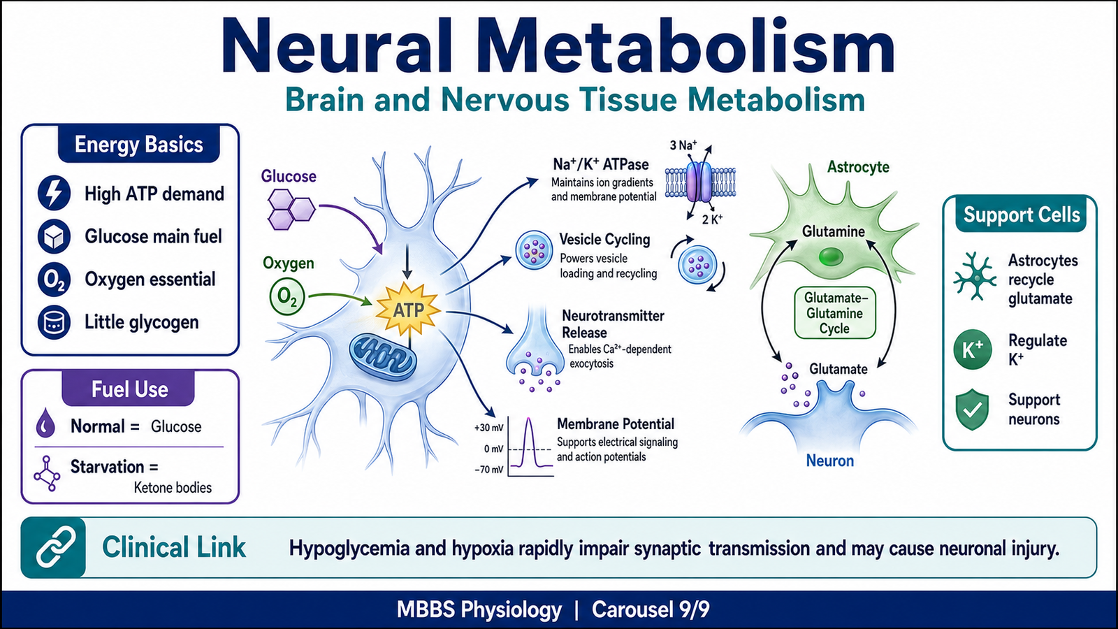
- The brain has very high energy demand.
- Glucose is the main fuel under normal conditions.
- Oxygen is essential for aerobic ATP production.
- ATP supports ion pumps, neurotransmitter cycling, vesicle transport, and membrane potential.
- The brain stores very little glycogen.
- Astrocytes support neurons metabolically and help recycle neurotransmitters.
- Amino acid metabolism is closely linked to neurotransmitter synthesis.
- Hypoxia or hypoglycemia rapidly impairs synaptic transmission.
🔬 CONCEPT EXPLAINED
Neurons are highly active cells. Even at rest, they must maintain ionic gradients across their membranes. During synaptic activity, they must restore sodium, potassium, calcium, and chloride gradients; synthesize and recycle neurotransmitters; transport vesicles; and maintain membrane excitability. All these processes require ATP.
Under normal conditions, the brain depends mainly on glucose and oxygen. Glucose is metabolized through glycolysis, the citric acid cycle, and oxidative phosphorylation to produce ATP. Oxygen is essential because most ATP in neurons comes from aerobic metabolism. Although the brain represents only a small percentage of body weight, it consumes a large proportion of body oxygen and glucose.
The purpose of neural metabolism is not only survival; it directly supports communication. The sodium–potassium ATPase uses ATP to maintain resting membrane potential. Calcium pumps and exchangers restore low intracellular calcium after neurotransmitter release. Vesicle loading of neurotransmitters depends on proton gradients generated by ATP-dependent pumps. Therefore, synaptic transmission is an energy-dependent process.
Astrocytes play a major supportive role. They help regulate extracellular potassium, take up neurotransmitters such as glutamate, convert glutamate into glutamine, and supply metabolic substrates to neurons. The glutamate–glutamine cycle is especially important. After glutamate is released and taken up by astrocytes, it is converted to glutamine. Glutamine returns to neurons, where it is converted back to glutamate. Glutamate can also be converted into GABA in inhibitory neurons.
This shows a strong biochemical link:
Glucose metabolism → ATP production → ion gradient maintenance → neurotransmitter release and recycling → normal synaptic function.
Because the brain has limited energy stores, interruption of glucose or oxygen rapidly impairs neuronal activity. First, synaptic transmission fails. Then membrane potentials collapse. Finally, neurons may be injured or die, especially if excessive glutamate causes calcium overload and excitotoxicity.
During prolonged starvation, the brain can use ketone bodies to some extent. This is a physiological adaptation that reduces dependence on glucose, but under normal feeding conditions glucose remains the major fuel.
⚠️ Clinical Importance
Hypoglycemia causes confusion, seizures, coma, and neuronal injury because neurons cannot generate enough ATP. Hypoxia or ischemia causes failure of ion pumps, depolarization, excessive glutamate release, calcium overload, and neuronal damage.
⚙️ 4️⃣ Functional Flow
| Structure / Biochemical Element | Function | Outcome |
|---|---|---|
| Presynaptic terminal | Stores vesicles and releases neurotransmitter | Converts electrical signal into chemical signal |
| Voltage-gated Ca²⁺ channels | Allow Ca²⁺ entry during depolarization | Trigger vesicle fusion |
| Synaptic vesicles | Store neurotransmitter | Permit quantal release |
| Synaptic cleft | Separates pre- and postsynaptic membranes | Allows regulated chemical communication |
| Postsynaptic receptors | Detect neurotransmitters | Produce excitation, inhibition, or modulation |
| Dendrites and soma | Receive synaptic inputs | Collect graded potentials |
| Axon initial segment | Integrates inputs and initiates AP | Converts graded signals into all-or-none output |
| Na⁺/K⁺ ATPase | Maintains ionic gradients | Supports resting membrane potential and excitability |
| Glucose and oxygen | Produce ATP | Maintain synaptic activity |
| Astrocytes | Support metabolism and neurotransmitter recycling | Protect synaptic environment |
| Glutamate–glutamine cycle | Recycles excitatory transmitter | Maintains neurotransmitter supply |
| GABA synthesis from glutamate | Produces inhibitory transmitter | Maintains excitation–inhibition balance |
Synaptic transmission is therefore an integrated process. Anatomically, specialized synaptic structures bring neurons close enough to communicate. Physiologically, electrical signals are converted into chemical signals and then back into electrical or biochemical responses. Biochemically, neurotransmitter synthesis and ATP production support every step. Functionally, the balance of excitation and inhibition determines whether neural circuits remain silent, respond normally, or become overactive.
🩺 5️⃣ Clinical Correlation
Epilepsy
Epilepsy may occur when excitatory activity becomes excessive or inhibitory control becomes insufficient. If glutamatergic excitation dominates over GABAergic inhibition, neurons may fire repeatedly and synchronously. This abnormal discharge produces seizures. Therefore, many antiepileptic drugs either enhance inhibition, reduce excitation, or stabilize ion channels.
Parkinson Disease
Dopamine is essential for normal basal ganglia function and movement control. In Parkinson disease, dopamine deficiency in basal ganglia circuits causes abnormal motor control, leading to bradykinesia, rigidity, tremor, and postural instability. At this level, the key concept is that loss of a neurotransmitter disturbs circuit function.
Botulism
Botulinum toxin prevents acetylcholine release from presynaptic terminals by interfering with vesicle fusion. As a result, neuromuscular transmission fails and muscles become weak or paralyzed. The cause–effect relationship is: blocked transmitter release → failed muscle activation → flaccid paralysis.
Tetanus
Tetanus toxin reduces release of inhibitory neurotransmitters such as GABA and glycine from inhibitory interneurons. Without inhibition, motor neurons become overactive. This produces muscle stiffness and spasms. The key mechanism is loss of inhibition, not excessive direct excitation.
Hypoxia and Ischemia
When oxygen supply decreases, ATP production falls. Ion pumps fail, membrane potentials collapse, glutamate accumulates, calcium enters neurons excessively, and neuronal injury occurs. This explains why the brain is highly sensitive to interruption of blood flow.
Hypoglycemia
Glucose is the main fuel of the brain. Severe hypoglycemia reduces ATP production and impairs synaptic transmission. Early features include sweating, confusion, irritability, and weakness; severe cases may cause seizures, coma, and neuronal injury.
Excitotoxicity
Excess glutamate overactivates excitatory receptors, especially those allowing calcium entry. Increased intracellular calcium activates damaging enzymes and injures neurons. This is important in ischemia, trauma, and some neurodegenerative processes.
📌 6️⃣ Summary Points
- Chemical synapses are the main communication points in the CNS because they allow regulation, integration, inhibition, and plasticity.
- Action potential arrival at the presynaptic terminal causes Ca²⁺ entry, which triggers vesicle fusion and neurotransmitter release.
- Neurotransmitter action depends on the receptor type, not only on the neurotransmitter itself.
- Ionotropic receptors produce fast responses; metabotropic receptors produce slower but longer-lasting effects through second messengers.
- EPSPs depolarize the postsynaptic neuron and bring it closer to threshold.
- IPSPs hyperpolarize or stabilize the postsynaptic neuron and make firing less likely.
- The axon initial segment is the main site of action potential initiation because it has many voltage-gated Na⁺ channels.
- Glutamate is the major excitatory neurotransmitter in the CNS.
- GABA is the major inhibitory neurotransmitter in the brain, while glycine is important in the spinal cord and brainstem.
- Brain metabolism is essential for synaptic function because ATP maintains ion gradients, transmitter cycling, and vesicle processes.
- Hypoxia and hypoglycemia first disturb synaptic transmission and then may cause neuronal injury.
- Many neurological disorders can be understood as problems of neurotransmitter release, receptor function, excitation–inhibition balance, or neural metabolism.