
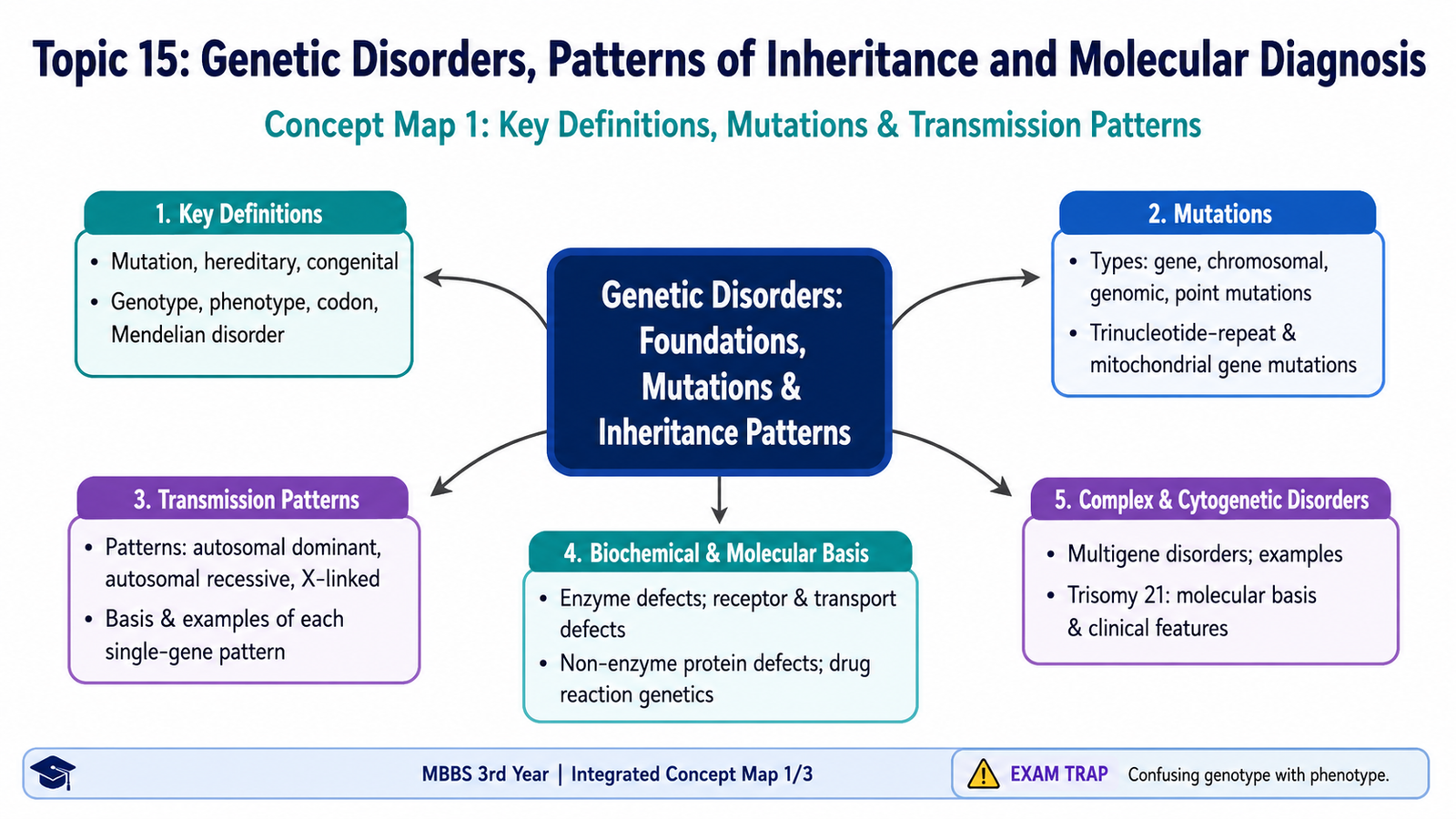
This chapter follows the supplied KMU learning outcomes and builds genetics from basic terminology to inheritance patterns and molecular diagnosis. First understand how genetic changes produce disease, then use the final high-yield review for revision.
Topic 15 — Genetic Disorders, Patterns of Inheritance and Molecular Diagnosis
Module/Theme: Infection and Inflammation · Pathology
Understand how mutations alter genes and proteins, how single-gene and complex disorders are transmitted, why Trisomy 21 develops, and how PCR, FISH and blotting techniques help in molecular diagnosis.
Topic Introduction
Genetic disorders develop when an abnormality in genetic material alters the structure, quantity or function of a gene product. The abnormality may involve a single nucleotide, a larger part of a gene, mitochondrial DNA, several interacting genes, or whole chromosomes. Some genetic disorders are inherited from parents, whereas others arise as new mutations. In this chapter, you will first learn the basic genetic terms and types of mutation, then study autosomal dominant, autosomal recessive and X-linked inheritance. You will also see how genetic defects disturb enzymes, receptors, transport proteins and other proteins, how complex disorders and Trisomy 21 arise, and how important molecular techniques are used to detect genetic abnormalities.
A. Genetic Foundations: Essential Terms and Gene Expression
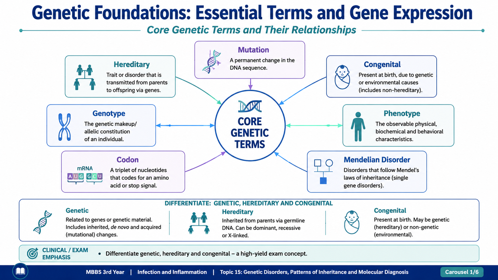
Before studying genetic disease, several basic terms must be clearly understood. A gene contains information that ultimately contributes to the production of a functional RNA or protein. A change in this information can alter cellular function and produce disease. However, the presence of a genetic abnormality does not always mean that the abnormality was inherited from a parent, and a disorder present at birth is not automatically hereditary.
Mutation
A mutation is a permanent alteration in the DNA sequence. A mutation may involve a single nucleotide, several nucleotides, part of a gene or a larger region of genetic material. Its effect depends on where the change occurs and how it influences the gene product.
Hereditary
A hereditary disorder is transmitted genetically from a parent to offspring. Therefore, hereditary disease implies transmission through germ cells. A newly arising mutation in a child may be genetic without initially being hereditary from either parent.
Congenital
Congenital means present at birth. A congenital disorder may result from a genetic abnormality, but it may also result from environmental influences during fetal development. Therefore, the terms congenital and hereditary are not interchangeable.
Genotype and Phenotype
The genotype is the genetic constitution of an individual at a particular locus or group of loci. The phenotype is the observable expression of that genotype, such as a physical feature, biochemical abnormality or disease manifestation.
Phenotype is influenced not only by the genotype but also by factors such as penetrance, expressivity and environmental influences. Therefore, people carrying the same genetic mutation may not always show exactly the same clinical severity.
Codon
A codon is a sequence of three nucleotides in messenger RNA that specifies an amino acid or provides a signal for termination of translation. Because protein synthesis depends on the sequence of codons, a mutation that changes a codon may change the amino-acid sequence of a protein or prematurely terminate protein synthesis.
Mendelian Disorder
A Mendelian disorder is usually caused by a pathogenic variant in a single gene and follows a recognizable inheritance pattern based on Mendelian principles. Important patterns include autosomal dominant, autosomal recessive and X-linked inheritance.
B. Mutations: Types, Trinucleotide Repeats and Mitochondrial Gene Defects
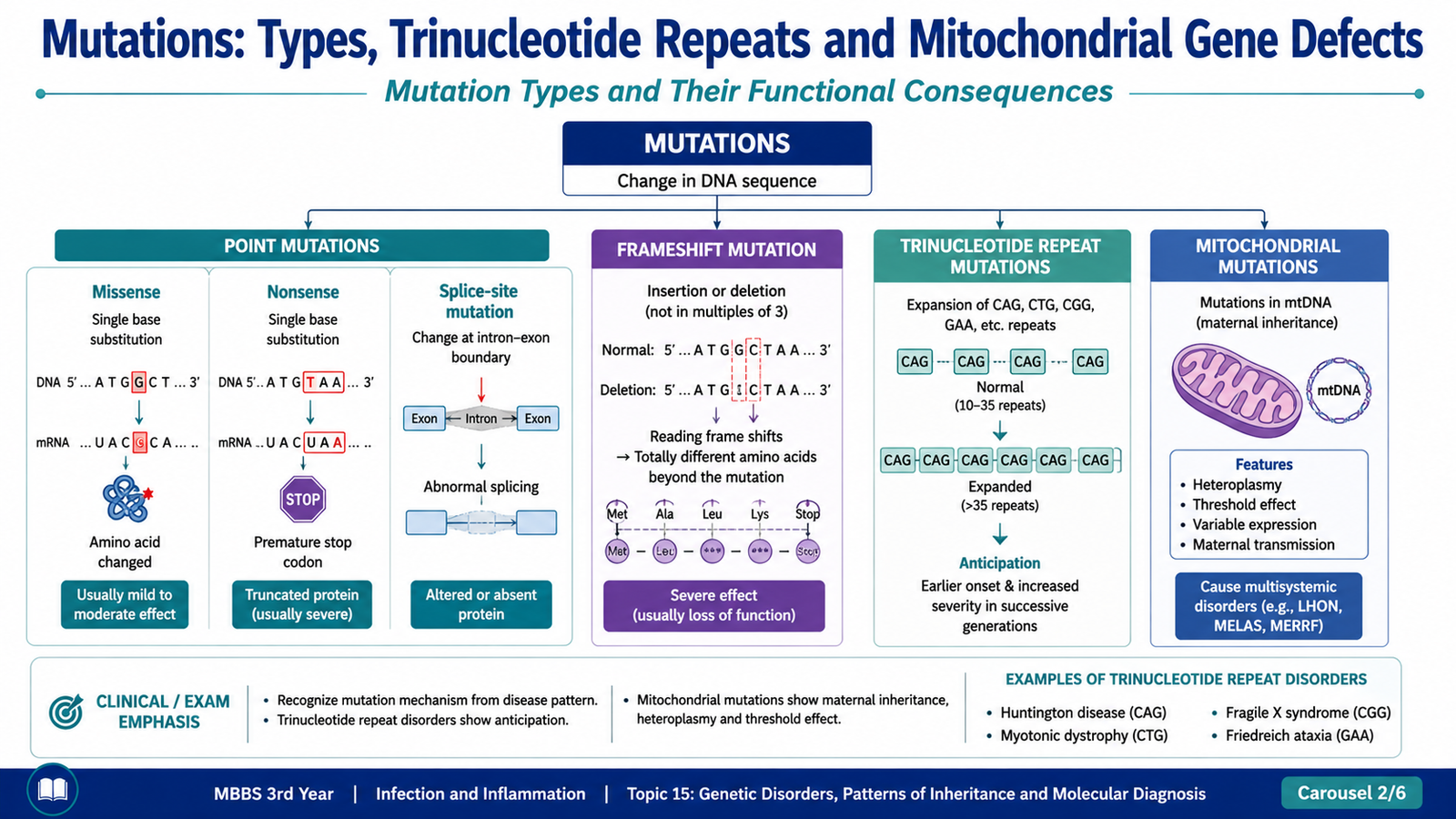
Mutations produce disease when they alter the amount or function of an important gene product. Some mutations change only one nucleotide, whereas others insert or remove several nucleotides. The functional consequence ranges from no significant effect to complete absence of a protein or production of an abnormal protein.
Point Mutations
A point mutation involves alteration of a single nucleotide. The effect depends on how the altered nucleotide changes the genetic code.
- Missense mutation: a nucleotide substitution changes one codon so that a different amino acid is incorporated into the protein. The altered amino acid may change protein structure or function.
- Nonsense mutation: a nucleotide substitution converts an amino-acid codon into a stop codon. Translation stops prematurely, usually producing a shortened and unstable protein.
- Mutation affecting splicing: a change at or near a splice site may interfere with normal removal of introns and joining of exons, producing abnormal messenger RNA.
Mechanism:
DNA nucleotide alteration → altered codon or RNA processing → abnormal protein structure or quantity → disturbed cellular function → disease phenotype
Insertions and Deletions
An insertion adds one or more nucleotides, whereas a deletion removes them. When the number of inserted or deleted nucleotides is not a multiple of three, the reading frame of the gene changes. This is called a frameshift mutation.
A frameshift changes the way subsequent codons are read. As a result, many downstream amino acids may be altered and a premature stop codon may appear. The resulting protein is often severely abnormal or absent.
Trinucleotide-Repeat Mutations
In trinucleotide-repeat disorders, a sequence of three nucleotides is repeated an abnormally large number of times. The repeat may expand when transmitted from one generation to the next. When the number of repeats crosses a critical level, gene expression or protein function becomes abnormal and disease develops.
An important feature of several trinucleotide-repeat disorders is anticipation. This means the disorder may appear at an earlier age or become more severe in successive generations because the repeat sequence expands further during transmission.
Examples include:
- Huntington disease
- Fragile X syndrome
- Myotonic dystrophy
- Friedreich ataxia
Mutations in Mitochondrial Genes
Mitochondria contain their own DNA. Mutations affecting mitochondrial DNA can disturb oxidative phosphorylation and therefore reduce cellular energy production. Tissues with high energy requirements, particularly the nervous system and skeletal or cardiac muscle, are commonly affected.
Mitochondrial DNA is transmitted mainly through the ovum. Therefore, mitochondrial disorders show maternal inheritance: an affected mother may transmit the mutation to her children, whereas an affected father does not normally transmit mitochondrial DNA to his offspring.
The severity of mitochondrial disease may vary among tissues and individuals because cells can contain mixtures of normal and mutant mitochondrial DNA. Different proportions of mutant mitochondria can therefore produce different levels of functional impairment.
C. Transmission Patterns of Single-Gene Disorders
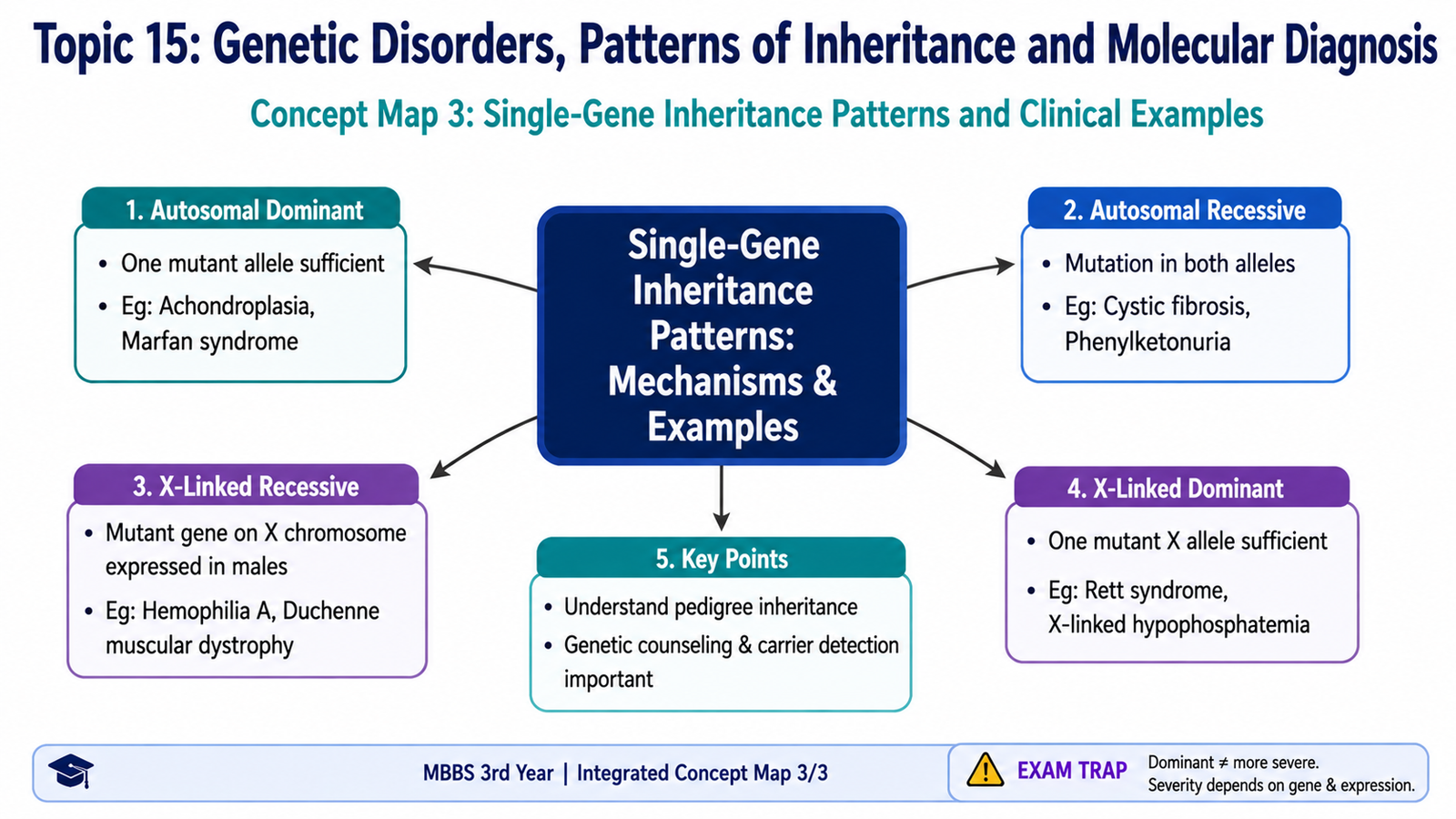
Single-gene disorders are caused mainly by pathogenic variants in individual genes. Their pattern of appearance within a family depends on whether the affected gene is located on an autosome or sex chromosome and whether one or two abnormal alleles are required for the phenotype.
Autosomal Dominant Disorders
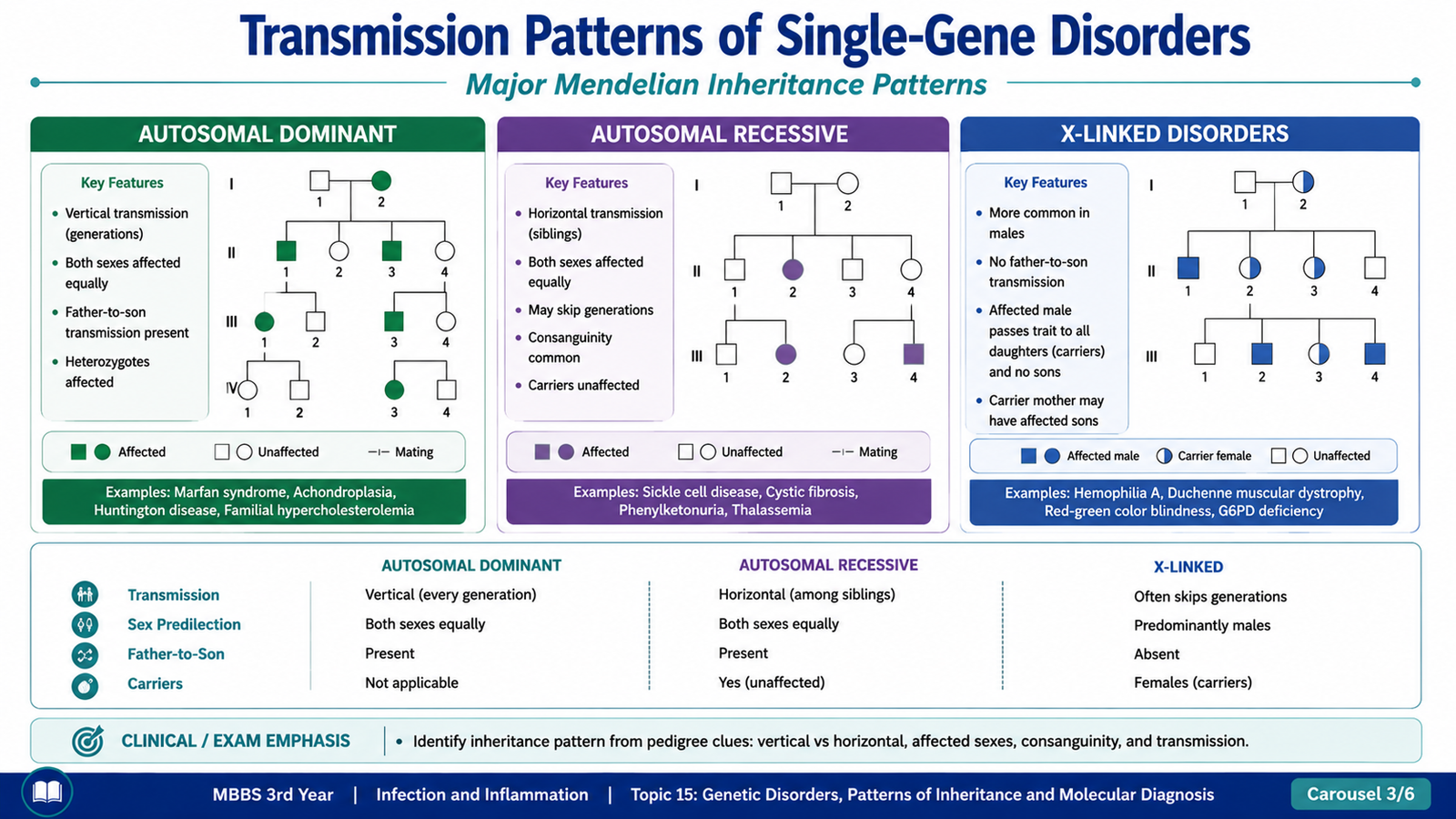
In an autosomal dominant disorder, one abnormal allele is sufficient to produce the disease phenotype. An affected person is commonly heterozygous and may transmit the abnormal allele to either sons or daughters.
Autosomal dominant disorders often show a vertical pattern in a pedigree because affected individuals may be seen in successive generations. Males and females are generally affected with similar frequency because the gene is located on an autosome.
The molecular abnormality frequently involves proteins that perform structural, regulatory or receptor functions rather than enzymes in metabolic pathways. Disease may arise through several mechanisms:
- Reduced quantity of a normal protein: one functioning allele may not produce enough protein for normal function.
- Dominant-negative effect: the abnormal protein interferes with the function of the normal protein produced by the other allele.
- Gain-of-function mutation: the mutated protein develops an abnormal or excessive activity.
Examples of autosomal dominant disorders include:
- Marfan syndrome
- Familial hypercholesterolemia
- Huntington disease
- Neurofibromatosis
- Adult polycystic kidney disease
Autosomal Recessive Disorders
In an autosomal recessive disorder, disease usually occurs when both alleles at a gene locus are abnormal. Parents of an affected individual are commonly clinically unaffected carriers, each possessing one normal and one abnormal allele.
Autosomal recessive disorders often appear among siblings rather than in successive generations. Males and females are affected equally. The pattern may be more evident in families in which the parents share common ancestry because relatives are more likely to carry the same uncommon pathogenic allele.
Many autosomal recessive disorders are caused by loss-of-function mutations affecting enzymes. A carrier often retains enough enzyme activity from the normal allele to prevent disease. An affected individual has marked reduction or absence of functional enzyme activity, leading to metabolic disturbance.
Examples include:
- Cystic fibrosis
- Phenylketonuria
- Sickle cell disease
- Many lysosomal storage diseases
- Albinism
X-Linked Disorders
In X-linked disorders, the abnormal gene is located on the X chromosome. The inheritance pattern differs between males and females because males have one X chromosome while females have two.
In an X-linked recessive disorder, a male who inherits the pathogenic allele on his single X chromosome usually expresses the disease because there is no second X chromosome carrying a normal allele to compensate. A heterozygous female is usually a carrier, although the degree of expression may vary because of X-chromosome inactivation.
An important pedigree rule is that father-to-son transmission does not occur for X-linked traits because a father gives his Y chromosome, not his X chromosome, to a son.
Examples of X-linked disorders include:
- Hemophilia A
- Duchenne muscular dystrophy
- Glucose-6-phosphate dehydrogenase deficiency
- Fragile X syndrome
| Feature | Autosomal Dominant | Autosomal Recessive | X-Linked Recessive |
|---|---|---|---|
| Alleles usually required | One abnormal allele | Two abnormal alleles | One abnormal X is sufficient in a male |
| Pedigree pattern | Often successive generations | Often affected siblings | Predominantly affected males |
| Sex distribution | Both sexes | Both sexes | Males more commonly affected |
| Father-to-son transmission | Possible | Possible | Absent |
D. Biochemical and Molecular Basis of Single-Gene Disorders
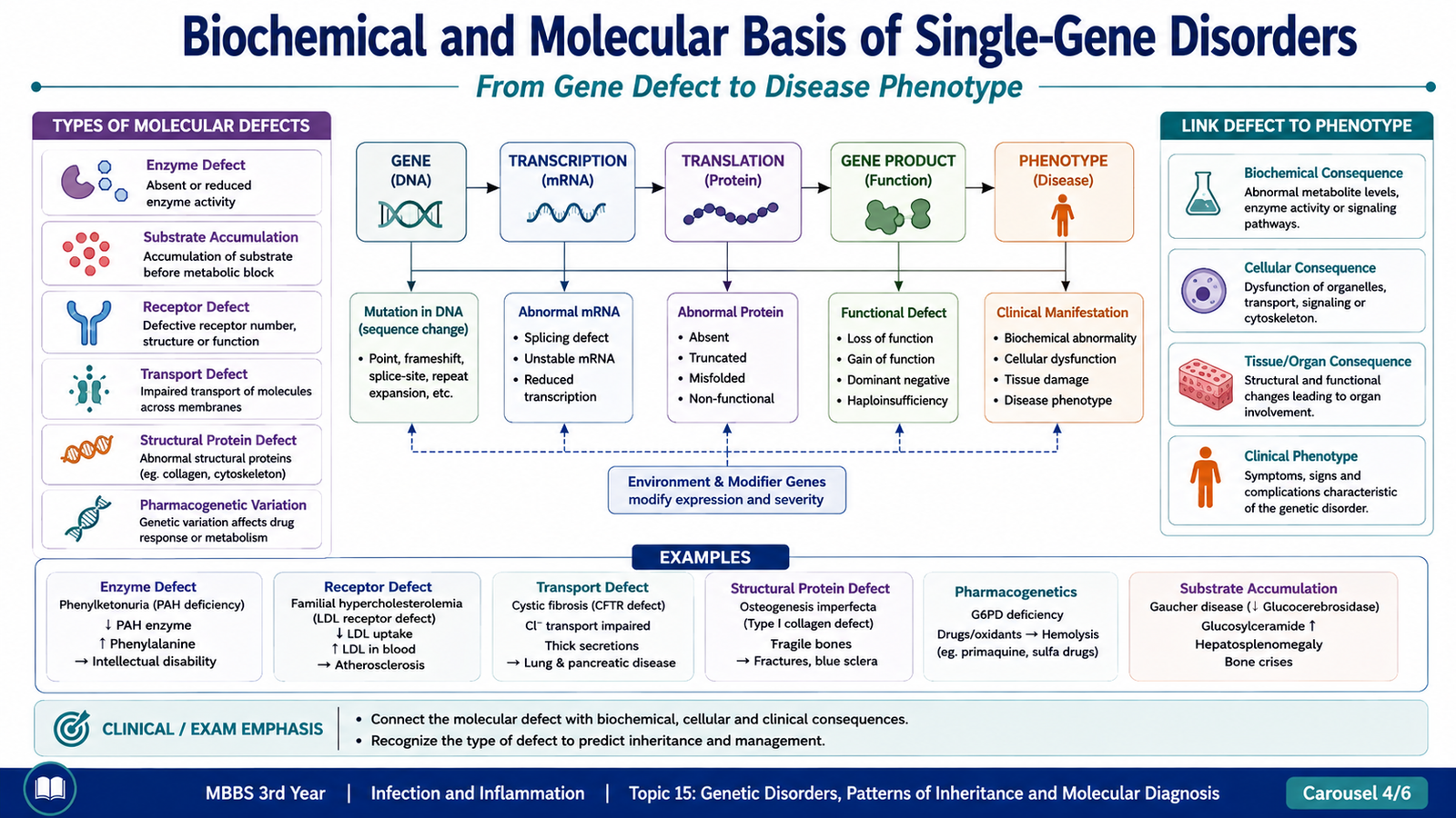
A mutation causes disease because it changes a functional product of the gene. The affected product may be an enzyme, receptor, transporter, structural protein or another regulatory protein. Understanding which type of protein is defective helps explain the biochemical abnormality and the resulting clinical phenotype.
Enzyme Defects and Their Consequences
Enzyme defects are particularly common in autosomal recessive disorders. When an enzyme in a metabolic pathway is markedly reduced or absent, several consequences may follow.
Normal pathway:
Substrate → enzyme → product
With enzyme deficiency:
Enzyme defect → substrate cannot be processed normally → substrate or its derivatives accumulate → required product may become deficient → abnormal metabolites may be produced → cellular or tissue dysfunction
The disease may therefore result from:
- Accumulation of a toxic substrate
- Accumulation of stored material within cells
- Deficiency of an essential end product
- Diversion of substrate into an abnormal metabolic pathway
For example, many inborn errors of metabolism occur because a particular enzyme is deficient. Similarly, lysosomal enzyme deficiencies can cause accumulation of undegraded material within lysosomes.
Defects in Receptors
Receptors allow cells to recognize and respond to extracellular molecules. A receptor defect can prevent a ligand from binding normally or interfere with the cellular response after binding.
A classic example is familial hypercholesterolemia, in which abnormalities affecting the LDL receptor pathway reduce normal cellular uptake and removal of LDL from the circulation. The result is elevation of circulating LDL cholesterol and increased deposition of cholesterol in tissues and arterial walls.
Defects in Transport Systems
Transport proteins move substances across cell membranes or between cellular compartments. A mutation affecting a transporter can therefore alter the movement of ions or molecules and disturb tissue function.
Cystic fibrosis illustrates this principle. Abnormal CFTR protein alters chloride transport across epithelial surfaces. This changes movement of salt and water and produces abnormally thick secretions in affected organs.
Alterations in Non-Enzyme Proteins
Some genetic disorders result from abnormalities in the structure, function or quantity of non-enzyme proteins. These proteins may form structural tissues, carry molecules, regulate growth or participate in cell signaling.
Examples of mechanisms include:
- Abnormal structural protein: tissue loses normal mechanical strength or elasticity.
- Abnormal functional protein: the protein is present but performs its task poorly.
- Reduced protein quantity: insufficient normal protein is produced.
- Abnormal regulatory protein: signaling or control of cellular activity becomes disturbed.
For example, abnormal fibrillin in Marfan syndrome alters connective-tissue integrity, while abnormal globin chains in hemoglobin disorders alter the structure or function of hemoglobin.
Genetically Determined Adverse Reactions to Drugs
Inherited genetic variation can modify the way an individual handles or responds to a drug. This field is commonly described as pharmacogenetics. A genetically determined deficiency or abnormality in an enzyme may make a person unusually susceptible to a drug-related adverse effect.
Important undergraduate examples include:
- G6PD deficiency: exposure to certain oxidant drugs can produce oxidative damage to red cells and precipitate hemolysis.
- Inherited abnormality of plasma cholinesterase: breakdown of certain neuromuscular blocking drugs may be prolonged, resulting in prolonged neuromuscular paralysis.
E. Complex Genetic Disorders and Trisomy 21
Not all genetic disorders follow simple Mendelian inheritance. Many common diseases result from the combined effects of several genes together with environmental influences. In addition, abnormalities may involve the number or structure of whole chromosomes rather than a mutation in a single gene.
Multigenic and Multifactorial Disorders
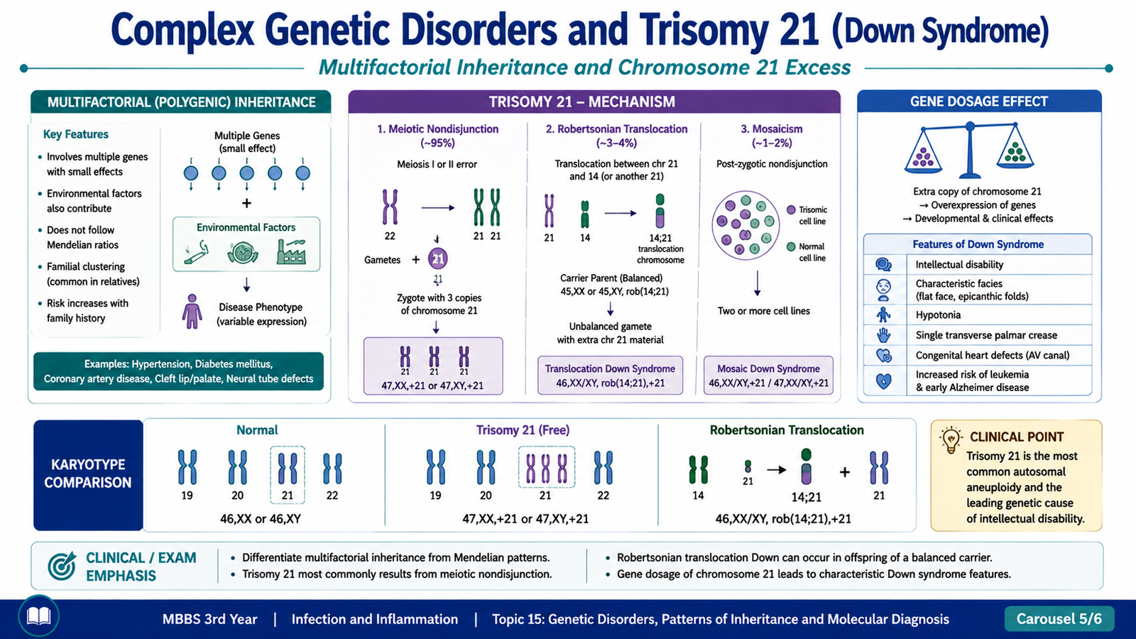
A multigenic disorder results from the combined influence of variants in multiple genes. In many such disorders, environmental factors also contribute significantly; these conditions are therefore often described as multifactorial disorders.
Each individual susceptibility gene may have only a relatively small effect. Disease develops when the combined genetic susceptibility and relevant environmental influences cross a threshold sufficient to disturb normal function.
Examples include common disorders such as:
- Hypertension
- Type 2 diabetes mellitus
- Coronary artery disease
- Some congenital malformations
Unlike classical Mendelian disorders, these conditions do not usually produce a simple dominant or recessive pedigree pattern.
Trisomy 21: Molecular and Cytogenetic Basis
Trisomy 21, or Down syndrome, is a chromosomal disorder caused by the presence of additional genetic material from chromosome 21. The increased dosage of genes located on chromosome 21 alters normal development and produces a recognizable group of physical and functional abnormalities.
The most common mechanism is meiotic nondisjunction. During formation of a gamete, chromosome 21 fails to separate normally. A gamete therefore receives an extra chromosome 21. After fertilization with a normal gamete, the embryo has three copies of chromosome 21.
Nondisjunction
→ gamete receives an extra chromosome 21
→ fertilization
→ three copies of chromosome 21
→ increased gene dosage
→ abnormal development and Down syndrome phenotype
Other mechanisms include Robertsonian translocation, in which additional chromosome 21 material is attached to another chromosome, and mosaicism, in which some cells have the abnormal chromosome number while others have a normal chromosome complement.
Clinical Recognition of Trisomy 21
The phenotype results from altered development produced by excess chromosome 21 genetic material. No single physical finding alone is sufficient to establish the diagnosis, but a characteristic combination raises clinical suspicion and should be confirmed by chromosome analysis.
Important clinical features include:
- Developmental and intellectual impairment of variable degree
- Generalized hypotonia, particularly evident in infancy
- Characteristic facial appearance with a relatively flat facial profile
- Upslanting palpebral fissures and epicanthic folds
- Small ears and a relatively short neck
- Single transverse palmar crease in some individuals
- Wide separation between the first and second toes may be present
- Congenital cardiac abnormalities occur with increased frequency
F. Molecular Genetic Diagnosis: PCR, FISH and Blotting Techniques
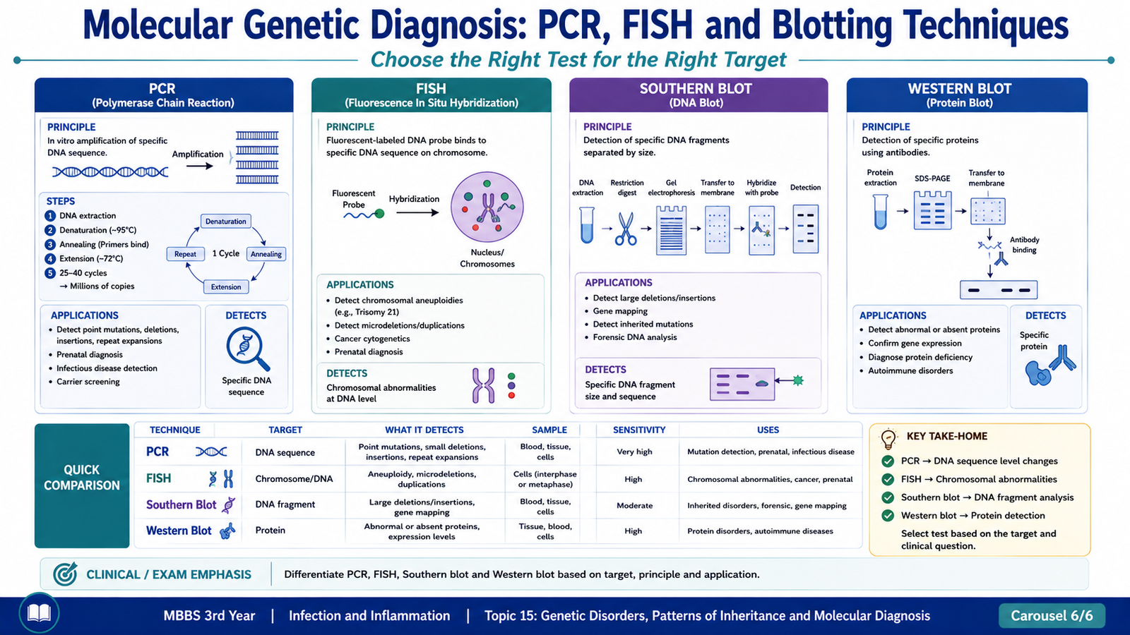
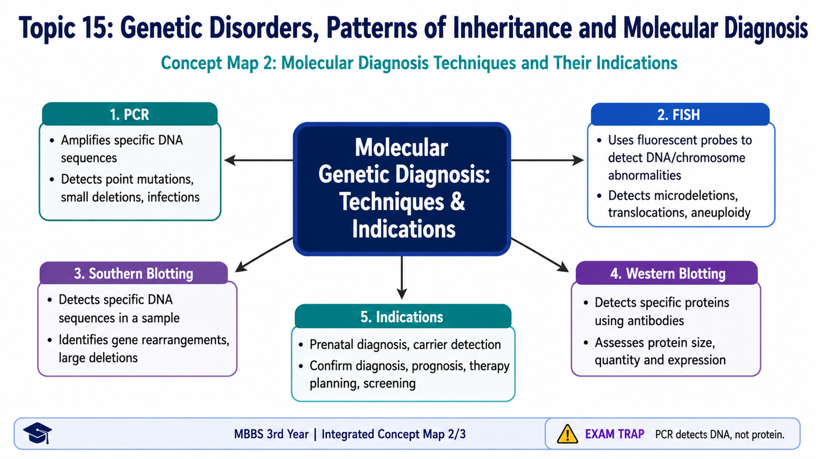
Molecular and cytogenetic techniques allow genetic abnormalities to be detected at different levels. Some methods identify or amplify a specific DNA sequence, some show the location or number of a chromosome region within cells, and others detect particular DNA fragments or proteins. The correct investigation therefore depends on what abnormality is suspected.
Polymerase Chain Reaction — PCR
Polymerase chain reaction (PCR) is a technique used to selectively amplify a specific DNA sequence. Starting with a very small amount of DNA, repeated cycles produce a large number of copies of the target region so that it can be studied more easily.
The basic cycle consists of:
- Denaturation: double-stranded DNA separates into single strands.
- Annealing: short DNA primers bind to complementary sequences surrounding the target region.
- Extension: a thermostable DNA polymerase synthesizes new DNA from the primers.
Repeating these steps results in exponential amplification of the selected sequence.
Principle:
Target DNA → specific primers bind → DNA polymerase copies target → repeated cycles → large amount of target DNA available for analysis
Important indications include detection or analysis of known gene sequences and mutations, analysis of small amounts of DNA, and preparation of DNA for further molecular study.
Fluorescence In Situ Hybridization — FISH
FISH uses fluorescently labeled DNA probes that bind to complementary DNA sequences in chromosomes or cell nuclei. The bound probe can then be visualized with fluorescence microscopy.
Because the probe binds to a specific chromosome region, FISH can demonstrate the presence, absence, location or abnormal number of that region. It is particularly useful when a specific chromosomal abnormality is suspected.
Important applications include:
- Detection of selected chromosomal numerical abnormalities
- Detection of specific deletions or duplications
- Demonstration of selected translocations or rearrangements
- Assessment of specific gene amplification in appropriate settings
Southern Blotting
Southern blotting is used to detect a specific DNA sequence within a mixture of DNA fragments. DNA is cut into fragments, separated according to size by electrophoresis, transferred to a membrane and exposed to a labeled probe that binds to the complementary DNA sequence.
The method therefore answers a basic question: Is a particular DNA sequence present, and what is the size or pattern of the DNA fragment containing it?
Southern blotting has been used to analyze DNA rearrangements, selected gene abnormalities and expanded DNA repeat regions.
Western Blotting
Western blotting detects specific proteins rather than DNA. Proteins are separated, transferred to a membrane and detected using antibodies that specifically bind the protein of interest.
This is important because a genetic mutation may ultimately alter the quantity, size or presence of a protein. Western blotting can therefore provide information about the protein product resulting from gene expression.
| Technique | Main Target | Basic Principle | Typical Use |
|---|---|---|---|
| PCR | DNA | Amplifies a selected DNA sequence | Analysis of specific DNA sequences or mutations |
| FISH | Chromosomal DNA | Fluorescent probe binds its complementary sequence in cells | Specific chromosome number or structural abnormality |
| Southern blot | DNA | Probe detects a selected DNA fragment after separation and transfer | Selected DNA rearrangements or repeat abnormalities |
| Western blot | Protein | Antibody detects a selected protein after separation | Assessment of presence, size or amount of a protein |
Integrated Mechanism Flow
↓
2. Gene sequence, gene dosage or gene expression is altered
↓
3. Quantity, structure or function of a gene product changes
↓
4. Enzyme, receptor, transporter or structural function becomes abnormal
↓
5. Cellular and tissue homeostasis is disturbed
↓
6. A recognizable biochemical, structural or clinical phenotype develops
↓
7. Molecular or cytogenetic testing can identify the underlying abnormality
⭐ AIM High-Yield Review
- A mutation is a permanent alteration in DNA sequence.
- Hereditary means genetically transmitted from parent to offspring, whereas congenital means present at birth.
- A missense mutation changes an amino acid; a nonsense mutation introduces a premature stop codon.
- Insertion or deletion not involving a multiple of three nucleotides can produce a frameshift.
- Trinucleotide-repeat expansion can produce anticipation.
- Mitochondrial gene disorders characteristically show maternal transmission.
- Autosomal dominant disease requires one abnormal allele and commonly shows affected individuals in successive generations.
- Autosomal recessive disorders commonly involve enzyme deficiencies and may occur among siblings of unaffected carrier parents.
- ⭐ No father-to-son transmission occurs in X-linked inheritance.
- Single-gene disorders may result from defects in enzymes, receptors, transport proteins or non-enzyme structural and regulatory proteins.
- Genetic variation can alter drug response; G6PD deficiency is an important example of genetically determined susceptibility to drug-induced hemolysis.
- Multifactorial disease reflects interaction between multiple susceptibility genes and environmental factors.
- Down syndrome results from excess chromosome 21 genetic material, most commonly due to meiotic nondisjunction.
- ⭐ PCR amplifies DNA; FISH localizes selected DNA sequences; Southern blot detects DNA fragments; Western blot detects proteins.
- Clinical recognition of a genetic syndrome suggests the diagnosis, while molecular or cytogenetic testing can identify its underlying genetic abnormality.
Genetic Disorders, Patterns of Inheritance and Molecular Diagnosis
Use these videos after reading the AIM chapter to reinforce mutations, Mendelian inheritance and molecular diagnostic techniques.
▶ Video 1 — Genetic Mutations and Patterns of Inheritance
Recommended for revising mutation types, autosomal dominant, autosomal recessive and X-linked inheritance.
▶ Video 2 — PCR, FISH, Southern Blot and Western Blot
Recommended for understanding the basic principles and differences between the major molecular diagnostic techniques covered in this topic.
While watching, focus on the mutation → abnormal gene product → disease sequence, pedigree clues for inheritance, and the key distinction that PCR amplifies DNA, FISH localizes specific DNA sequences, Southern blot detects DNA fragments and Western blot detects proteins.