
Topic 9 — Death, Brain Death and Early Postmortem Changes
Topic Introduction
Thanatology is the branch of forensic medicine concerned with death and the changes that occur after death. In medical practice, death must be recognized accurately because it has important clinical, legal and social consequences. A doctor may need to distinguish actual death from apparent death, recognize brain death, document the time and circumstances of death, and interpret changes that develop in the body afterward. Postmortem changes occur in a sequence, although their timing is influenced by environmental and individual factors. Understanding these changes—especially cooling, lividity, rigor mortis and decomposition—helps the medical practitioner describe the state of the body and make cautious medico-legal interpretations.
A. Death, Thanatology and the Phases of Death
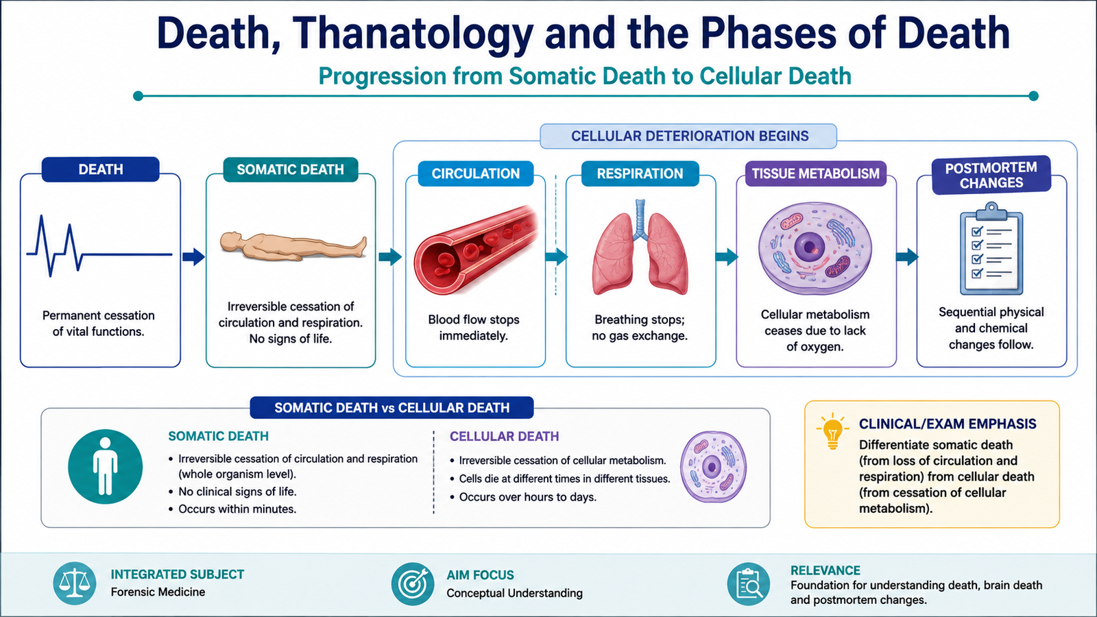
Death is not simply one instantaneous cellular event. In forensic medicine, it is understood as a process in which vital functions cease irreversibly and the tissues of the body subsequently lose their individual cellular activity. This distinction helps explain why some organs and tissues may remain viable for a limited period even after the person as a whole has died.
Definition of death
Death is the irreversible cessation of the functions necessary to sustain the organism as an integrated whole. Traditionally, this was recognized by permanent cessation of circulation and respiration. Modern medicine also recognizes death on neurological grounds when there is irreversible loss of all functions of the brain, including the brainstem.
Phases of death
The transition from life to complete cellular death can be understood in two major stages.
- Somatic death: irreversible death of the individual as an integrated organism. There is permanent loss of consciousness, spontaneous respiration and effective circulation, or irreversible loss of all brain functions depending on the criterion used.
- Molecular or cellular death: individual cells and tissues die progressively after somatic death. Different tissues survive for different periods because their oxygen and metabolic requirements differ.
Irreversible failure of vital integrated functions → somatic death → continuing loss of oxygen and metabolism in tissues → progressive cellular death → postmortem changes
B. Brain Death and Its Criteria
Modern intensive care can artificially maintain ventilation and circulation even when the brain has irreversibly ceased functioning. Therefore, brain death has become an important medical and medico-legal concept. Brain death is different from coma or a persistent disorder of consciousness because the loss of brain function in brain death is irreversible.
Definition of brain death
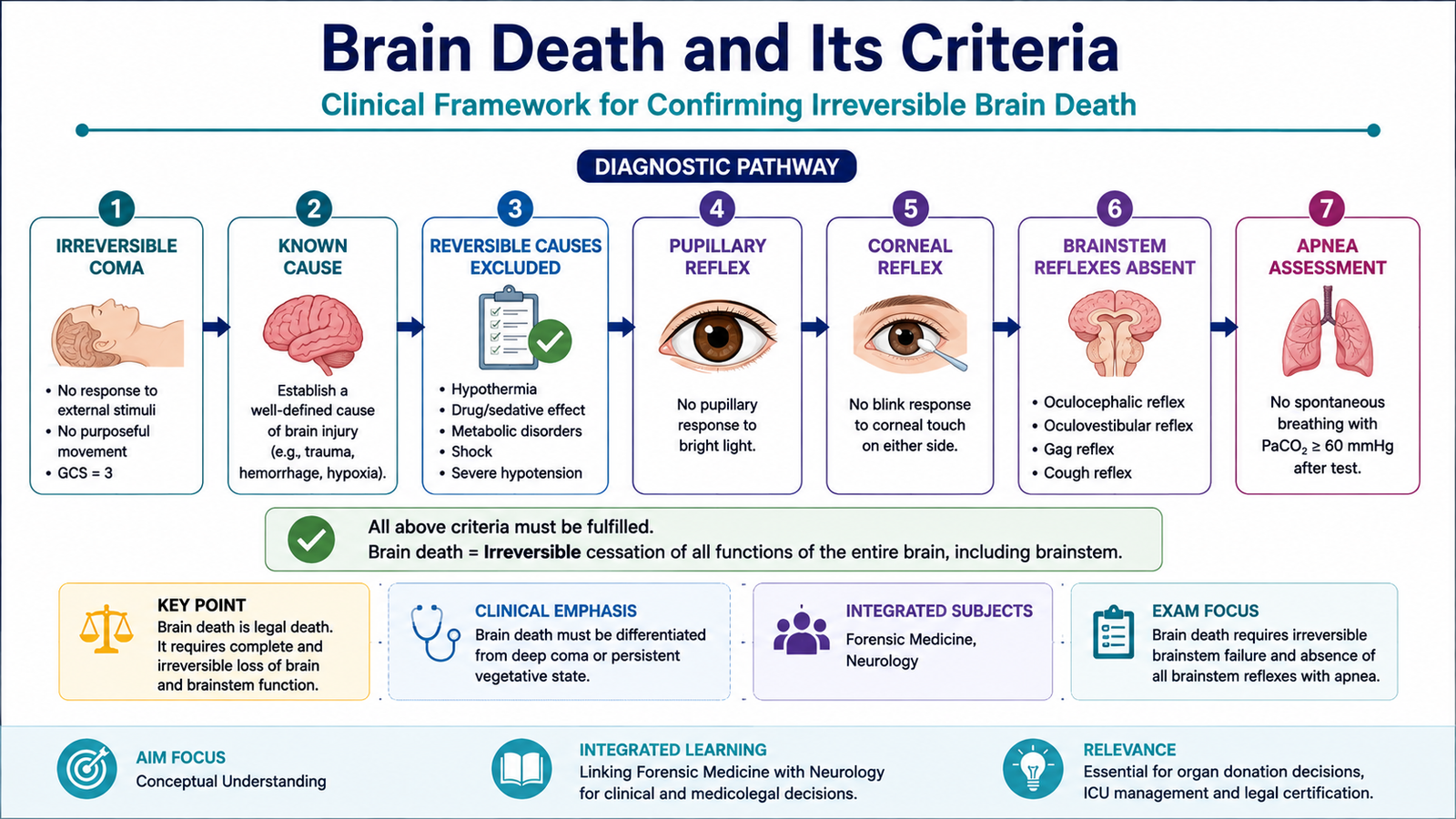
Brain death is the irreversible cessation of all functions of the entire brain, including the brainstem. Once properly established according to accepted medical criteria, it represents death of the individual.
Essential principles before testing
Before brain death is diagnosed, the doctor must first establish that the patient has suffered a catastrophic brain injury capable of causing irreversible loss of brain function. Reversible conditions that can suppress neurological responses must be excluded.
- The cause of the severe brain injury should be known and sufficient to explain the clinical state.
- The condition should be considered irreversible.
- Central nervous system depressant drugs and significant intoxication must be excluded as explanations.
- Major metabolic or endocrine disturbances capable of suppressing brain function must be corrected or excluded.
- Severe hypothermia must not be responsible for the absent neurological responses.
- Neuromuscular paralysis must not be mistaken for absence of motor responses.
Clinical criteria
Once the prerequisites are satisfied, the diagnosis depends principally on demonstrating irreversible coma, absence of brainstem reflexes and absence of spontaneous respiratory drive.
| Component | Finding in brain death | Meaning |
|---|---|---|
| Consciousness | Deep unresponsive coma | No cerebral response to external stimulation |
| Pupillary response | Absent response to light | Loss of relevant brainstem reflex pathways |
| Corneal reflex | Absent | Indicates severe brainstem failure |
| Oculocephalic/vestibulo-ocular responses | Absent where testing is appropriate | Supports loss of brainstem function |
| Gag and cough reflexes | Absent | Loss of medullary brainstem reflex activity |
| Spontaneous respiration | Absent on appropriate apnea assessment | No effective respiratory drive from the brainstem |
C. EEG, ECG, Apparent Death and Human Tissue Considerations
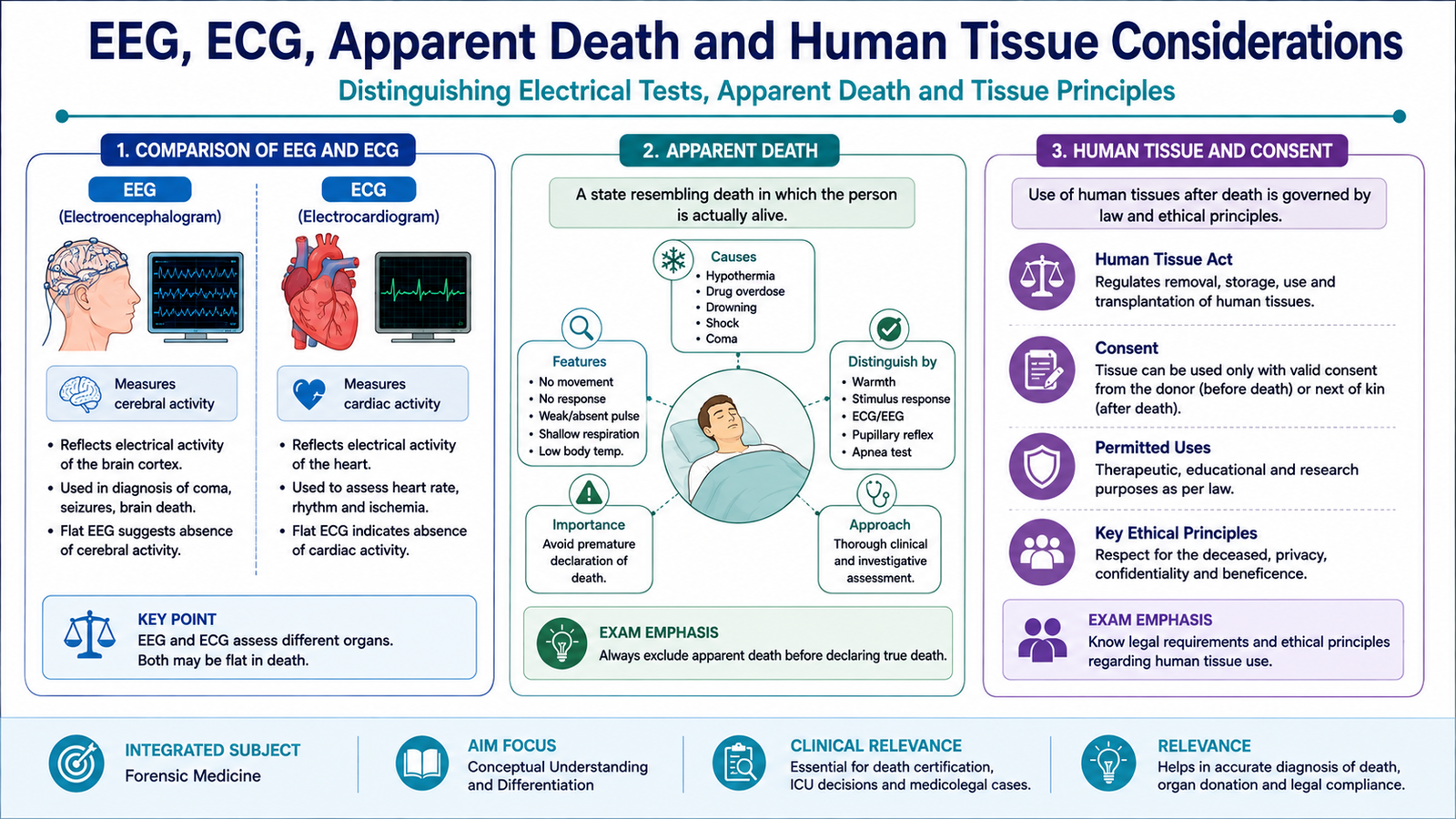
Electrical investigations can support the assessment of death, but they must be interpreted according to what they actually measure. An electroencephalogram records electrical activity of the brain, whereas an electrocardiogram records electrical activity of the heart. Neither should be confused with a complete clinical assessment of the patient.
Role of EEG
Electroencephalography (EEG) records cortical electrical activity. Absence of detectable cerebral electrical activity may be used as an ancillary finding in selected circumstances. However, brain death is fundamentally a clinical diagnosis when the required clinical examination can be completed reliably. EEG also mainly reflects cortical activity and does not by itself demonstrate every aspect of brainstem function.
Role of ECG
Electrocardiography (ECG) records the electrical activity of the heart. It may help demonstrate cardiac electrical activity or its absence during assessment of circulatory arrest. However, ECG alone is not a test for brain death because cardiac electrical activity may continue even when brain function has irreversibly ceased.
Apparent death
Apparent death is a state in which the signs of life are so greatly reduced that the person may appear dead although life persists. Respiration and circulation may be extremely difficult to detect. The concept is important because conditions producing profound physiological suppression must not be mistaken for irreversible death.
Situations capable of producing a death-like appearance may include severe hypothermia, profound drug-induced central nervous system depression, severe shock and other states in which vital activity becomes markedly depressed. The essential principle is to establish death carefully and exclude potentially reversible conditions.
Human tissue legislation: core principle
Human tissue legislation provides an ethical and legal framework for the removal, storage and use of human tissues and organs. Its central concerns include lawful authorization, valid consent where applicable, respect for the deceased and appropriate medical governance. Exact statutory procedures vary according to the jurisdiction and must follow the applicable local law.
In relation to death and organ or tissue donation, accurate determination and documentation of death are essential. Decisions regarding declaration of death must remain medically independent from decisions concerning retrieval or use of organs and tissues.
D. Medico-Legal Importance of Death and Classification of Postmortem Changes
Recognition and documentation of death have major medico-legal consequences. Once death has occurred, questions may arise regarding identification of the deceased, cause and circumstances of death, possible time since death, disposal of the body, inheritance, insurance and criminal or civil investigation. For this reason, observations made by the doctor should be objective and clearly documented.
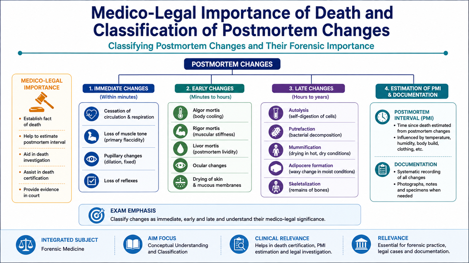
Medico-legal importance of death
- Establishing that death has occurred.
- Helping determine the identity of the deceased where required.
- Assisting investigation of the cause and circumstances of death.
- Providing findings that may contribute to an estimation of the postmortem interval.
- Distinguishing postmortem changes from injuries or disease occurring during life.
- Providing appropriate medical documentation for legal and administrative purposes.
- Allowing lawful procedures relating to the body, including autopsy or tissue-related processes where applicable.
Postmortem findings should not be interpreted as an exact clock. Environmental temperature, clothing, body habitus, illness, activity before death and many other factors alter their development.
Definition of postmortem changes
Postmortem changes are the physical, chemical and biological changes that occur in the body after death. They develop because circulation and respiration have ceased, metabolism stops, muscles undergo biochemical changes, blood becomes redistributed by gravity and tissues eventually undergo decomposition.
Classification
| Group | Main changes | Basic explanation |
|---|---|---|
| Immediate changes | Loss of consciousness, respiration, circulation and voluntary muscular activity; primary muscular flaccidity | Result directly from cessation of vital integrated functions |
| Early changes | Cooling, postmortem lividity, rigor mortis and early ocular/body changes | Develop as metabolism ceases and blood and tissues respond to loss of circulation |
| Late changes | Putrefaction and other forms of decomposition or preservation | Result mainly from tissue breakdown, enzymatic processes and microbial activity |
E. Early Postmortem Changes: Cooling and Rigor Mortis
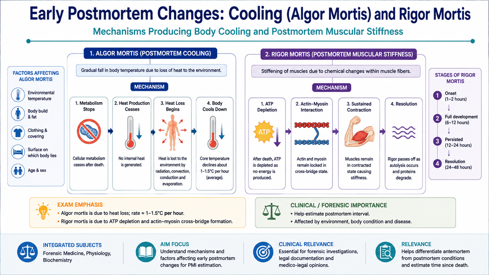
After death, the body no longer regulates temperature or maintain normal muscle energy metabolism. These failures produce two important early postmortem changes: cooling of the body and rigor mortis. Both can contribute to medico-legal assessment, but neither develops at an identical rate in every body.
Algor mortis — postmortem cooling
Algor mortis is the gradual cooling of the body after death until its temperature approaches that of the surrounding environment. Heat production falls because metabolism has ceased, while heat continues to be lost to the environment.
The rate of cooling varies considerably. Important influences include environmental temperature, air movement, clothing, body size and fat, contact with surrounding surfaces and whether the body is wet or dry. Therefore, body temperature is only one part of postmortem assessment and should not be used as an isolated exact measure of time since death.
Rigor mortis
Rigor mortis is postmortem stiffening of muscles. Immediately after death, muscles are initially flaccid. As oxygen-dependent metabolism stops, ATP can no longer be adequately regenerated. Actin and myosin become locked in a contracted state, producing muscular stiffness.
Rigor mortis involves both voluntary and involuntary muscles and generally becomes evident progressively throughout the body. It eventually passes off as muscle proteins undergo decomposition.
The onset and duration of rigor are influenced by temperature, muscular activity before death, body condition and the circumstances surrounding death. Because these factors vary, rigid timing formulas should be interpreted cautiously.
F. Postmortem Lividity
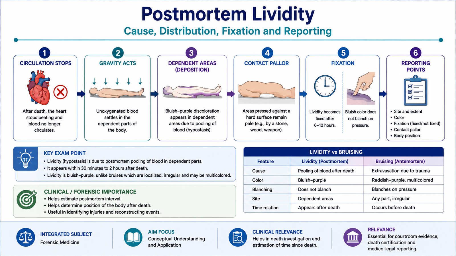
Postmortem lividity, also called livor mortis or hypostasis, is the purplish-red discoloration that develops in dependent areas of the body after circulation has stopped. It occurs because blood is no longer being propelled through the vessels and therefore moves under the influence of gravity into the vessels of the lowest parts of the body.
Mechanism
Areas under firm pressure may remain pale because the vessels there are compressed and cannot fill with blood. These pale areas are sometimes called areas of contact pallor.
Distribution
The distribution depends on the position of the body after death. For example, in a body lying on its back, lividity tends to develop in dependent posterior areas while pressure points remain relatively pale.
Fixation
During the earlier stage, the accumulated blood remains relatively mobile within the vessels. If the position of the body is changed, the distribution of lividity may therefore change. With increasing postmortem interval, lividity becomes progressively fixed and is less likely to redistribute completely.
This feature can sometimes provide information about whether the body may have been moved, but interpretation must remain cautious because fixation is gradual rather than an instantaneous event.
Medico-legal importance of lividity
- Supports the conclusion that death has occurred when interpreted with other postmortem findings.
- May indicate the dependent position of the body after death.
- May contribute to an assessment of whether the body was repositioned after lividity began to develop.
- May contribute, together with other findings, to an estimation of the postmortem interval.
- Its color may occasionally provide a clue to particular circumstances, but color alone is not sufficiently specific to establish a cause of death.
- It must be distinguished from bruising and other forms of discoloration.
How to report postmortem lividity
A report should describe observable features rather than jump directly to conclusions. A systematic description should include:
- Site and distribution: state which body surfaces show lividity and whether they correspond to dependent areas.
- Color: describe the visible color objectively.
- Extent: note whether the lividity is patchy, confluent or widespread.
- Pressure areas: describe pallor at contact or compressed areas when present.
- Response to pressure: note whether blanching occurs if clinically appropriate during examination.
- Degree of fixation: record whether the lividity appears mobile, partially fixed or fixed based on examination.
- Relation to body position: state whether the distribution is consistent with the position in which the body was found or examined.
| Feature | Postmortem lividity | Bruise |
|---|---|---|
| Basic process | Gravitational pooling of blood within vessels after death | Extravasation of blood into tissues due to trauma |
| Typical distribution | Dependent areas | Site of injury |
| Blood location | Predominantly intravascular | Blood infiltrates extravascular tissues |
| Significance | Postmortem positional change | Evidence of tissue injury |
G. Other Immediate, Early and Late Postmortem Changes
Postmortem examination requires the doctor to look at the entire pattern of changes rather than one isolated sign. Some changes appear immediately when vital functions stop, others develop during the early postmortem period, and later the tissues undergo decomposition or, under particular environmental conditions, preservation.
Immediate changes
- Loss of consciousness and responsiveness.
- Cessation of spontaneous respiration.
- Cessation of effective circulation.
- Loss of voluntary movement.
- Primary muscular flaccidity before rigor mortis develops.
- Loss of normal reflex activity as vital neurological function ceases.
Other early changes
In addition to cooling, lividity and rigor mortis, the body gradually loses normal tissue tone and moisture. Ocular changes may become evident because tear production, circulation and normal intraocular physiology have ceased.
- The cornea may gradually lose its normal clarity, particularly when the eyelids remain open.
- Loss of intraocular pressure contributes to softening of the eyeballs.
- Exposed surfaces gradually become dry.
- Skin and mucosal surfaces may show drying in areas exposed to air.
Late postmortem changes
Late changes occur when the tissues undergo progressive breakdown. The most important general process is decomposition. This is produced by a combination of self-digestion by cellular enzymes and bacterial action, especially from organisms already present within the body.
Putrefaction is decomposition associated particularly with bacterial activity. It progressively alters the color, structure and consistency of tissues and eventually destroys normal anatomical features.
Under particular environmental conditions, decomposition may be modified. Dry conditions can favor mummification, in which tissues become dehydrated and preserved. Moist conditions with limited oxygen may favor adipocere, in which body fat undergoes characteristic chemical alteration and tissue preservation. These represent modified postmortem processes rather than normal living responses.
Integrated Mechanism Flow
↓
Somatic death
↓
Circulation, respiration and normal metabolism stop
↓
Early postmortem changes: cooling + gravitational blood pooling + ATP depletion
↓
Algor mortis + postmortem lividity + rigor mortis
↓
Autolysis and increasing microbial tissue breakdown
↓
Late postmortem decomposition
⭐ AIM High-Yield Review
- Death involves irreversible loss of the functions required for the organism to remain integrated.
- Somatic death occurs before all individual cells and tissues have undergone molecular death.
- Brain death is irreversible cessation of all functions of the entire brain, including the brainstem.
- Brain death assessment requires an irreversible cause and exclusion of reversible confounders such as severe hypothermia, drugs and major metabolic disturbance.
- The clinical framework includes unresponsive coma, absent brainstem reflexes and absent spontaneous respiratory drive.
- EEG records cerebral electrical activity; ECG records cardiac electrical activity.
- Apparent death means vital activity is profoundly depressed and may be difficult to detect although the individual remains alive.
- Human tissue legislation centers on lawful authorization, consent where applicable, respect for the deceased and proper governance; specific statutory procedures depend on the applicable jurisdiction.
- Postmortem changes are broadly classified as immediate, early and late.
- Algor mortis occurs because heat production stops while heat loss continues.
- Rigor mortis develops mainly because ATP depletion prevents normal separation of actin and myosin.
- Postmortem lividity is gravitational pooling of blood in dependent vessels after circulation stops.
- Pressure areas may remain pale in lividity because vessels are compressed.
- Lividity should be reported by its site, distribution, color, extent, pressure pallor, fixation and relation to body position.
- ⭐ A bruise contains blood extravasated into tissues, whereas postmortem lividity is predominantly intravascular.
- Late decomposition results from autolysis and microbial activity; environmental conditions may modify this process.
- ⭐ No isolated postmortem change should be treated as an exact clock for determining time since death.
Thanatology & Postmortem Changes
Use this video after the learning material to reinforce death, immediate and early postmortem changes, algor mortis, livor mortis, rigor mortis and late decomposition.
This video specifically covers **Thanatology & Post-Mortem Changes** and is aimed at MBBS-level forensic medicine, including the major early postmortem changes in your topic. ([youtube.com][1]) [1]: https://www.youtube.com/watch?v=80MoiLZlLwI&utm_source=chatgpt.com “9. Thanatology & Post-Mortem Changes: The Timeline of Death | Forensic Medicine MBBS / NEET PG – YouTube”