
Topic 18 — Clinical Behavior, Spread, Diagnosis, Staging and Immunity of Tumors
Understand how malignant tumors affect the host, metastasize, are diagnosed and staged, and how the immune system recognizes or fails to eliminate tumor cells.
Topic Introduction
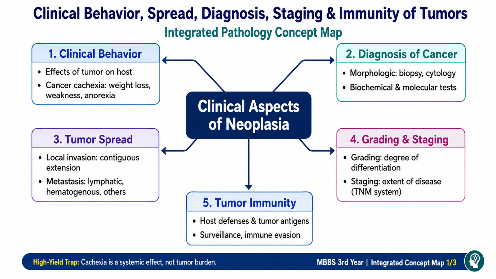
The clinical behavior of a tumor depends not only on where it arises but also on how it affects surrounding tissues, whether it spreads to distant sites, and how the host responds to it. Malignant tumors may produce local effects, systemic manifestations such as cachexia, and paraneoplastic syndromes. Their diagnosis requires examination of cells and tissues, supported when necessary by biochemical and molecular methods. Tumor grade describes the degree of differentiation and biological aggressiveness, while tumor stage describes the anatomical extent of disease. The immune system can recognize tumor-associated abnormalities and destroy malignant cells, but cancers may develop several mechanisms that allow them to escape immune surveillance.
A. Clinical Effects of Tumors on the Host
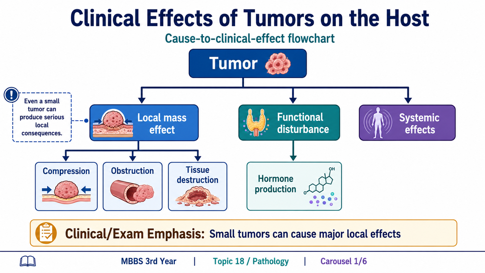
A neoplasm can produce illness through its physical presence, destruction of normal tissue, interference with organ function, abnormal production of biologically active substances, or systemic metabolic effects. The severity of symptoms does not always correspond directly to tumor size. A small tumor in a critical location may cause major symptoms, whereas a much larger tumor in a spacious body cavity may remain unnoticed for longer.
Local effects
As a tumor enlarges, it may compress, obstruct, ulcerate or invade nearby structures. Malignant tumors are particularly important because their infiltrative growth destroys surrounding tissues.
- Compression: a growing mass may compress nearby nerves, vessels or organs, producing pain or functional impairment.
- Obstruction: tumors arising in hollow organs may narrow the lumen and interfere with normal flow.
- Ulceration and bleeding: tumors involving epithelial surfaces may ulcerate and damage vessels.
- Tissue destruction: invasive malignant cells replace and destroy normal tissue, reducing organ function.
- Secondary infection: ulcerated or obstructed tissues may become infected.
Functional activity of tumors
Some tumors retain or acquire the ability to produce hormones or other biologically active substances. Tumors arising from endocrine tissues may therefore produce clinical manifestations through excessive hormone production. Importantly, some non-endocrine cancers may also produce hormone-like substances and cause paraneoplastic syndromes.
B. Cancer Cachexia and Paraneoplastic Syndromes
Two important systemic consequences of malignancy are cancer cachexia and paraneoplastic syndromes. These manifestations are clinically important because they may cause substantial morbidity, influence prognosis and, in some patients, provide an early clue to an underlying cancer.
Cancer cachexia
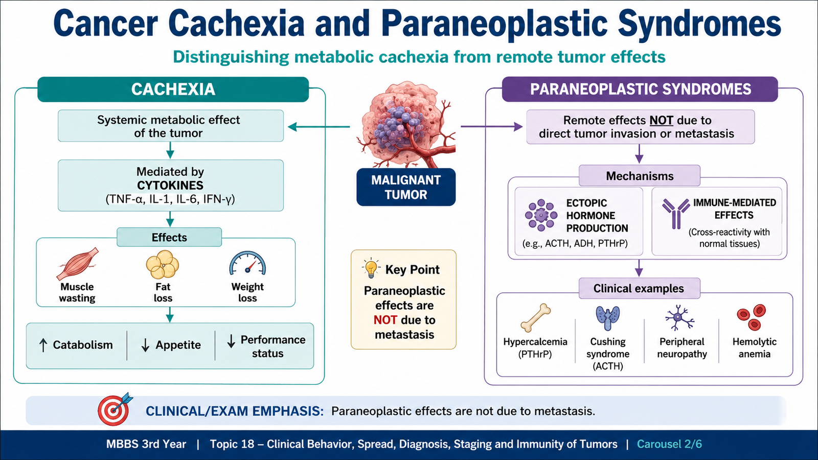
Cachexia is a syndrome characterized by progressive loss of body fat and lean muscle mass, accompanied by weakness and reduced functional capacity. It is common in advanced malignancy and cannot be explained simply by reduced food intake.
Cancer cells and host inflammatory cells produce mediators that alter metabolism. Pro-inflammatory cytokines such as tumor necrosis factor and other mediators increase catabolism and contribute to loss of muscle and fat. The patient therefore loses weight even when nutritional support is provided.
Tumor and host inflammatory response → release of metabolic and inflammatory mediators → increased protein and fat breakdown → loss of skeletal muscle and adipose tissue → weakness and progressive weight loss
Cachexia is important because severe loss of muscle mass reduces physical reserve and contributes to poor tolerance of advanced disease.
Paraneoplastic syndromes
Paraneoplastic syndromes are symptom complexes in patients with cancer that cannot be explained by local or distant spread of the tumor or by hormones normally produced by the tissue from which the tumor arose. They usually result from ectopic production of hormones, hormone-like substances, cytokines or immune-mediated reactions.
Their clinical significance is considerable. A paraneoplastic syndrome may be the earliest manifestation of an occult cancer, may produce serious illness independently of the primary tumor, and may mimic metastatic disease.
| Paraneoplastic syndrome | Main mechanism | Important associated cancers |
|---|---|---|
| Cushing syndrome | Ectopic ACTH or ACTH-like activity | Small-cell carcinoma of lung |
| Hypercalcemia | PTH-related protein or other tumor-associated mechanisms | Squamous cell carcinomas; some renal, breast and other cancers |
| Polycythemia | Erythropoietin production | Renal cell carcinoma and some other tumors |
| Nonbacterial thrombotic manifestations | Tumor-associated hypercoagulability | Especially some advanced adenocarcinomas |
| Neuromuscular syndromes | Immune cross-reaction with neural structures | Classically associated with some lung cancers |
C. Local Invasion and Metastatic Spread
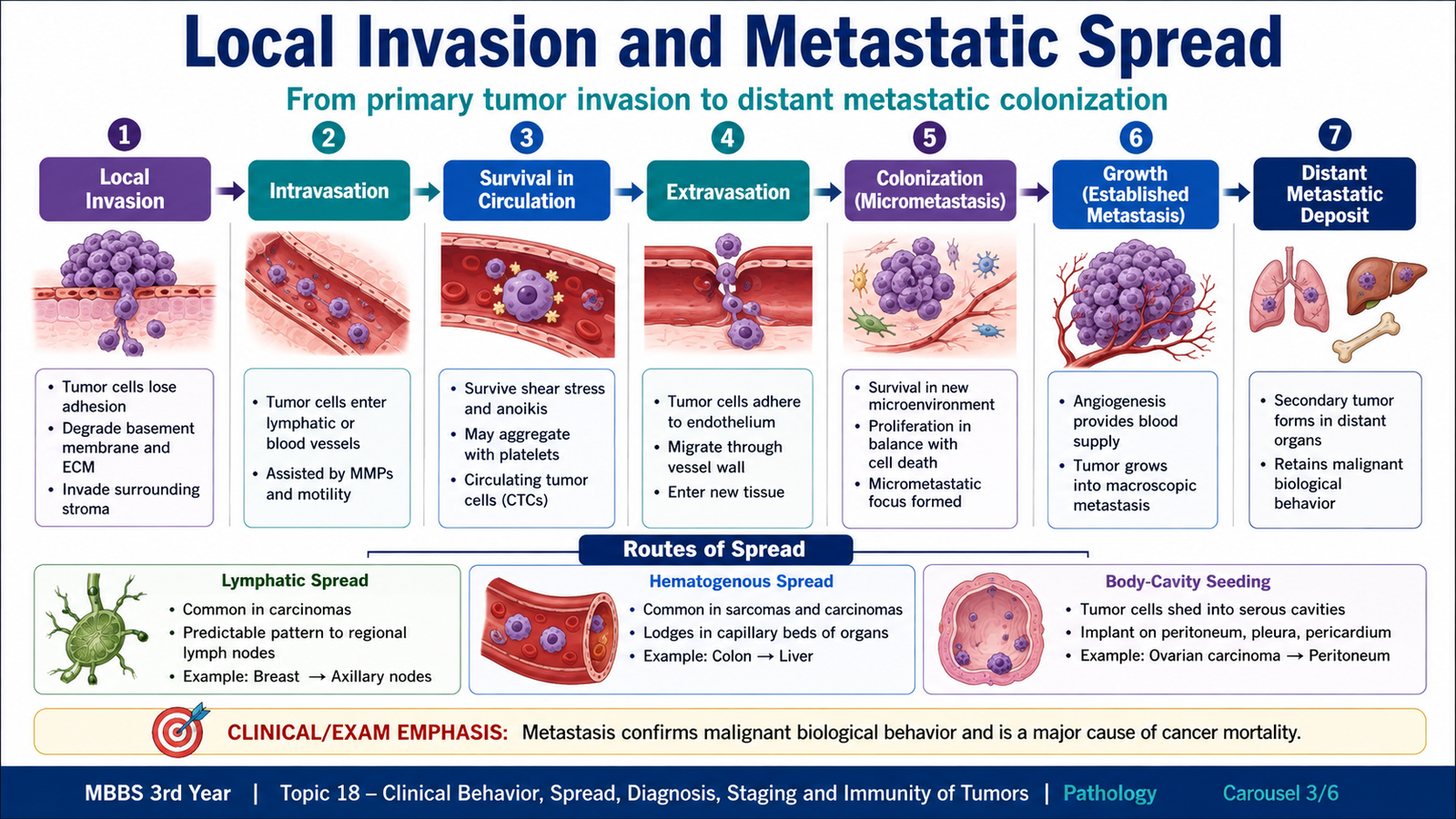
The ability to invade surrounding tissue and metastasize distinguishes malignant tumors from benign neoplasms. Local invasion means direct infiltration and destruction of adjacent tissue. Metastasis means development of secondary tumor deposits at sites that are physically discontinuous from the primary tumor.
Local invasion
Malignant cells do not remain confined by a capsule. They penetrate the extracellular matrix, move through surrounding tissue and may enter lymphatic channels, blood vessels or body cavities. Invasion requires changes in tumor-cell attachment, degradation of extracellular matrix and movement of malignant cells through the altered tissue.
Reduced normal cell adhesion → attachment to extracellular matrix → degradation of basement membrane and interstitial matrix → migration of tumor cells → entry into surrounding tissues or vessels
Major pathways of metastasis
Malignant tumors spread through three major routes. The preferred route is influenced partly by the type and anatomical location of the tumor.
- Lymphatic spread: particularly common in carcinomas. Tumor cells enter lymphatic channels and may reach regional lymph nodes.
- Hematogenous spread: common in sarcomas but also occurs in carcinomas. Tumor cells enter blood vessels, especially veins, and travel to distant organs.
- Seeding of body cavities and surfaces: occurs when a malignant tumor penetrates a natural body cavity and sheds cells across its surface.
The metastatic cascade
Metastasis is a multistep process. Tumor cells must leave the primary mass, survive transport, exit the circulation and establish growth in a new tissue environment. Only a fraction of disseminated malignant cells successfully complete all these steps.
Primary malignant tumor → local invasion → entry into lymphatic or blood vessels → survival in circulation → arrest at distant site → exit from vessels → growth of metastatic deposit
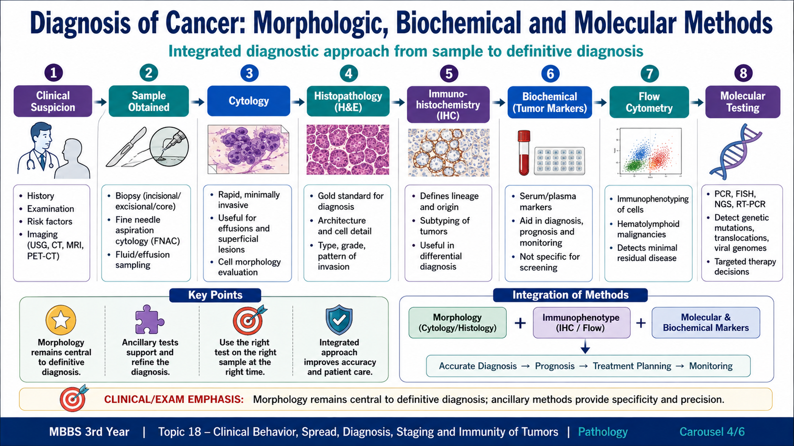
D. Diagnosis of Cancer: Morphologic, Biochemical and Molecular Methods
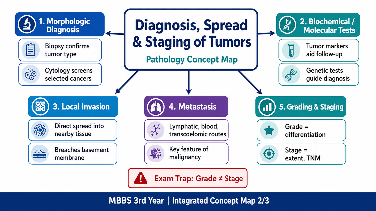
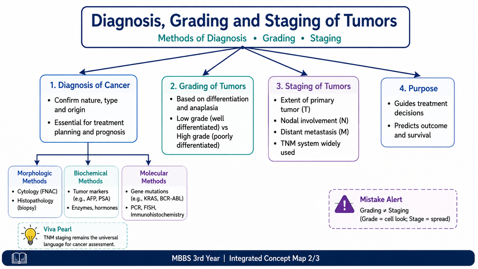
Accurate diagnosis of cancer requires confirmation that a lesion is neoplastic and determination of its type and biological characteristics. Morphological examination of cells and tissues remains central, while immunohistochemical, biochemical and molecular methods provide additional information when morphology alone is insufficient.
Morphologic diagnosis
Morphologic methods assess the appearance and arrangement of tumor cells. They allow the pathologist to identify differentiation, atypia, tissue architecture, invasion and other features that help classify a tumor.
- Histopathology: examination of tissue architecture and cellular morphology, usually after biopsy or surgical removal.
- Cytology: examination of individual or small groups of cells obtained from body fluids, surface scrapings, brushings or needle aspiration.
- Frozen section: provides rapid intraoperative morphological assessment in selected situations.
A biopsy specimen is particularly valuable because it preserves the relationship of tumor cells to surrounding tissue. This helps the pathologist assess invasion and classify the lesion more confidently.
Immunohistochemistry
Immunohistochemistry uses antibodies to detect specific antigens in tissue sections. It is especially useful when a poorly differentiated tumor cannot be confidently classified by routine morphology. Detection of lineage-associated proteins can help determine whether a tumor is epithelial, hematolymphoid or of another cellular origin. Selected markers may also provide prognostic or predictive information.
Biochemical methods and tumor markers
Some tumors release proteins, hormones, enzymes or other substances that can be measured in blood or other body fluids. These are commonly called tumor markers. Their major value is usually in supporting clinical assessment, evaluating tumor burden in selected cancers and following patients after diagnosis.
A tumor marker by itself usually cannot establish a definitive diagnosis because many markers lack complete specificity and may also increase in non-neoplastic conditions. Therefore, the result must be interpreted together with clinical findings and tissue diagnosis where appropriate.
Molecular methods
Molecular tests detect genetic or chromosomal abnormalities in tumor cells. They may identify mutations, gene rearrangements, amplifications or other alterations characteristic of particular neoplasms. These methods are valuable when a molecular abnormality helps confirm tumor classification, provides prognostic information or identifies a clinically relevant biological characteristic.
- Detection of specific DNA or RNA abnormalities.
- Identification of characteristic chromosomal changes.
- Assessment of gene amplification or mutation in selected tumors.
- Detection of small residual populations of malignant cells in appropriate hematological or other malignancies.
Flow cytometry
Flow cytometry analyzes characteristics of individual cells in suspension and can rapidly assess cell-surface or intracellular markers. It is particularly useful in the evaluation and classification of many hematolymphoid neoplasms.
E. Tumor Grading and Staging
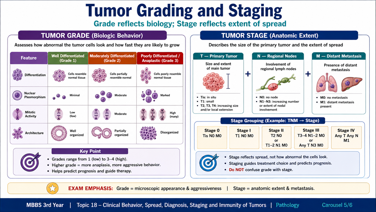
After a malignant tumor has been identified, its likely behavior and anatomical extent must be assessed. Grading and staging describe different aspects of a cancer and should not be confused. Grade is mainly based on microscopic appearance, whereas stage describes how far the tumor has spread in the patient.
Tumor grading
Grade reflects the degree of differentiation and other microscopic features associated with biological aggressiveness. A well-differentiated tumor resembles its normal tissue of origin more closely, whereas a poorly differentiated or anaplastic tumor shows greater loss of normal cellular structure and function.
Depending on the tumor, grading systems may consider:
- degree of differentiation;
- nuclear atypia;
- architectural abnormality;
- mitotic activity;
- other tumor-specific microscopic features.
In general, increasing loss of differentiation and greater proliferative activity are associated with more aggressive biological behavior.
Tumor staging
Stage describes the anatomical extent of malignant disease. It considers the size or local extent of the primary tumor, spread to regional lymph nodes and presence or absence of distant metastasis.
The commonly used TNM concept organizes these components as:
- T — Primary tumor: size and/or degree of local extension.
- N — Regional lymph nodes: involvement of regional lymph nodes.
- M — Distant metastasis: presence or absence of distant spread.
Stage has major clinical importance because increasing anatomical spread generally indicates a less favorable prognosis and strongly influences the overall assessment of disease.
| Feature | Tumor Grade | Tumor Stage |
|---|---|---|
| Main question | How abnormal and aggressive does the tumor look microscopically? | How far has the tumor spread anatomically? |
| Basis | Differentiation, atypia, mitoses and tumor-specific microscopic features | Primary tumor, lymph nodes and distant metastasis |
| Typical assessment | Pathological examination | Clinical, radiological and pathological assessment |
| Clinical significance | Indicates biological aggressiveness | Usually has major prognostic importance |
F. Tumor Immunity, Immune Surveillance and Immune Evasion
Tumor cells arise from the body’s own cells, but malignant transformation may produce abnormal proteins or abnormal expression of cellular proteins. These changes can make tumor cells recognizable to the immune system. The concept that the immune system identifies and destroys newly arising malignant cells is called immune surveillance.
Tumor antigens
Tumor cells may express antigens that differ quantitatively or qualitatively from those of normal cells. These antigens can arise from mutated proteins, abnormal expression of cellular proteins, proteins produced by oncogenic viruses, or proteins that are normally expressed only in restricted tissues or stages of development.
At undergraduate level, the important concept is that tumor antigens provide targets through which the immune system can distinguish at least some malignant cells from normal tissues.
Anti-tumor effector mechanisms
Several components of immunity can participate in the destruction of tumor cells. Cell-mediated mechanisms are particularly important.
- Cytotoxic T lymphocytes: recognize tumor-derived antigens presented on major histocompatibility complex class I molecules and can directly kill tumor cells.
- Natural killer cells: can destroy abnormal cells, particularly when normal inhibitory signals associated with MHC class I expression are reduced.
- Activated macrophages: may contribute to tumor-cell killing through inflammatory mediators and cytotoxic mechanisms.
- Antibodies: may recognize surface tumor antigens and contribute to immune-mediated destruction in some circumstances.
Tumor-associated antigen → recognition by host immune cells → activation of effector mechanisms → attack on malignant cells → elimination or restriction of tumor growth
Immune surveillance
Immune surveillance proposes that transformed cells arise periodically but may be recognized and eliminated before they form clinically apparent tumors. The increased occurrence of certain malignancies in states of impaired immunity supports the importance of host immune defenses against some cancers.
How tumors evade immunity
A clinically detectable malignancy has successfully avoided or resisted host immune control. Tumor cells may achieve this in several ways.
- Loss or reduced expression of tumor antigens: less antigenic variants may escape immune recognition.
- Reduced antigen presentation: tumor cells may decrease expression of molecules needed for effective T-cell recognition.
- Expression of inhibitory signals: tumors may suppress activation of anti-tumor lymphocytes.
- Production of immunosuppressive factors: the tumor microenvironment may inhibit effective immune responses.
- Selection of resistant tumor-cell populations: immune pressure may eliminate sensitive cells while resistant variants survive.
Therefore, the interaction between cancer and immunity is dynamic. Host defenses may eliminate some malignant cells, but surviving clones can progressively develop features that permit immune escape and continued growth.
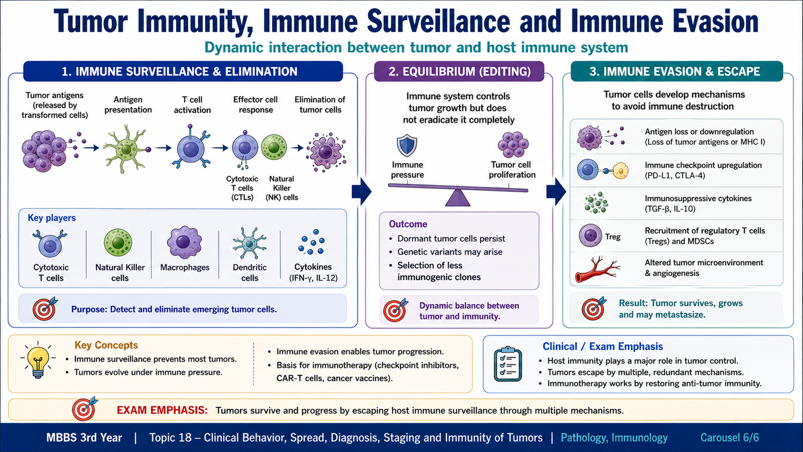
Integrated Mechanism Flow
⭐ AIM High-Yield Review
- Tumors affect the host through local mass effects, tissue destruction, functional disturbance and systemic manifestations.
- Cachexia is progressive loss of fat and skeletal muscle associated with altered metabolism in cancer.
- ⭐ Paraneoplastic syndromes cannot be explained by direct tumor spread or normal hormone production of the tissue of origin.
- Paraneoplastic syndromes may be an early clue to an otherwise occult malignancy.
- Malignant tumors spread by lymphatics, blood vessels and seeding of body cavities or surfaces.
- Lymphatic spread is classically important in carcinomas; hematogenous spread is prominent in sarcomas but also occurs in carcinomas.
- Morphologic examination of cells and tissues remains central to cancer diagnosis.
- Immunohistochemistry helps determine tumor lineage and characterize poorly differentiated neoplasms.
- Tumor markers are supportive tools and usually should not be interpreted as standalone proof of malignancy.
- Molecular methods can identify characteristic mutations, rearrangements, amplifications or other genetic abnormalities.
- ⭐ Grade describes microscopic differentiation and aggressiveness; stage describes anatomical extent.
- TNM evaluates the primary tumor, regional lymph nodes and distant metastasis.
- Cytotoxic T cells, NK cells and activated macrophages are important anti-tumor effector cells.
- Immune surveillance refers to host recognition and elimination of newly transformed cells.
- Tumors may escape immunity through antigen loss, impaired antigen presentation and immunosuppressive mechanisms.