
This chapter follows the KMU learning outcomes and builds the autonomic nervous system step by step before introducing cholinomimetic drugs. First understand the receptor–organ relationships and drug mechanisms, then use the high-yield review for revision.
Topic 1 — Autonomic Nervous System Foundations and Cholinergic Pharmacology
Multisystem Module • Pharmacology with essential autonomic physiology
Topic Introduction
The autonomic nervous system, or ANS, controls many involuntary functions such as heart rate, vascular tone, glandular secretion, gastrointestinal activity, pupil size and bladder function. It works mainly through specific neurotransmitters acting on cholinergic, adrenergic and dopaminergic receptors. Understanding where these receptors are located and what happens when they are activated provides the foundation for autonomic pharmacology. Cholinomimetic drugs increase the effects of acetylcholine either by directly stimulating cholinergic receptors or by preventing acetylcholine breakdown. Their actions explain both useful therapeutic effects, such as improving bladder contraction or lowering intraocular pressure, and predictable adverse effects such as diarrhea, bronchospasm and bradycardia.
A. Functional Organization, Neurotransmitters and Autonomic Receptors
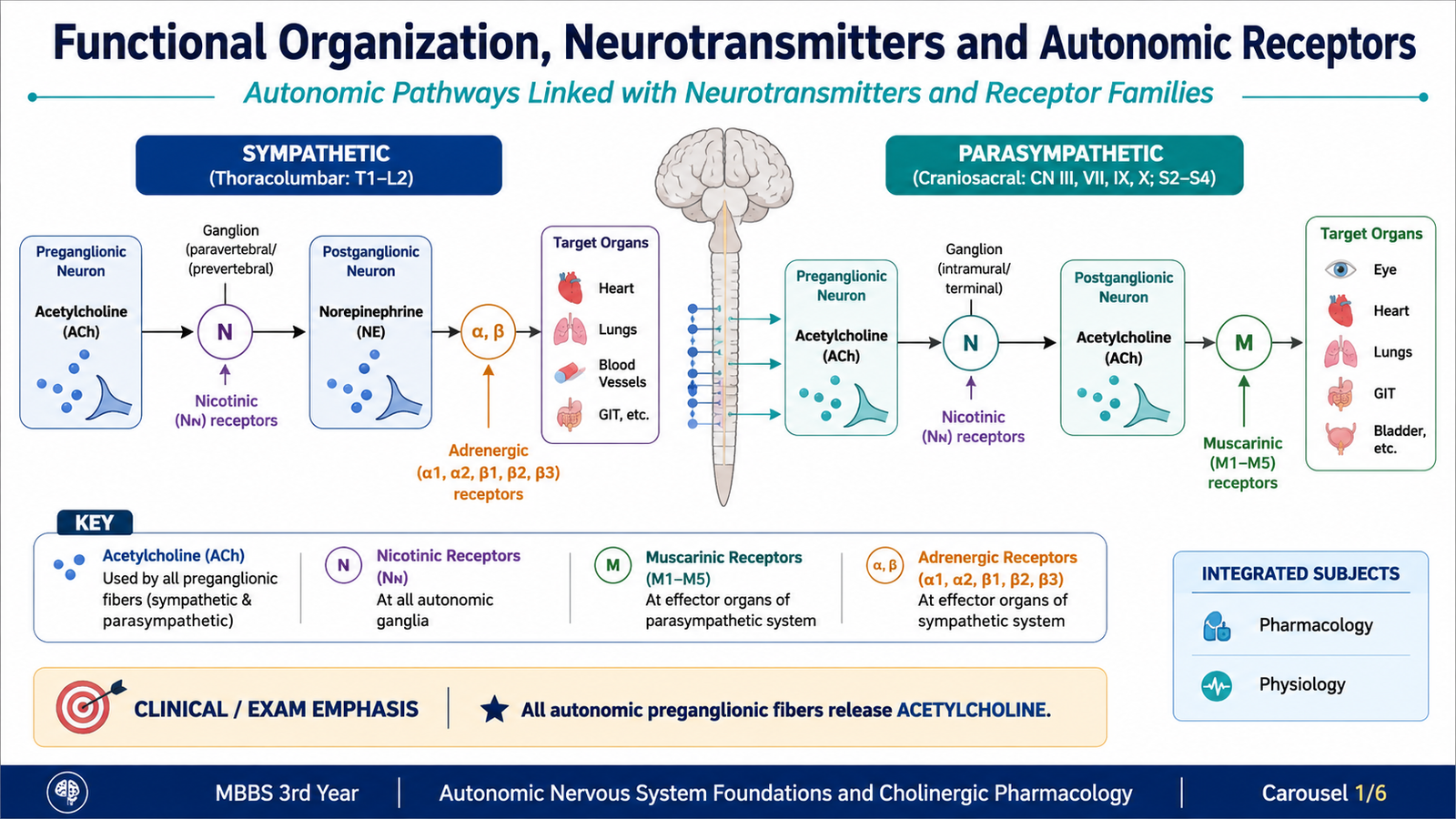
The ANS is divided mainly into the sympathetic and parasympathetic divisions. Both usually use a two-neuron pathway. A preganglionic neuron begins in the central nervous system and synapses in an autonomic ganglion with a postganglionic neuron, which then reaches the target organ.
The major difference between the divisions is not simply where they originate, but also which neurotransmitter is released at the neuroeffector junction and which receptor receives that signal.
Major autonomic neurotransmitters
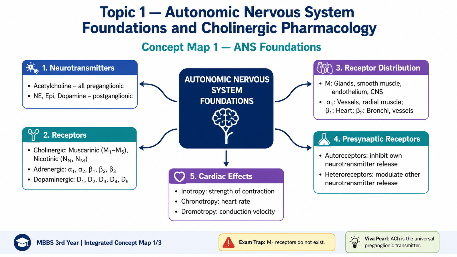
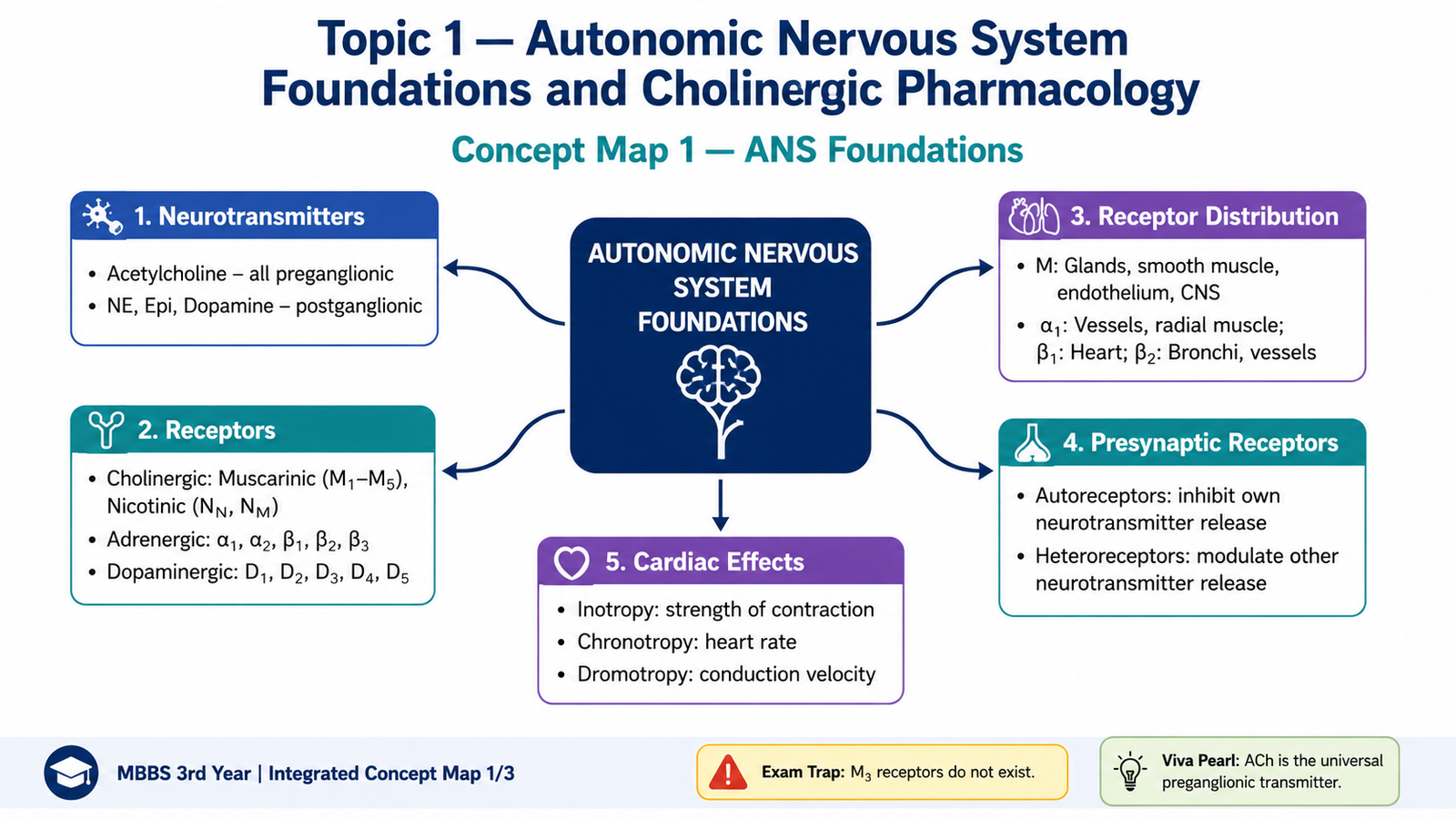
- Acetylcholine (ACh): released by all autonomic preganglionic fibers, parasympathetic postganglionic fibers and sympathetic fibers supplying eccrine sweat glands.
- Norepinephrine: released by most sympathetic postganglionic fibers.
- Epinephrine: released mainly from the adrenal medulla into the circulation.
- Dopamine: functions as a neurotransmitter in selected pathways and can act on specific dopamine receptors.
Major receptor families
Autonomic receptors can be organized into cholinergic, adrenergic and dopaminergic families. Recognizing their major subtypes allows the student to predict the effect of both endogenous neurotransmitters and autonomic drugs.
| Receptor family | Important subtypes | Main signalling principle | Representative locations |
|---|---|---|---|
| Muscarinic | M1, M2, M3, M4, M5 | G-protein-coupled receptors | Heart, smooth muscle, glands, eye, CNS |
| Nicotinic | NN, NM | Ligand-gated ion channels | Autonomic ganglia, adrenal medulla, neuromuscular junction |
| Adrenergic | α1, α2, β1, β2, β3 | G-protein-coupled receptors | Heart, vessels, bronchi, metabolic tissues and other organs |
| Dopaminergic | D1-like and D2-like families | G-protein-coupled receptors | CNS and selected peripheral tissues |
B. Organ Distribution and Functional Meaning of Autonomic Receptors
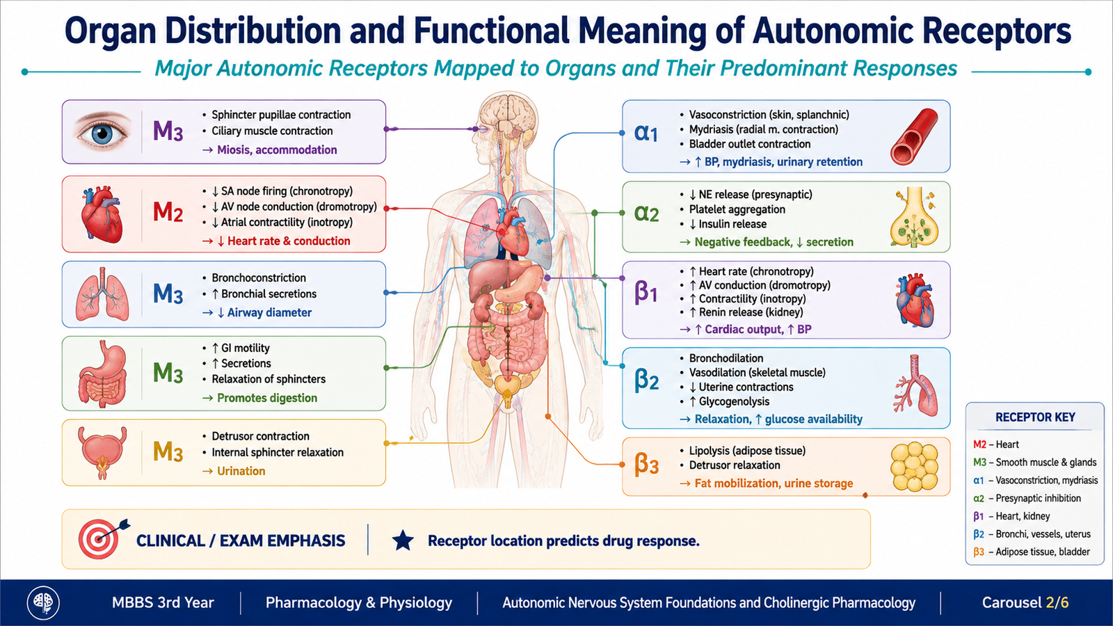
Knowing the receptor name alone is not enough. The important pharmacological skill is to connect the receptor with its organ location and physiological response. A drug that stimulates or blocks a receptor tends to reproduce or oppose the normal effect mediated through that receptor.
| Receptor | Important locations | Important effect of activation |
|---|---|---|
| M1 | CNS, autonomic ganglia, gastric-enteric sites | Neuronal excitation and facilitation of secretory activity |
| M2 | Heart, especially SA and AV nodal tissue | Decreases heart rate and AV conduction |
| M3 | Smooth muscle, glands, eye and vascular endothelium | Smooth-muscle contraction, secretion, miosis and endothelial nitric-oxide-mediated vasodilation |
| NN | Autonomic ganglia and adrenal medulla | Rapid neuronal depolarization |
| NM | Skeletal-muscle neuromuscular junction | Skeletal-muscle depolarization and contraction |
| α1 | Vascular smooth muscle, radial muscle of iris and several sphincters | Smooth-muscle contraction |
| α2 | Presynaptic sympathetic terminals and CNS | Reduces neurotransmitter release |
| β1 | Heart and juxtaglomerular cells | Increases cardiac activity and promotes renin release |
| β2 | Bronchial, vascular and uterine smooth muscle | Smooth-muscle relaxation |
| β3 | Adipose tissue and urinary bladder detrusor | Metabolic effects and detrusor relaxation |
Presynaptic receptors
Some autonomic receptors are located on the nerve terminal itself rather than only on the target organ. They regulate how much neurotransmitter is released.
- Autoreceptors respond to the same neurotransmitter released by that nerve terminal. For example, presynaptic α2 receptors can reduce further norepinephrine release, creating negative feedback.
- Heteroreceptors respond to a different transmitter and modify release from the terminal on which they are located.
This presynaptic regulation allows autonomic signalling to be adjusted before the neurotransmitter reaches the postsynaptic receptor.
C. Inotropy, Chronotropy and Dromotropy
Autonomic drugs frequently alter cardiac function. Three terms are especially important because they describe different aspects of cardiac performance rather than the same effect.
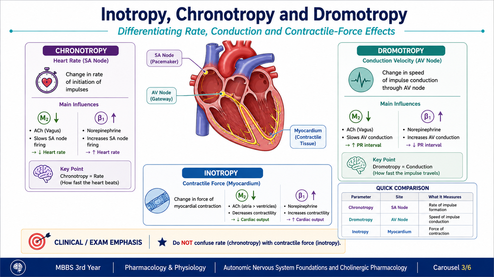
- Chronotropy refers to the rate of cardiac impulse generation, mainly at the sinoatrial node. Positive chronotropy increases heart rate; negative chronotropy decreases it.
- Dromotropy refers to the speed of electrical conduction, particularly through the atrioventricular node. Positive dromotropy increases conduction velocity; negative dromotropy slows conduction.
- Inotropy refers to myocardial contractile force. Positive inotropy increases the force of contraction, while negative inotropy decreases it.
Parasympathetic stimulation through cardiac M2 receptors produces important negative chronotropic and negative dromotropic effects. In contrast, sympathetic stimulation of β1 receptors increases heart rate, conduction and myocardial contractility.
| Term | What changes? | Main cardiac site |
|---|---|---|
| Chronotropy | Heart rate | SA node |
| Dromotropy | Conduction velocity | Especially AV node |
| Inotropy | Force of contraction | Myocardium |
D. Cholinomimetic Drugs: Classification, Mechanism and Pharmacokinetics
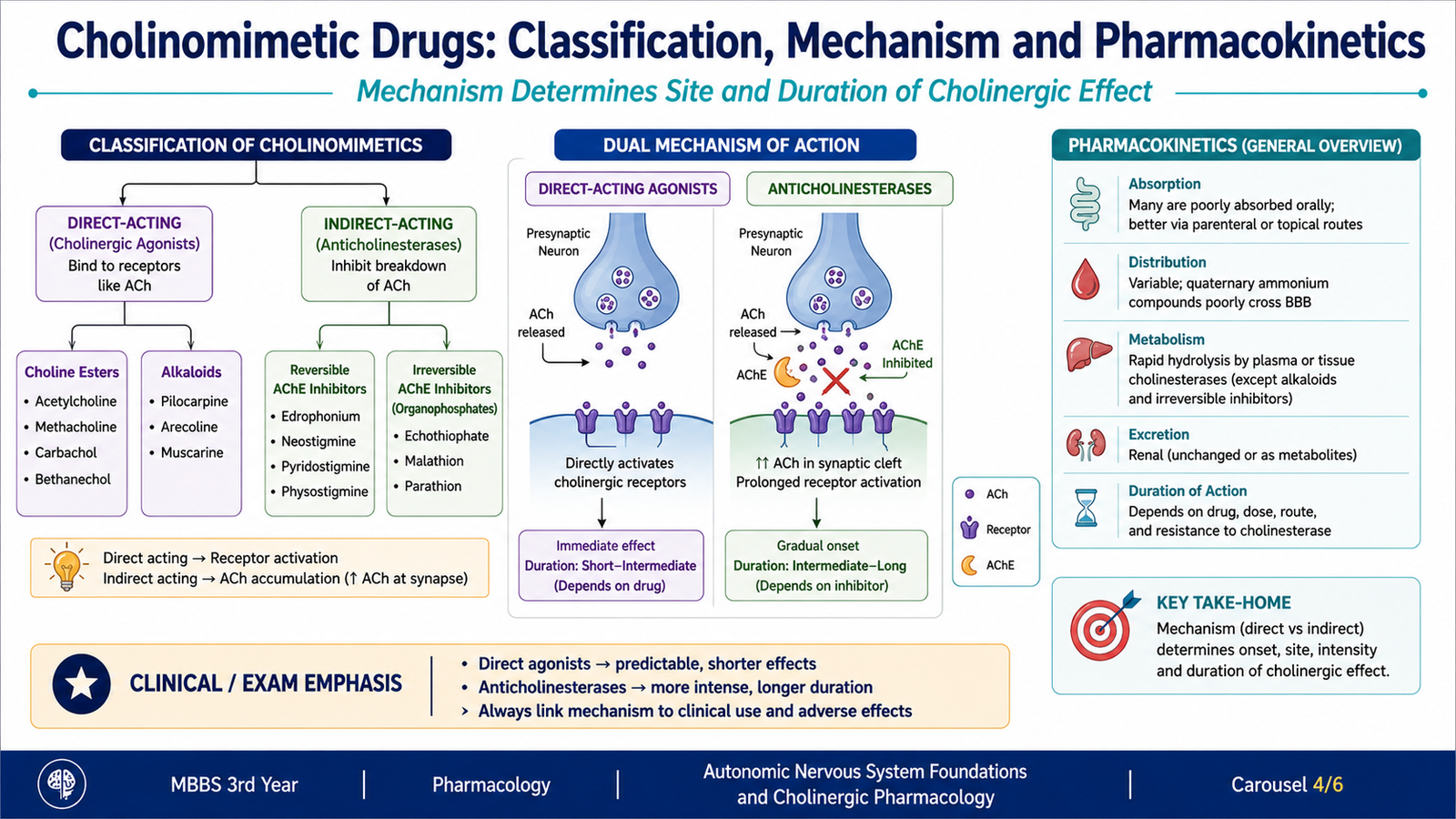
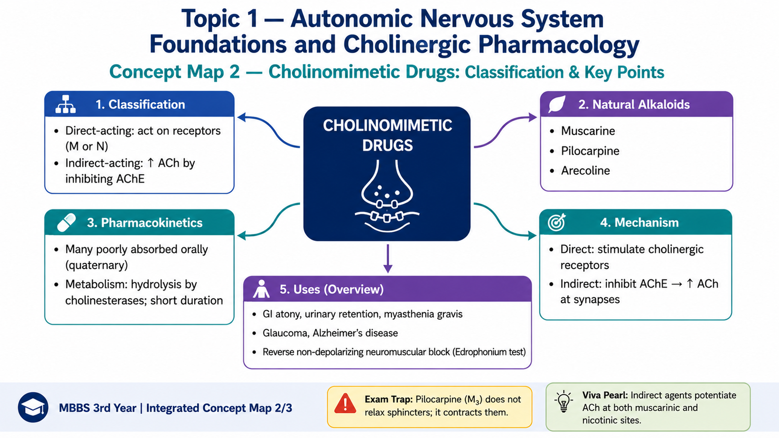
Cholinomimetic drugs reproduce or enhance the effects of acetylcholine. They do this in two major ways. Direct-acting drugs bind to and activate cholinergic receptors, whereas indirect-acting drugs inhibit acetylcholinesterase and therefore allow endogenous acetylcholine to accumulate at cholinergic synapses.
Classification
| Group | Subgroup | Important examples |
|---|---|---|
| Direct-acting | Choline esters | Acetylcholine, methacholine, carbachol, bethanechol |
| Naturally occurring alkaloids | Pilocarpine, muscarine, arecoline | |
| Indirect-acting | Reversible acetylcholinesterase inhibitors | Edrophonium, neostigmine, pyridostigmine, physostigmine, donepezil, rivastigmine, galantamine |
| Long-lasting/irreversible organophosphate inhibitors | Organophosphate compounds |
Mechanism of direct-acting cholinomimetics
Direct agonists bind directly to muscarinic and/or nicotinic receptors. Their organ effects therefore depend on which receptors they stimulate and where those receptors are located.
Mechanism of indirect-acting cholinomimetics
Acetylcholinesterase normally terminates cholinergic transmission by rapidly hydrolyzing acetylcholine. When the enzyme is inhibited, acetylcholine accumulates at sites where it is normally released. Therefore, both muscarinic and nicotinic cholinergic transmission can be enhanced.
Pharmacokinetic relevance: metabolism and duration
The duration of action of cholinomimetics depends greatly on how easily the drug is hydrolyzed or metabolized. Acetylcholine itself is rapidly destroyed and therefore has an extremely brief action. Modification of the molecule can make a choline ester more resistant to cholinesterase and prolong its action.
- Acetylcholine: rapidly hydrolyzed and very short acting.
- Methacholine: more resistant to hydrolysis than acetylcholine.
- Carbachol and bethanechol: relatively resistant to acetylcholinesterase and therefore act longer.
- Edrophonium: reversible enzyme inhibitor with a very brief duration of action.
- Neostigmine and pyridostigmine: longer acting than edrophonium and predominantly peripheral because they cross the blood–brain barrier poorly.
- Physostigmine: a tertiary compound that can enter the CNS.
- Donepezil, rivastigmine and galantamine: act within the CNS and are used to enhance cholinergic transmission in Alzheimer disease.
- Organophosphates: produce very prolonged acetylcholinesterase inhibition because enzyme recovery is slow after strong covalent inhibition.
E. Organ-System Effects and Clinical Uses of Cholinomimetics
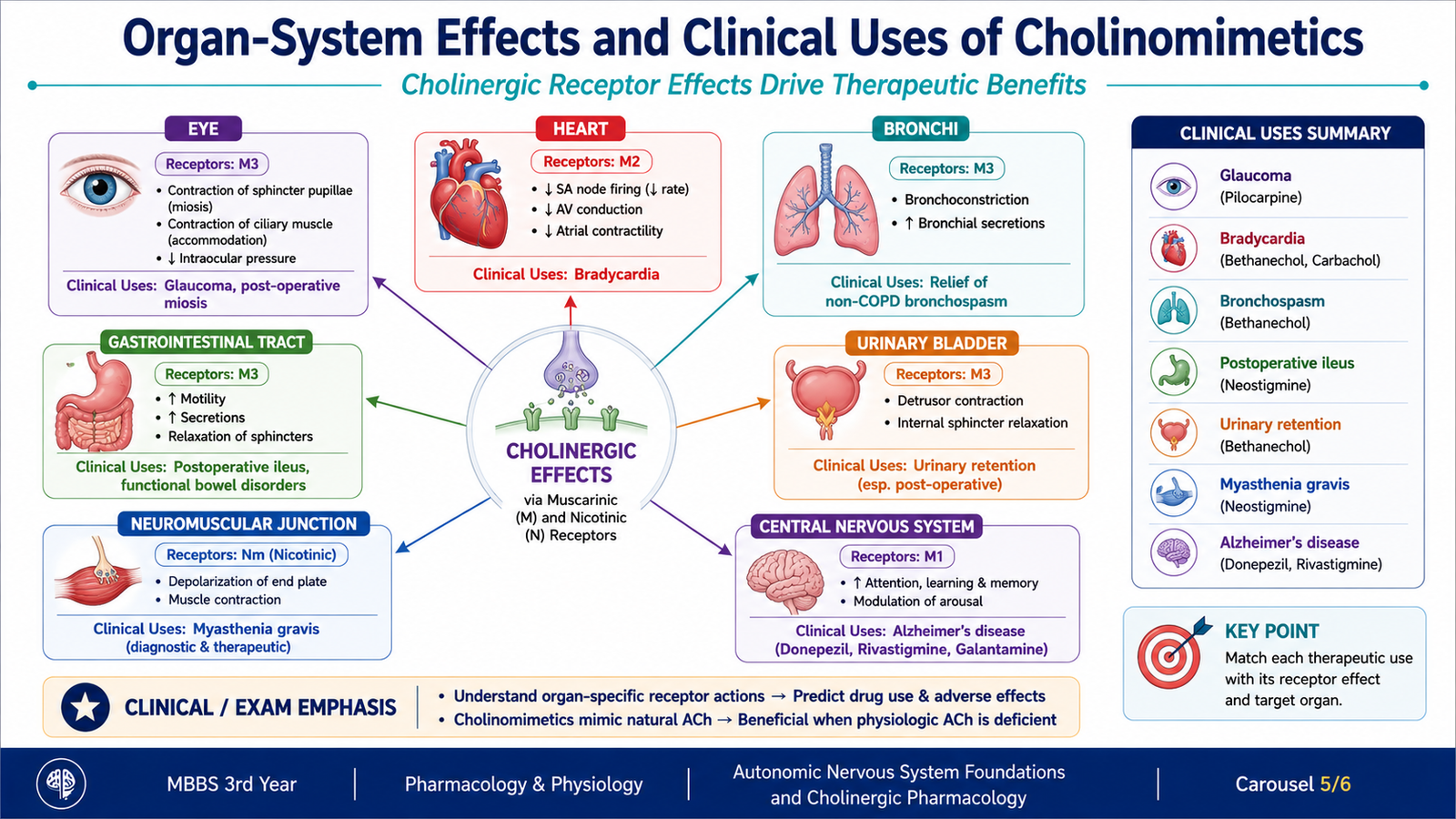
The effects of cholinomimetic drugs can largely be predicted from normal parasympathetic physiology. Increased muscarinic activity generally promotes glandular secretion, smooth-muscle contraction, slowing of the heart and functions associated with digestion, urination and near vision.
Cardiovascular system
Activation of cardiac M2 receptors slows SA nodal firing and AV nodal conduction. Therefore, excessive cholinergic activity may produce bradycardia. Muscarinic stimulation of intact vascular endothelium can release nitric oxide and produce vasodilation.
Eye
M3 receptor stimulation contracts the sphincter pupillae, causing miosis. Contraction of the ciliary muscle facilitates accommodation for near vision and increases access of aqueous humor to the trabecular outflow pathway. This explains the usefulness of muscarinic agonists such as pilocarpine in glaucoma.
Respiratory system
Muscarinic stimulation contracts bronchial smooth muscle and increases bronchial secretions. These effects explain why excessive cholinergic stimulation can be dangerous in patients with obstructive airway disease.
Gastrointestinal tract
Cholinergic stimulation increases gastrointestinal smooth-muscle activity and secretions. Increased motility may be useful when gastrointestinal movement needs to be restored, but excessive stimulation causes abdominal cramps and diarrhea.
Urinary bladder
Muscarinic stimulation promotes detrusor contraction and facilitates bladder emptying. Bethanechol can therefore be useful in selected forms of non-obstructive urinary retention.
Exocrine glands
Muscarinic receptors increase salivary, lacrimal and other glandular secretions. Pilocarpine can therefore be used when increased salivary secretion is therapeutically desirable.
Neuromuscular junction
Acetylcholinesterase inhibitors increase acetylcholine concentration at the neuromuscular junction. This increases the opportunity for acetylcholine to activate remaining nicotinic receptors and can improve neuromuscular transmission in myasthenia gravis.
Central nervous system and Alzheimer disease
Cholinergic neurotransmission contributes to cognitive functions. Centrally acting acetylcholinesterase inhibitors such as donepezil, rivastigmine and galantamine increase acetylcholine availability in the CNS. They are used in Alzheimer disease to provide symptomatic improvement in cognitive function; they do not reverse the underlying neurodegenerative process.
F. Adverse Effects, Contraindications and Edrophonium in Myasthenic vs Cholinergic Crisis
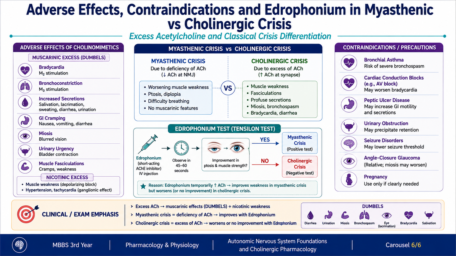
Because cholinomimetics amplify normal cholinergic physiology, their adverse effects are usually exaggerated versions of muscarinic or nicotinic responses. Understanding the receptor effect makes these reactions easier to predict than memorizing an isolated list.
Important adverse effects
- Salivation and lacrimation from increased glandular secretion.
- Sweating due to cholinergic stimulation of sympathetic eccrine sweat glands.
- Abdominal cramps and diarrhea from increased gastrointestinal motility and secretion.
- Urinary urgency from increased detrusor activity.
- Miosis and blurred vision due to contraction of ocular smooth muscle.
- Bradycardia and hypotension from cardiac slowing and vasodilator effects.
- Bronchoconstriction and increased bronchial secretion, which can compromise respiration in susceptible patients.
- Marked acetylcholinesterase inhibition can also produce nicotinic manifestations, including muscle fasciculations followed by weakness when neuromuscular transmission becomes severely disturbed.
Major contraindications and cautions
A cholinomimetic should be avoided or used cautiously when its normal physiological action could worsen an existing problem.
- Asthma or significant obstructive airway disease: bronchoconstriction and secretions may worsen airflow.
- Marked bradycardia or important conduction disturbance: muscarinic stimulation may further reduce cardiac rate or conduction.
- Peptic ulcer disease: cholinergic activity can increase gastrointestinal secretion and motility.
- Mechanical gastrointestinal or urinary obstruction: increasing smooth-muscle contraction against a physical obstruction may be harmful.
Edrophonium and myasthenic versus cholinergic crisis
Edrophonium is a very short-acting acetylcholinesterase inhibitor. Classically, its brief action was used to assess whether worsening muscle weakness in a patient receiving anticholinesterase therapy represented insufficient cholinergic stimulation or excessive cholinergic stimulation.
| Situation | Underlying problem | Classical response to brief AChE inhibition |
|---|---|---|
| Myasthenic crisis | Insufficient effective neuromuscular transmission | Transient improvement in muscle strength |
| Cholinergic crisis | Excessive cholinergic stimulation from anticholinesterase effect | Weakness does not improve and may worsen; muscarinic excess may also be present |
Integrated Mechanism Flow
↓
Direct receptor stimulation OR acetylcholinesterase inhibition
↓
Increased cholinergic receptor activation
↓
M2 cardiac effects + M3 smooth-muscle/glandular effects + nicotinic effects where applicable
↓
Therapeutic responses such as miosis, increased bladder contraction and improved neuromuscular transmission
↓
Excessive stimulation produces bradycardia, secretions, diarrhea, bronchoconstriction and possible muscle weakness
Important Comparison — Direct vs Indirect Cholinomimetics
| Feature | Direct-acting cholinomimetics | Indirect-acting cholinomimetics |
|---|---|---|
| Primary target | Muscarinic and/or nicotinic receptor | Acetylcholinesterase |
| How ACh is affected | Does not require increased endogenous ACh | Increases endogenous ACh by reducing breakdown |
| Examples | Bethanechol, pilocarpine, carbachol | Neostigmine, pyridostigmine, physostigmine, donepezil |
| Typical reasoning | Drug directly activates a desired receptor response | Useful when enhancement of naturally released ACh is desired |
⭐ AIM High-Yield Review
- All autonomic preganglionic fibers release acetylcholine.
- Most parasympathetic postganglionic fibers also release acetylcholine, whereas most sympathetic postganglionic fibers release norepinephrine.
- Muscarinic receptors are G-protein-coupled; nicotinic receptors are ligand-gated ion channels.
- M2 receptors are especially important in the heart and reduce heart rate and AV nodal conduction.
- M3 receptors mediate smooth-muscle contraction and increased glandular secretion; endothelial M3 activation can cause nitric-oxide-mediated vasodilation.
- Chronotropy = heart rate; dromotropy = conduction velocity; inotropy = contractile force.
- An autoreceptor responds to the transmitter released from the same nerve terminal; a heteroreceptor responds to another transmitter.
- Direct cholinomimetics activate cholinergic receptors; indirect cholinomimetics inhibit acetylcholinesterase.
- ⭐ Bethanechol promotes bladder contraction and is useful in selected non-obstructive urinary retention.
- ⭐ Pilocarpine causes miosis and ciliary-muscle contraction and can increase aqueous-humor outflow in glaucoma.
- Donepezil, rivastigmine and galantamine enhance central cholinergic transmission in Alzheimer disease.
- Acetylcholinesterase inhibitors can enhance neuromuscular transmission by increasing acetylcholine at the neuromuscular junction.
- Excess cholinergic activity commonly produces salivation, lacrimation, sweating, diarrhea, miosis, bradycardia and bronchoconstriction.
- Mechanical gastrointestinal or urinary obstruction is an important reason to avoid drugs that strongly increase smooth-muscle contraction.
- ⭐ In the classical edrophonium distinction, transient improvement favors myasthenic crisis; failure to improve or worsening favors cholinergic crisis.
Autonomic Pharmacology — Cholinergic Agonists
Use this video to reinforce cholinergic receptors, direct- and indirect-acting cholinomimetics, mechanisms of action, clinical uses and important adverse effects.