
Topic 17 — Molecular Basis of Cancer and Carcinogenesis
Topic Introduction
Cancer develops when a normal cell accumulates abnormalities that disturb the genes controlling cell growth, survival, differentiation and genomic stability. These abnormalities may activate genes that promote proliferation, disable genes that normally restrain growth, impair programmed cell death or prevent repair of damaged DNA. The resulting cell gains a survival and growth advantage and, through additional changes, may eventually become malignant. Carcinogenesis is therefore a multistep process rather than a single genetic event. In this chapter, you will learn the molecular and genetic basis of cancer, the role of oncogenes and p53, the major hallmarks acquired by cancer cells, and the mechanisms by which chemical agents, radiation, viruses and bacteria contribute to malignant transformation.
A. Molecular and Genetic Basis of Cancer
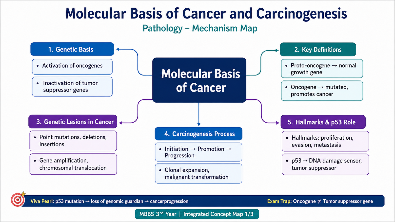
Cancer is fundamentally a disorder of abnormal cell growth caused by accumulated molecular abnormalities. A normal cell does not become malignant merely because it divides rapidly. Malignant transformation occurs when the regulatory systems that control proliferation, survival, differentiation and genome integrity are progressively disrupted.
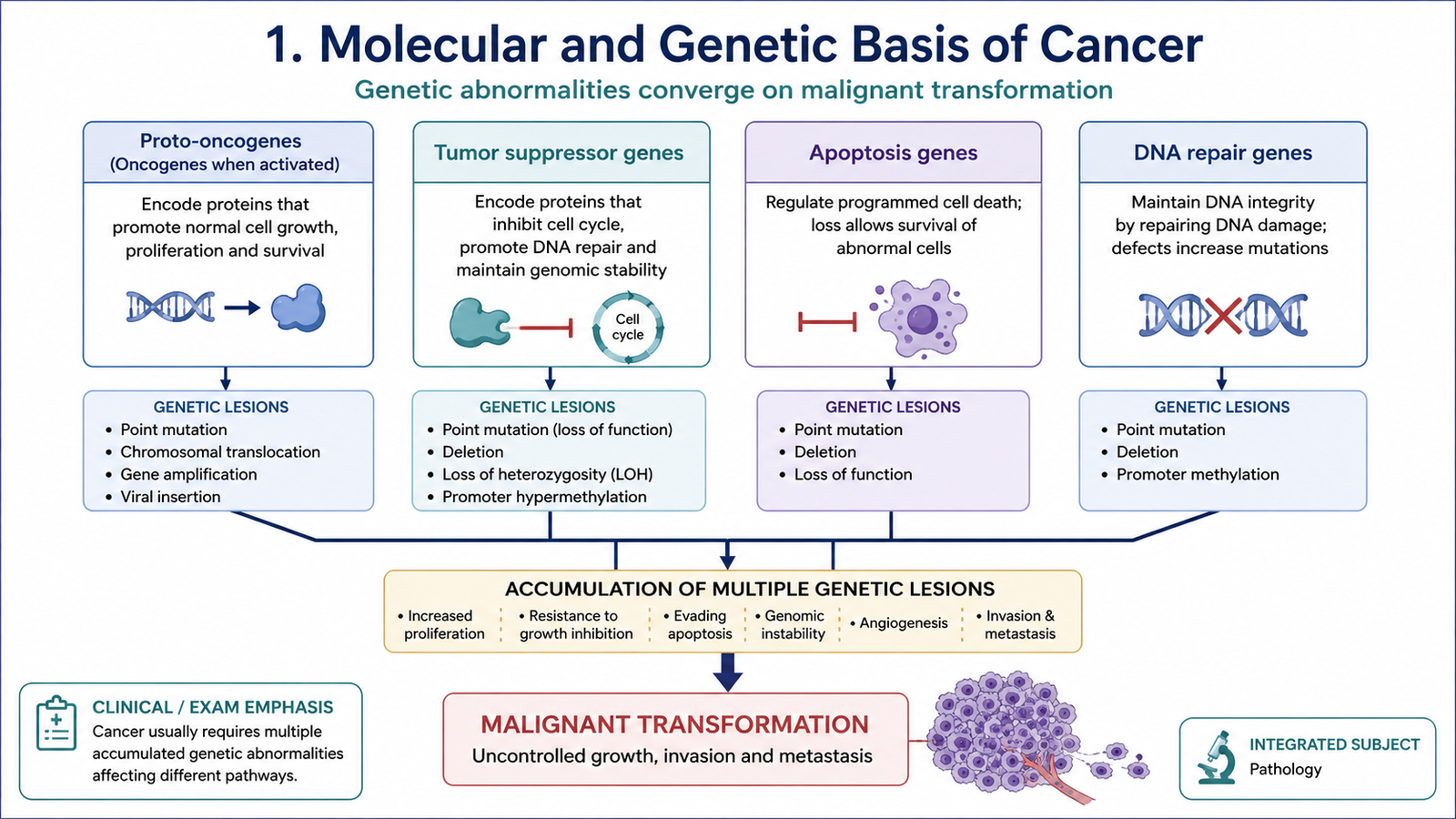
The important molecular targets of genetic damage can be grouped into four functional categories. These categories work together, so alteration of several pathways is usually necessary before a fully malignant phenotype develops.
Major genetic targets in carcinogenesis
- Growth-promoting proto-oncogenes: normally stimulate controlled cell growth. Activating abnormalities convert them into oncogenes.
- Growth-inhibiting tumor suppressor genes: normally restrain proliferation. Loss or inactivation removes important growth-control mechanisms.
- Genes regulating apoptosis: determine whether severely damaged cells survive or undergo programmed cell death.
- DNA repair genes: maintain genomic integrity. Their failure allows mutations to accumulate more rapidly.
Types of genetic lesions found in cancer
Cancer-associated genes may be altered by several types of acquired or inherited lesions. The biological effect depends more on which gene and pathway are affected than on the physical form of the lesion.
- Point mutations may activate growth-promoting proteins or inactivate growth-suppressing proteins.
- Chromosomal translocations may create a new fusion gene or place a growth-promoting gene under the control of a highly active promoter.
- Gene amplification produces multiple copies of a gene and excessive production of its protein product.
- Deletions may remove tumor suppressor genes.
- Abnormal chromosome number or structure may alter the dosage or regulation of multiple genes.
- Inherited pathogenic variants can provide the first genetic abnormality in hereditary cancer syndromes, after which additional acquired changes are required.
B. Proto-Oncogenes, Oncogenes and Oncoproteins
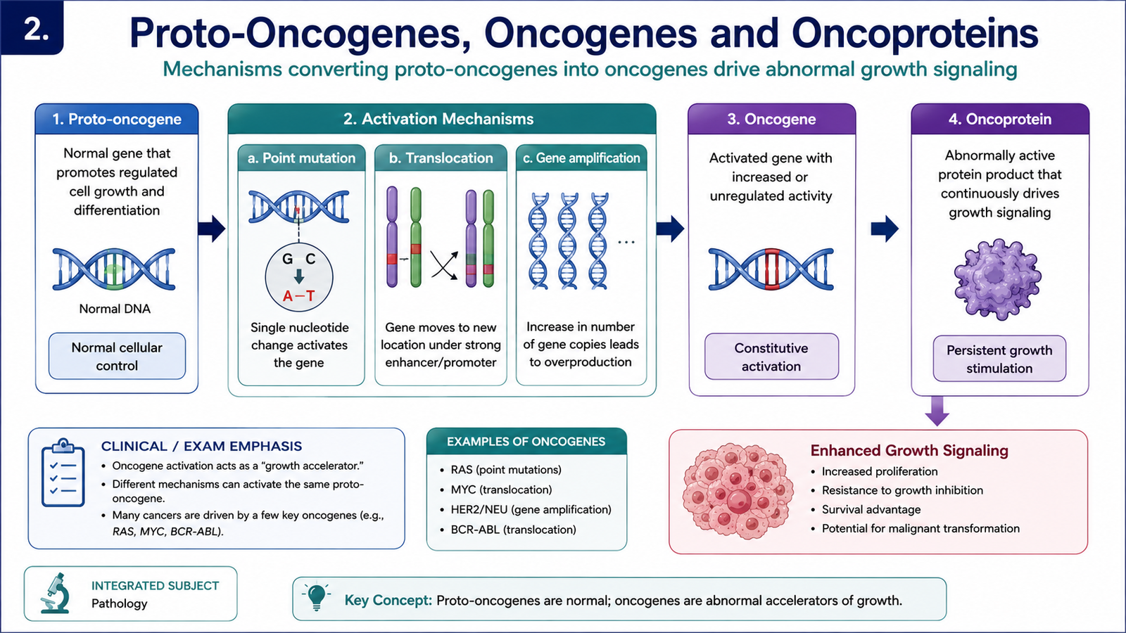
Proto-oncogenes are normal cellular genes whose products participate in regulated growth, proliferation and survival. When a proto-oncogene is abnormally activated, it becomes an oncogene. The proteins produced by oncogenes are called oncoproteins. These proteins promote growth or survival even when normal regulatory signals are absent or inappropriate.
Activation of an oncogene is a gain-of-function abnormality. Therefore, alteration of one copy of the gene may be sufficient to produce an abnormal growth-promoting effect at the cellular level.
How proto-oncogenes become oncogenes
1. Point mutation: A mutation can produce a protein that remains continuously active. A classic example involves RAS, a signaling protein that normally cycles between active and inactive states. Mutated RAS may remain active, continuously transmitting proliferative signals.
Growth signal → RAS activation → intracellular signaling → cell proliferation
Mutated RAS: persistent signaling → excessive proliferation
2. Chromosomal translocation: A gene may be moved next to a highly active regulatory sequence, causing excessive expression. Alternatively, two genes may join and form a fusion gene whose protein has abnormal activity.
For example, the MYC gene can be activated by chromosomal translocation. MYC encodes a transcription factor that promotes expression of genes required for cell growth and proliferation.
3. Gene amplification: Multiple copies of a proto-oncogene may be produced. This increases the amount of its growth-promoting protein. Amplification of genes such as ERBB2/HER2 can therefore provide a strong proliferative advantage to tumor cells.
Functional classes of oncogene products
- Growth factors may allow tumor cells to stimulate their own growth.
- Growth-factor receptors may become overexpressed or active without appropriate stimulation.
- Signal-transducing proteins such as RAS may continuously transmit growth signals.
- Nuclear transcription factors such as MYC may increase expression of genes required for proliferation.
- Cell-cycle regulatory proteins may push cells excessively through the cell cycle.
C. Hallmarks of Cancer and the Role of p53
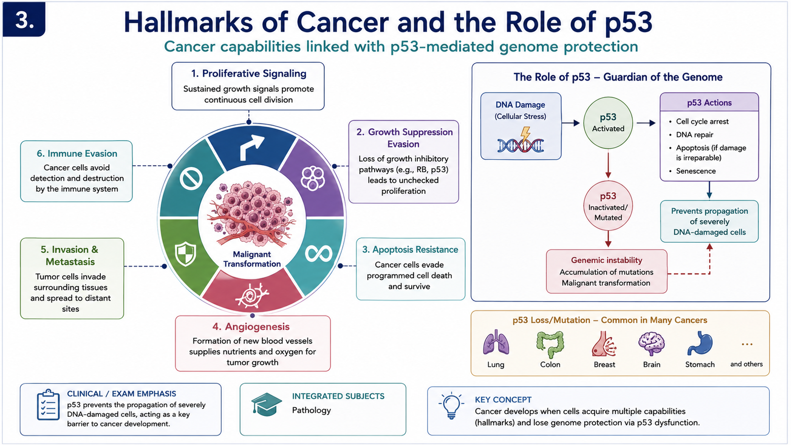
During carcinogenesis, tumor cells gradually acquire biological properties that allow them to grow despite normal controls, survive under unfavorable conditions and eventually invade and spread. These acquired functional capabilities are called the hallmarks of cancer. They are useful because they connect individual molecular abnormalities with the behavior of malignant tumors.
Major hallmarks of cancer cells
- Sustained proliferative signaling: cancer cells maintain signals that promote continued cell division.
- Evading growth suppressors: inhibitory pathways that normally stop proliferation are lost or bypassed.
- Resistance to cell death: cells with serious abnormalities avoid apoptosis and remain alive.
- Replicative immortality: tumor cells acquire the ability to continue dividing far beyond the normal replicative limit.
- Induction of angiogenesis: tumors promote formation of blood vessels that provide oxygen and nutrients.
- Invasion and metastasis: malignant cells acquire the ability to invade surrounding tissue and spread to distant sites.
- Altered cellular metabolism: tumor cells modify energy production to support rapid growth and biosynthesis.
- Escape from immune destruction: malignant cells develop mechanisms that reduce effective elimination by the immune system.
Two important processes help cancer cells acquire these hallmarks. Genomic instability increases the rate at which additional mutations arise, while tumor-promoting inflammation may provide growth factors, survival signals and tissue changes that support tumor development.
p53 — an important tumor suppressor
The TP53 gene encodes the p53 protein, one of the most important safeguards against malignant transformation. p53 responds to cellular stress, especially DNA damage. Instead of allowing a damaged cell to continue dividing, p53 can temporarily stop the cell cycle so that DNA repair can occur. If the damage cannot be adequately repaired, p53 can promote permanent growth arrest or apoptosis.
↓
p53 stabilization and activation
↓
Cell-cycle arrest → time for DNA repair
↓
Repair successful → cell may re-enter the cycle
OR
Severe/irreparable damage → senescence or apoptosis
When p53 function is lost, cells carrying damaged DNA may continue through the cell cycle. These cells can acquire additional mutations, increasing the chance of malignant transformation. For this reason, loss of p53 removes a major protective checkpoint against cancer.
D. The Multistep Process of Carcinogenesis
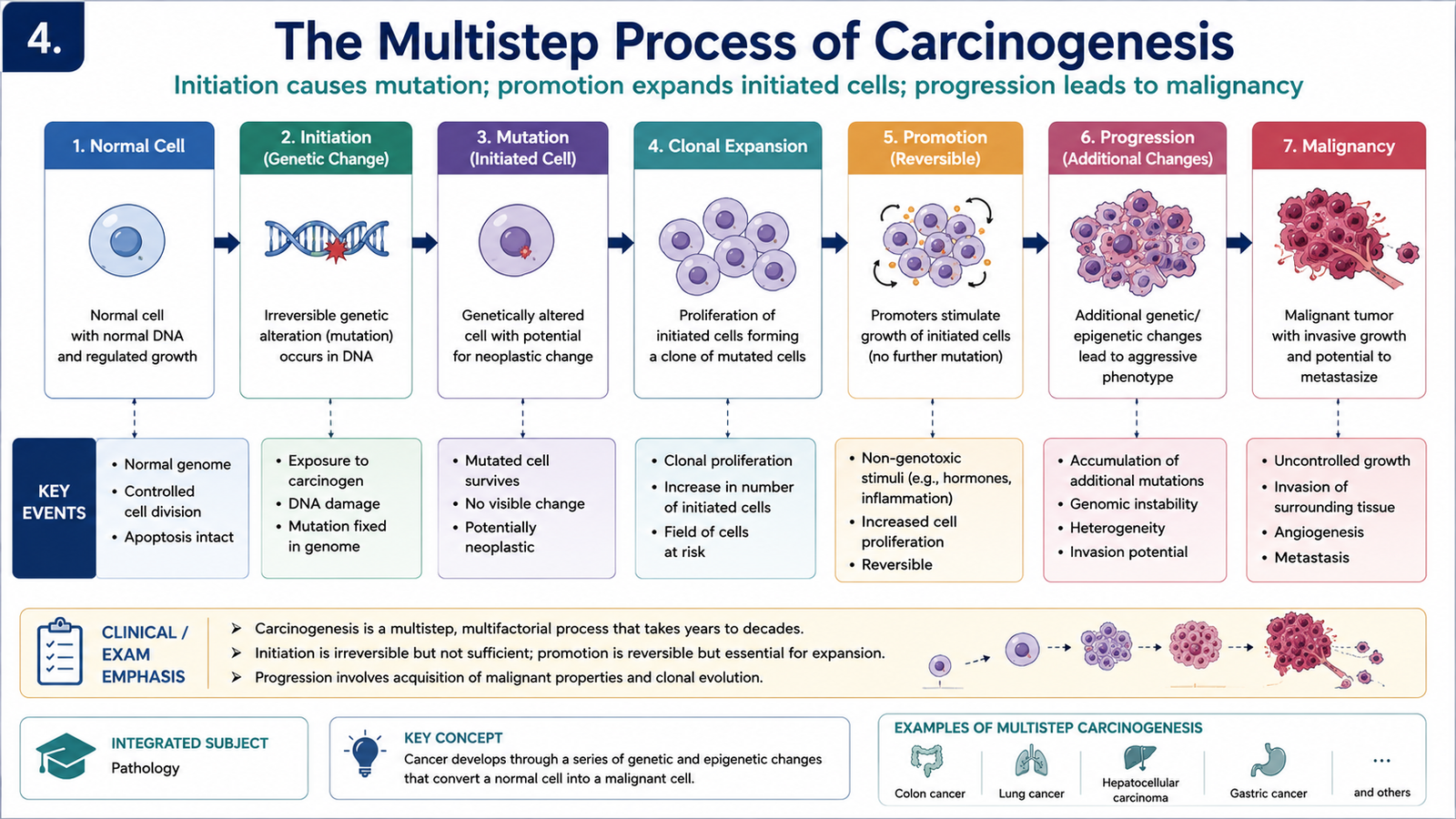
Carcinogenesis is the process through which a normal cell progressively becomes malignant. It usually occurs through repeated cycles of genetic damage, selection and clonal expansion. A cell that acquires a growth or survival advantage produces daughter cells carrying the same abnormality. If one of these descendants develops another advantageous change, that subclone expands further. Over time, increasingly aggressive subclones may emerge.
Clonal evolution
Most cancers arise from a single transformed precursor cell and are therefore clonal in origin. However, as the tumor grows, new mutations occur in different groups of tumor cells. This produces tumor heterogeneity, meaning that different subclones within the same tumor may have different growth rates, invasive abilities or responses to therapy.
→ initial genetic alteration
→ selective growth advantage
→ clonal expansion
→ additional genetic abnormalities
→ increasingly aggressive subclones
→ malignant tumor
Initiation and promotion
In experimental and chemical carcinogenesis, the process is commonly described using the concepts of initiation and promotion.
Initiation occurs when a carcinogenic agent causes permanent DNA damage in a cell. The initiated cell may look normal, but it carries a heritable genetic alteration.
Promotion involves stimulation of proliferation of initiated cells. Promoters usually do not directly damage DNA. By increasing cell division, however, they expand the population of initiated cells and increase the opportunity for additional abnormalities to accumulate.
Further genetic and epigenetic changes can then produce progression, during which the tumor acquires increasingly malignant characteristics such as rapid growth, invasion and metastatic ability.
E. Chemical Carcinogenesis
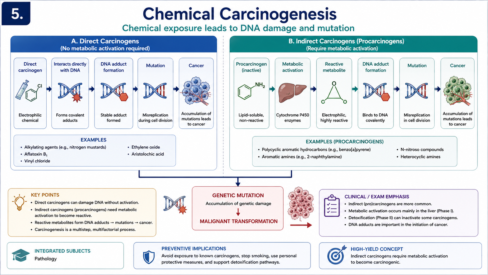
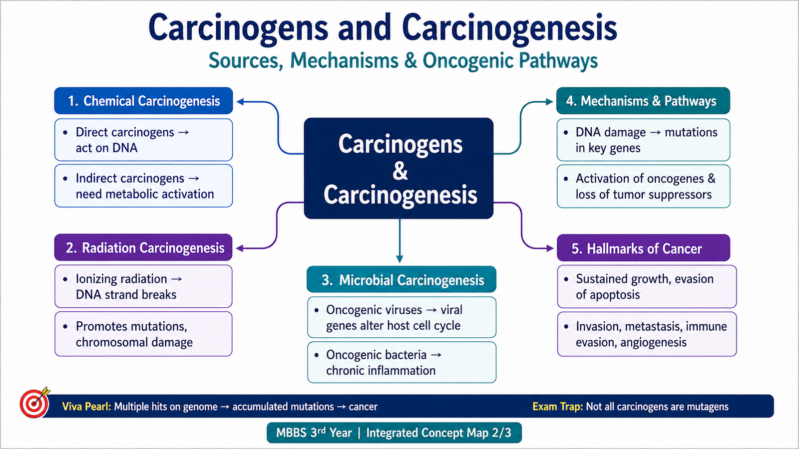
Chemical carcinogens promote cancer mainly by producing DNA damage or by generating reactive metabolites that interact with DNA. The important distinction is between direct-acting carcinogens, which are intrinsically reactive, and indirect-acting carcinogens, which require metabolic conversion before they become carcinogenic.
Direct-acting carcinogens
Direct-acting agents do not require metabolic activation. They can react directly with cellular molecules, including DNA. Some alkylating agents used as anticancer drugs belong to this group. Their therapeutic benefit comes from damaging rapidly dividing malignant cells, but their mutagenic effect on normal cells can also contribute to later development of secondary malignancy.
Indirect-acting carcinogens
Indirect carcinogens are often called procarcinogens because the original compound is not the final reactive carcinogen. It is metabolized, commonly by cytochrome P450-dependent enzymes, into an active electrophilic metabolite. This metabolite can bind covalently to DNA and produce mutations if the damage is not repaired.
→ metabolic activation
→ reactive electrophilic metabolite
→ DNA damage
→ mutation in growth-control genes
→ clonal expansion
→ cancer
Important examples
- Polycyclic aromatic hydrocarbons are produced during incomplete combustion and can be metabolically converted to DNA-reactive compounds.
- Aromatic amines can undergo metabolic activation and are associated with carcinogenic effects in exposed tissues.
- Aflatoxin B1, produced by certain fungi contaminating stored food, is metabolically activated and can damage DNA in hepatocytes.
- Nitrosamines and related compounds can form reactive metabolites capable of DNA injury.
- Vinyl chloride is a recognized industrial chemical carcinogen associated particularly with hepatic vascular malignancy.
Why exposure does not produce cancer in every person
The effect of a chemical carcinogen depends on the dose and duration of exposure, the ability of the body to activate or detoxify the agent, efficiency of DNA repair, and whether mutations occur in genes that provide a selective growth advantage. Therefore, exposure increases risk but does not guarantee that cancer will develop.
| Feature | Direct-acting carcinogens | Indirect-acting carcinogens |
|---|---|---|
| Metabolic activation | Not required | Required |
| Reactive form | Agent itself is reactive | Reactive metabolite is produced |
| Basic mechanism | Direct molecular/DNA injury | Metabolism → DNA-reactive intermediate |
F. Radiation and Microbial Carcinogenesis
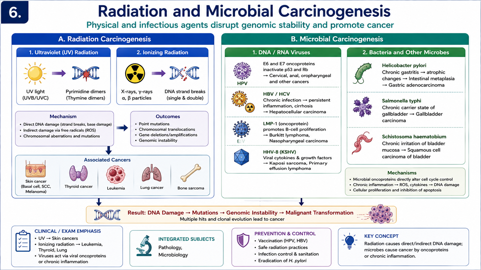
Carcinogenesis can also be produced by physical agents such as radiation and by certain infectious agents. Although the initiating factors differ, they ultimately disturb the same fundamental systems that regulate the genome, proliferation and survival of cells.
Ultraviolet radiation
Ultraviolet radiation from sunlight can damage DNA in epidermal cells. UVB is particularly important because it produces abnormal covalent bonds between adjacent pyrimidine bases, often described as pyrimidine dimers. Normally, these lesions are removed by DNA repair mechanisms. If repair fails, mutations accumulate in genes controlling cell growth and survival.
→ pyrimidine DNA damage
→ inadequate repair
→ mutation accumulation
→ malignant transformation
Ionizing radiation
Ionizing radiation includes X-rays, gamma rays and particulate radiation. It can injure DNA directly and can also generate reactive oxygen species that damage DNA. Important consequences include single- and double-strand DNA breaks, chromosomal abnormalities and mutations. A surviving cell that carries unrepaired growth-promoting genetic damage may later undergo malignant transformation.
Viral carcinogens
Oncogenic viruses contribute to cancer through several mechanisms. Some encode proteins that directly interfere with tumor suppressor pathways. Others produce persistent infection, chronic inflammation or prolonged cell proliferation, increasing the opportunity for additional mutations to accumulate.
- Human papillomavirus (HPV): high-risk types produce viral proteins that interfere with major growth-suppressing pathways. The E6 protein promotes loss of p53 activity, while E7 interferes with the RB growth-control pathway. The infected cell therefore loses important restraints on proliferation and survival.
- Epstein–Barr virus (EBV): infects B lymphocytes and can promote their survival and proliferation. Additional cellular abnormalities are required before malignant transformation occurs.
- Hepatitis B virus (HBV) and hepatitis C virus (HCV): persistent hepatic infection can produce chronic inflammation, cell injury, repeated regeneration and genomic damage, creating an environment favorable for hepatocellular carcinoma.
- Human T-cell leukemia virus type 1 (HTLV-1): stimulates proliferation and survival of infected T cells. Accumulation of additional genetic abnormalities may eventually produce malignant transformation.
- Kaposi sarcoma-associated herpesvirus (HHV-8): contributes to Kaposi sarcoma through viral proteins that promote cellular proliferation and survival.
- Merkel cell polyomavirus: is associated with Merkel cell carcinoma and can contribute to dysregulated cellular proliferation.
Bacterial carcinogen: Helicobacter pylori
Helicobacter pylori is the major bacterial example of microbial carcinogenesis. Long-standing infection produces chronic gastritis. Persistent inflammation causes repeated epithelial injury and regeneration and may lead to progressive mucosal changes and accumulation of genetic abnormalities. This can contribute to gastric adenocarcinoma. Chronic antigenic stimulation can also promote B-cell proliferation and contribute to gastric MALT lymphoma.
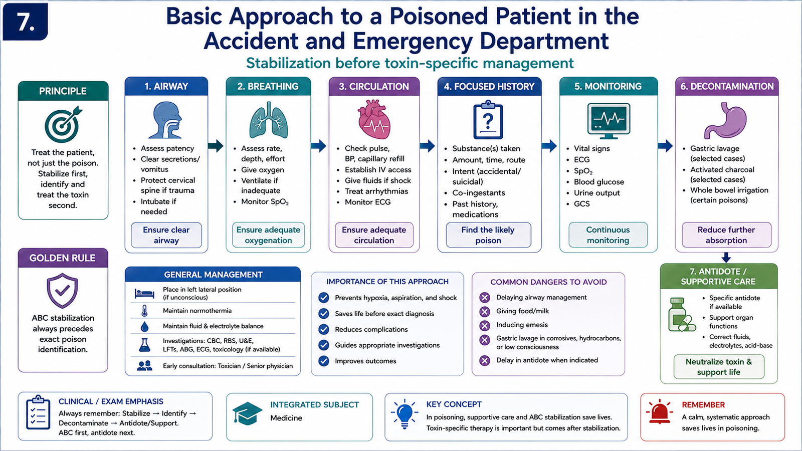
G. Basic Approach to a Poisoned Patient in the Accident and Emergency Department
The immediate approach to a poisoned patient is based on stabilization before identification of the exact poison. A severely poisoned patient may deteriorate because of airway obstruction, respiratory failure, shock, altered consciousness, seizures or cardiac arrhythmia. Therefore, the first priority is to identify and treat life-threatening physiological problems while simultaneously obtaining information about the possible toxic exposure.
1. Initial stabilization
The patient should first be assessed systematically for airway, breathing and circulation. Airway protection becomes especially important when consciousness is impaired. Breathing should be assessed for respiratory depression, hypoxia or excessive secretions. Circulatory assessment includes pulse, blood pressure and evidence of poor perfusion.
2. Rapid neurological and metabolic assessment
Level of consciousness, pupil size, seizures and abnormal behavior can provide useful toxicological clues. Blood glucose should be considered early in an unconscious or confused patient because hypoglycemia is an immediately treatable cause of altered mental status and may coexist with poisoning.
3. Focused history and examination
Once immediate threats are being managed, obtain information about the suspected substance, route of exposure, approximate time of exposure, circumstances and any available drug containers or labels. Examination should look for characteristic patterns such as pupil changes, abnormal sweating, dry skin, excessive secretions, altered bowel activity, abnormal temperature and distinctive cardiovascular or neurological findings.
4. Investigations and monitoring
Investigations are selected according to the clinical situation and suspected poison. Important basic monitoring may include vital signs, oxygenation and cardiac rhythm. Laboratory assessment is directed by the patient’s physiological disturbance and the suspected toxic agent rather than by indiscriminate testing.
5. Reduce further absorption when appropriate
Decontamination depends on the route, substance and clinical circumstances. Contaminated clothing may need removal and exposed skin or eyes may require irrigation. Gastrointestinal decontamination is not automatically required in every poisoning and should be considered only when appropriate and safe.
6. Antidotes and supportive care
A specific antidote is used when the responsible poison is known or strongly suspected and an appropriate antidote exists. However, supportive care remains central to poisoning management because many toxins have no specific antidote. Treatment therefore focuses on maintaining oxygenation, circulation and organ function while the toxin is metabolized or eliminated.
↓
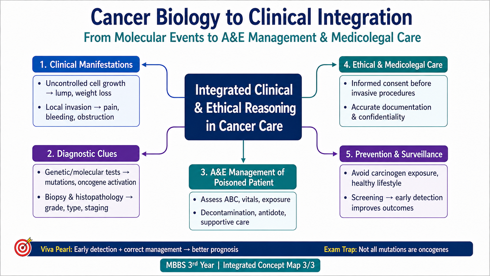
DNA damage or abnormal regulation of growth-control genes
↓
Oncogene activation + tumor suppressor loss + impaired apoptosis/DNA repair
↓
Selective growth and survival advantage
↓
Clonal expansion with accumulation of additional abnormalities
↓
Acquisition of cancer hallmarks
↓
Malignant growth, invasion and possible metastasis
⭐ AIM High-Yield Review
- Cancer develops through accumulated abnormalities in genes controlling proliferation, survival and genomic stability.
- Proto-oncogene → oncogene represents abnormal activation of a growth-promoting gene.
- Oncoproteins promote cell growth or survival even when normal regulatory signals are absent.
- Proto-oncogenes may be activated by point mutation, chromosomal translocation or gene amplification.
- Tumor suppressor genes normally restrain growth; their loss removes an important cellular “brake.”
- ⭐ p53 responds to DNA damage by promoting cell-cycle arrest, DNA repair, senescence or apoptosis.
- Loss of p53 permits cells carrying damaged DNA to survive and continue dividing.
- Major cancer hallmarks include sustained proliferation, escape from growth suppression and apoptosis, replicative immortality, angiogenesis, invasion/metastasis, altered metabolism and immune evasion.
- Carcinogenesis is a multistep clonal process; successive abnormalities select increasingly aggressive subclones.
- ⭐ An initiator produces permanent genetic damage, whereas a promoter mainly stimulates proliferation of initiated cells.
- Direct-acting chemical carcinogens do not require metabolic activation; indirect carcinogens do.
- UV radiation causes characteristic DNA injury including pyrimidine dimers; ionizing radiation can cause DNA breaks and free-radical injury.
- High-risk HPV proteins interfere with major tumor suppressor pathways, particularly p53 and RB.
- HBV, HCV and H. pylori illustrate the importance of persistent infection and chronic inflammation in carcinogenesis.
- In acute poisoning, stabilization of airway, breathing and circulation takes priority over exact toxin identification.
Topic 17 — Molecular Basis of Cancer and Carcinogenesis
Use these videos after reading the AIM learning material to reinforce the major mechanisms of carcinogenesis.
Carcinogens & Carcinogenesis — Chemical, Radiation and Biological Carcinogens
Covers the multistep process of carcinogenesis, initiation, mutations, clonal evolution, direct and indirect chemical carcinogens, radiation carcinogenesis and microbial carcinogenesis including HPV, EBV, HBV/HCV and H. pylori.
Chemical & Radiation Carcinogenesis
Focuses specifically on direct-acting and indirect-acting chemical carcinogens, metabolic activation of procarcinogens, ultraviolet radiation and ionizing radiation.