
Topic 6 — Cellular Foundations, Bacterial Classification, Structure, Growth and Genetics
Topic Introduction
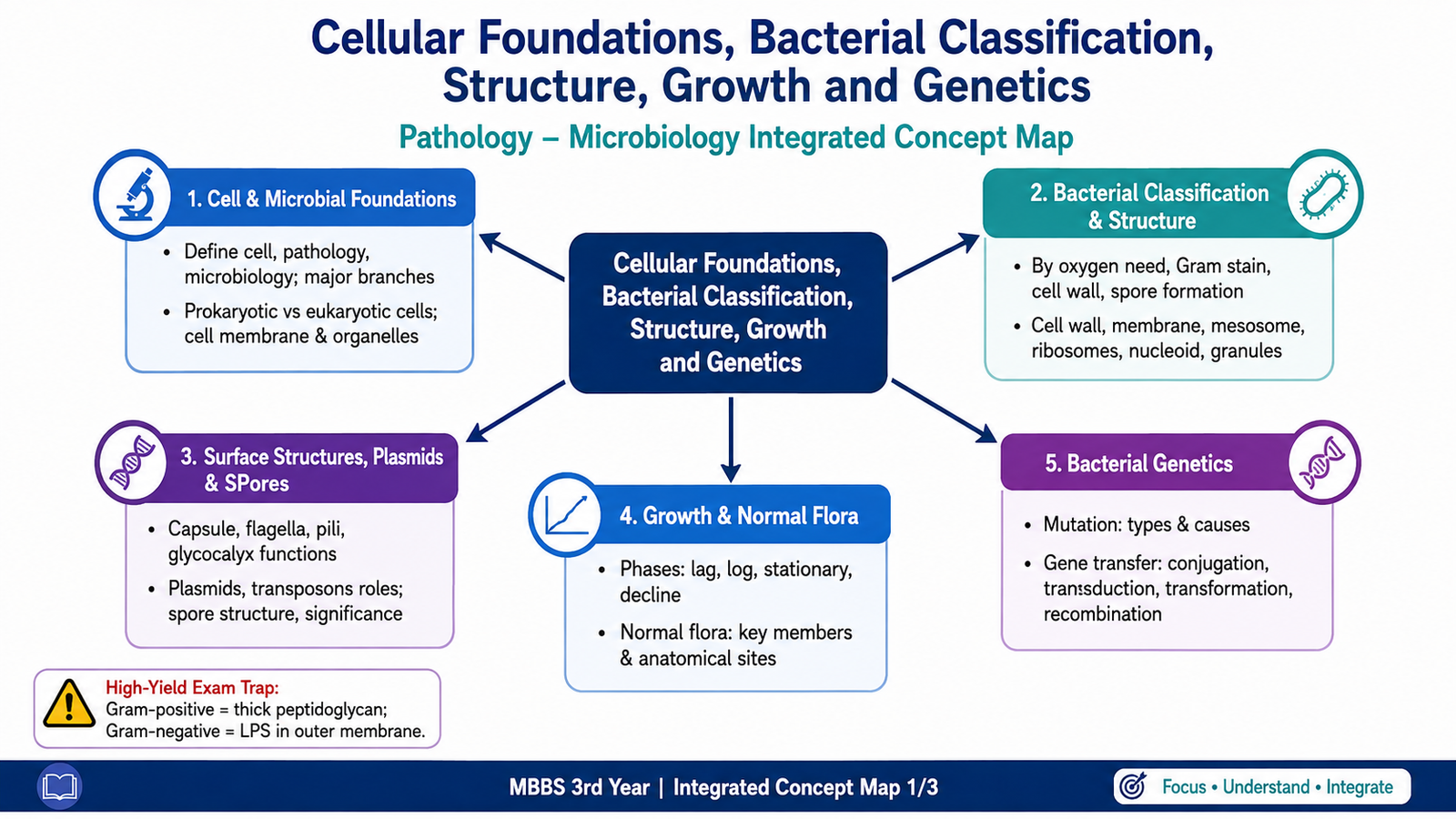
Pathology is the study of disease, while microbiology is the study of microorganisms. Understanding both begins with knowing how normal cells are organized and how microorganisms differ from human cells. Bacteria are particularly important because their structure, growth and genetic behaviour determine many of their biological characteristics. In this chapter, you will first understand the major groups of microorganisms and the differences between prokaryotic and eukaryotic cells. You will then study bacterial classification, bacterial cellular structures, plasmids, transposons and spores. Finally, you will learn the bacterial growth curve, important normal flora and the major mechanisms of mutation and genetic exchange.
A. Foundations of Pathology and Microbiology
Pathology and microbiology provide two closely related foundations for understanding disease. Pathology explains the structural, biochemical and functional changes that occur in cells and tissues during disease, whereas microbiology studies organisms that may interact with humans and, in some situations, cause disease.
Pathology
Pathology is the scientific study of disease. It examines the causes of disease, the mechanisms through which disease develops, the structural changes produced in cells and tissues, and the functional consequences of these changes.
Major branches include:
- General pathology: studies basic reactions of cells and tissues to injury that are common to many diseases.
- Systemic pathology: studies diseases as they affect particular organs or organ systems.
- Anatomical pathology: examines structural changes in tissues and organs, including histopathology and cytology.
- Clinical pathology: uses laboratory examination of blood, urine and other body fluids to assist diagnosis.
Microbiology
Microbiology is the study of microscopic organisms and infectious agents. Medical microbiology focuses on organisms relevant to human health and disease.
Major branches commonly encountered in medical microbiology include:
- Bacteriology: study of bacteria.
- Virology: study of viruses.
- Mycology: study of fungi.
- Parasitology: study of medically important protozoa and helminths.
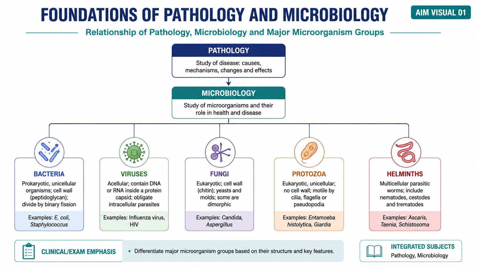
Five Major Groups of Microorganisms
The major microbial groups differ in cellular organization, structure and mode of reproduction. Recognizing these differences is important because the structure of an organism determines many of its biological properties.
| Group | Essential Characteristic |
|---|---|
| Bacteria | Single-celled prokaryotic organisms lacking a true membrane-bound nucleus. |
| Viruses | Acellular infectious agents containing nucleic acid and requiring living host cells for replication. |
| Fungi | Eukaryotic organisms occurring mainly as yeasts or moulds. |
| Protozoa | Unicellular eukaryotic organisms, some of which are medically important parasites. |
| Helminths | Multicellular parasitic worms whose eggs or larvae may also be microscopic. |
B. Cell Organization: Prokaryotic and Eukaryotic Cells
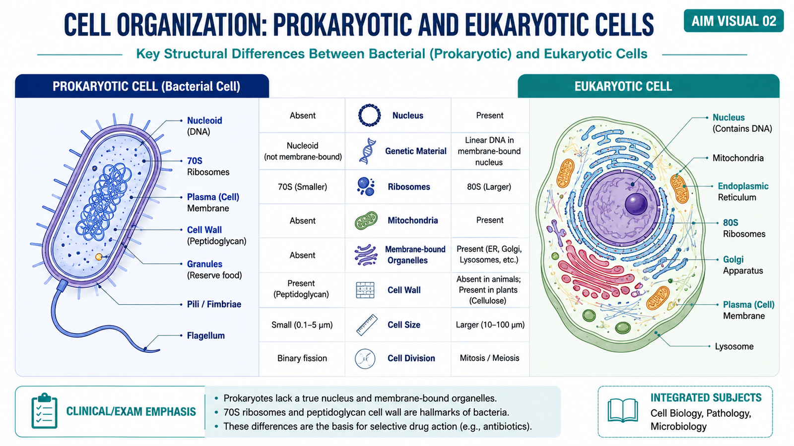
A cell is the basic structural and functional unit of living organisms. Cells contain genetic material and cellular machinery that allow them to perform essential functions. The most important distinction for this topic is between prokaryotic cells, represented by bacteria, and eukaryotic cells, which make up the human body and organisms such as fungi and protozoa.
Prokaryotic Cells
Prokaryotic cells are simpler and generally smaller. Their DNA is not enclosed inside a membrane-bound nucleus. Instead, the main bacterial chromosome occupies a region called the nucleoid. They also lack membrane-bound organelles such as mitochondria, lysosomes and endoplasmic reticulum.
Eukaryotic Cells
Eukaryotic cells contain a true nucleus surrounded by a nuclear membrane. They also possess specialized membrane-bound organelles that divide cellular functions into separate compartments.
| Feature | Prokaryotic Cell | Eukaryotic Cell |
|---|---|---|
| Nucleus | No true membrane-bound nucleus | True membrane-bound nucleus present |
| DNA | Usually a circular chromosome in nucleoid | Linear chromosomes within nucleus |
| Membrane-bound organelles | Absent | Present |
| Ribosomes | 70S | 80S cytoplasmic ribosomes |
| Typical example | Bacterium | Human cell, fungus, protozoan |
Cell Membrane
The cell membrane forms a selective boundary between the cell and its environment. In eukaryotic cells it consists mainly of a phospholipid bilayer containing proteins, cholesterol and carbohydrate-containing molecules. The phospholipid arrangement creates a barrier through which some substances can pass more easily than others.
Membrane proteins perform important functions such as transport, receptors, cell adhesion and enzymatic activity. The membrane therefore does much more than simply surround the cell; it actively regulates communication and movement of substances between the cell and its surroundings.
Major Eukaryotic Cell Organelles
- Nucleus: contains cellular DNA and regulates gene expression.
- Mitochondria: generate most cellular ATP through oxidative metabolism.
- Rough endoplasmic reticulum: contains ribosomes and synthesizes proteins destined for secretion or membranes.
- Smooth endoplasmic reticulum: participates in lipid synthesis and other metabolic functions.
- Golgi apparatus: modifies, sorts and packages proteins.
- Lysosomes: contain hydrolytic enzymes that digest cellular material.
- Peroxisomes: participate in oxidative reactions and metabolism of certain lipids.
- Ribosomes: synthesize proteins from messenger RNA.
- Cytoskeleton: provides structural support and contributes to movement and intracellular transport.
C. Bacterial Classification and Basic Cellular Structure
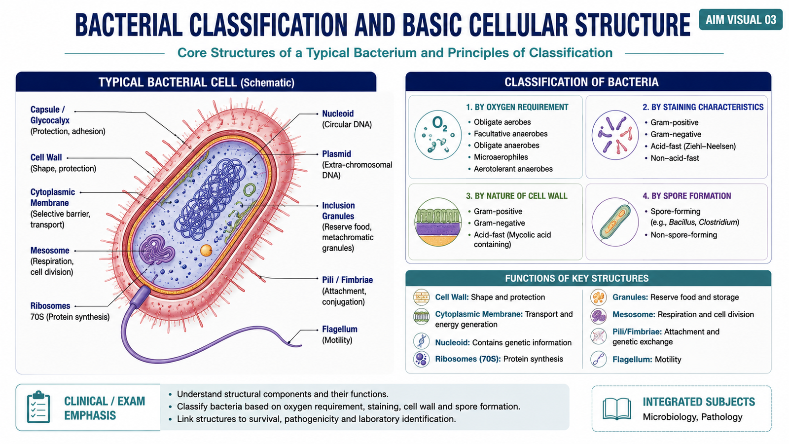
Bacteria can be classified using several practical characteristics. These classifications are useful because they describe major differences in bacterial biology. Important approaches include oxygen requirements, staining characteristics, cell-wall properties and the ability to form spores.
Classification According to Oxygen Requirement
- Obligate aerobes: require oxygen for growth.
- Obligate anaerobes: grow in the absence of oxygen and may be harmed by oxygen.
- Facultative anaerobes: can grow with or without oxygen.
- Microaerophiles: require oxygen at lower concentrations than that found in normal atmospheric air.
Classification According to Staining and Cell Wall
The Gram stain separates many bacteria into Gram-positive and Gram-negative groups according to differences in cell-wall structure. These structural differences determine how the organisms retain the staining dyes.
Classification According to Spore Formation
Some bacteria can produce highly resistant dormant structures called spores, whereas others cannot. Bacteria can therefore be described as spore-forming or non-spore-forming.
Important Internal Bacterial Structures
Although bacteria lack membrane-bound organelles, they contain specialized structures required for survival, metabolism, protein production and reproduction.
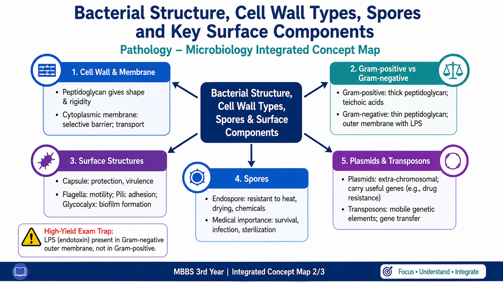
- Cell wall: provides shape and mechanical protection and helps prevent osmotic rupture.
- Cytoplasmic membrane: surrounds the cytoplasm and controls movement of substances into and out of the bacterium. It also participates in important metabolic processes.
- Mesosome: a term traditionally used to describe infoldings of the bacterial cytoplasmic membrane. It is mainly of historical importance in modern interpretation of bacterial ultrastructure.
- Ribosomes: bacterial 70S ribosomes synthesize proteins.
- Granules or inclusion bodies: may store nutrients or other substances needed by the bacterium.
- Nucleoid: contains the main bacterial chromosome and is not surrounded by a nuclear membrane.
D. Bacterial Surface Structures and Gram-Positive versus Gram-Negative Cell Walls
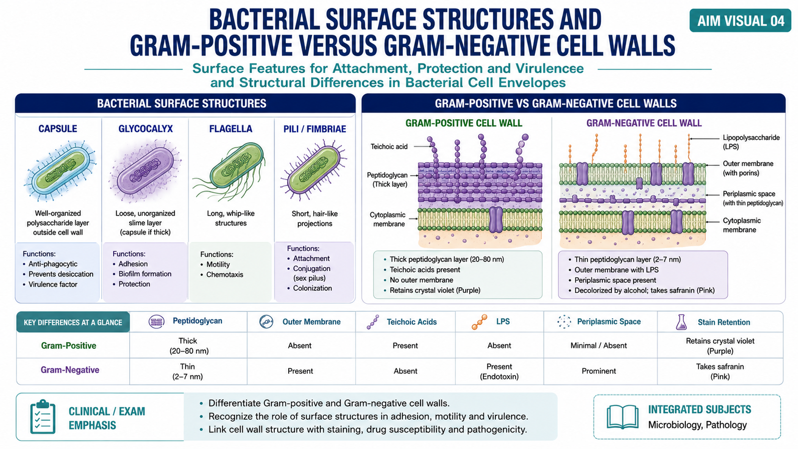
Several bacterial structures lie outside or project from the cytoplasmic membrane. They contribute to protection, attachment, movement and interaction with the environment. Their presence varies among bacterial species.
Capsule and Glycocalyx
The glycocalyx is an extracellular material surrounding some bacteria. When it is well organized and firmly attached, it is commonly described as a capsule. A capsule can help protect the organism from environmental threats and assists some bacteria in resisting phagocytosis. Less organized glycocalyx material can help organisms adhere to surfaces and to one another.
Flagella
Flagella are long filamentous structures responsible for bacterial motility. Their movement allows motile bacteria to change position in response to environmental conditions.
Pili
Pili are shorter surface appendages. Some participate in attachment to host cells or other surfaces, while specialized sex pili can bring bacterial cells together during conjugation and therefore participate in transfer of genetic material.
Gram-Positive and Gram-Negative Cell Walls
The major difference between Gram-positive and Gram-negative bacteria lies in the organization of their cell envelopes. Gram-positive bacteria have a thick peptidoglycan layer, whereas Gram-negative bacteria have a thinner peptidoglycan layer located between the cytoplasmic membrane and an additional outer membrane.
| Feature | Gram-Positive | Gram-Negative |
|---|---|---|
| Peptidoglycan | Thick | Thin |
| Outer membrane | Absent | Present |
| Teichoic acids | Present | Absent |
| Lipopolysaccharide | Absent | Present in outer membrane |
| Gram-stain appearance | Purple | Pink/red |
Gram-positive bacteria retain the primary crystal violet stain because their thick peptidoglycan layer traps the dye complex. Gram-negative bacteria lose the primary stain during decolorization and then take up the counterstain, producing a pink to red appearance.
E. Plasmids, Transposons and Bacterial Spores
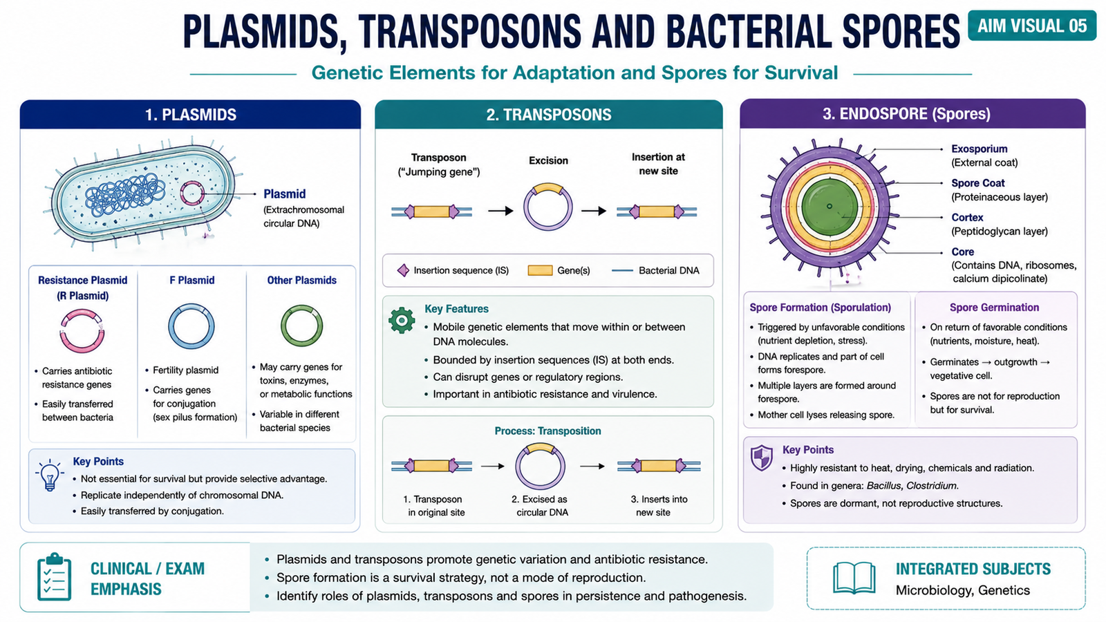
In addition to the main chromosome, bacteria may contain other genetic elements that can influence their characteristics. Plasmids and transposons are particularly important because they allow useful genetic traits to be carried or moved. Some bacteria can also form spores, which serve a completely different function: survival under unfavorable environmental conditions.
Plasmids
Plasmids are usually small, circular, double-stranded DNA molecules that exist separately from the bacterial chromosome and can replicate independently. They are not usually essential for basic bacterial survival, but they may provide an advantage under particular conditions.
Important functional categories include:
- Fertility or F plasmids: contain genes involved in conjugation.
- Resistance plasmids: may carry genes that provide resistance to antimicrobial agents.
- Virulence plasmids: may carry genes that enhance the ability of a bacterium to produce disease.
- Metabolic plasmids: may provide additional metabolic capabilities.
Transposons
Transposons are mobile DNA sequences capable of moving from one position in DNA to another. They may move within a chromosome or between chromosomal and plasmid DNA. Because they can carry genes, including resistance-related genes, they contribute to genetic variation within bacterial populations.
A simple arrangement may include a central gene or genes surrounded by DNA sequences required for movement. The movement process is called transposition.
Bacterial Spores
A bacterial endospore is a highly resistant dormant structure formed within certain bacteria when environmental conditions become unfavorable. Spore formation is a survival mechanism, not a method of reproduction. One vegetative bacterial cell forms one spore, and when conditions improve the spore can germinate back into one vegetative cell.
Spores possess multiple protective layers and contain a dehydrated, metabolically inactive core. These features allow them to tolerate conditions that would kill ordinary vegetative bacterial cells.
F. Bacterial Growth Curve and Normal Flora
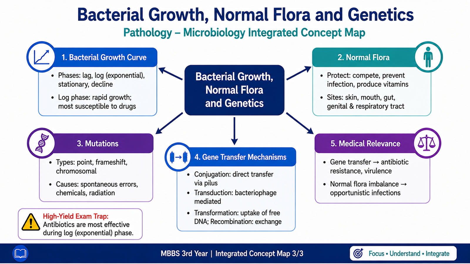
When bacteria are placed in a suitable closed culture system, their population does not increase at the same rate throughout the entire period. Instead, bacterial growth follows a characteristic sequence of phases. Understanding this growth curve explains how bacteria adapt, multiply, compete for nutrients and eventually decline.
Phases of the Bacterial Growth Curve
1. Lag Phase
Bacteria are adapting to the new environment. They are metabolically active and synthesize cellular components, but there is little or no increase in the number of organisms.
2. Log or Exponential Phase
Bacteria divide rapidly by binary fission. The population increases exponentially because nutrients are available and conditions are favorable. Cells are highly metabolically active during this phase.
3. Stationary Phase
The rate at which new bacteria are produced becomes approximately balanced by the rate at which bacteria die. This occurs because nutrients become limited and waste products accumulate.
4. Death or Decline Phase
The number of viable bacteria decreases because unfavorable conditions continue and bacterial death exceeds formation of new cells.
Lag phase → exponential multiplication → nutrient limitation and waste accumulation → stationary phase → decline in viable organisms.
Normal Flora
Normal flora, also called the normal microbiota, consists of microorganisms that normally colonize particular body sites without causing disease under ordinary circumstances. Different anatomical sites support different microbial populations because moisture, nutrients, oxygen tension and other local conditions vary.
Important sites and examples include:
- Skin: commonly contains coagulase-negative staphylococci such as Staphylococcus epidermidis and other resident organisms.
- Nose and anterior nares: may be colonized by staphylococci, including Staphylococcus aureus in some individuals.
- Mouth and oropharynx: contain viridans streptococci and numerous anaerobic organisms.
- Large intestine: contains a dense population of anaerobic bacteria together with organisms such as Escherichia coli and other enteric bacteria.
- Vagina: in many healthy women, lactobacilli are important members of the microbiota.
Normal flora can be beneficial because resident organisms occupy ecological niches and may limit colonization by competing microorganisms. However, organisms that are harmless at their usual site may cause disease if they enter a normally sterile tissue or if host defenses are impaired.

G. Bacterial Genetics: Mutation and Genetic Exchange
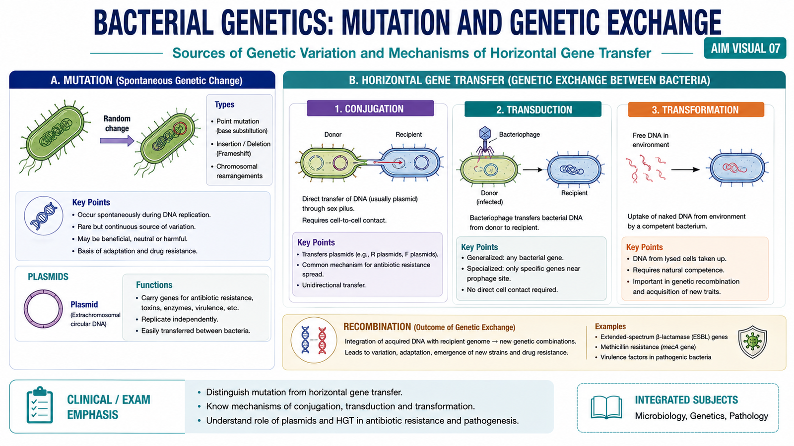
Bacterial characteristics are determined by genetic information. Changes in DNA can arise by mutation, while bacteria can also acquire genetic material from other sources. These mechanisms create genetic variation and may alter characteristics such as metabolism, virulence or antimicrobial resistance.
Mutation
A mutation is a stable change in the nucleotide sequence of genetic material. A mutation may arise spontaneously during DNA replication or may be induced by physical or chemical agents that damage DNA.
Major Types of Mutation
- Point mutation: alteration involving a single nucleotide or base pair.
- Substitution: one nucleotide is replaced by another.
- Insertion: one or more nucleotides are added.
- Deletion: one or more nucleotides are removed.
- Frameshift mutation: an insertion or deletion changes the reading frame when the number of nucleotides involved is not a multiple of three.
Mutations can be spontaneous, arising naturally during replication, or induced by mutagens such as radiation or certain chemicals. Their effect depends on where the mutation occurs and whether it alters an important gene product.
Conjugation
Conjugation is transfer of genetic material between bacteria through direct cell-to-cell contact. A donor bacterium carrying appropriate conjugative genes can form a connection with a recipient. Plasmid DNA can then be transferred.
Transduction
Transduction is transfer of bacterial DNA from one bacterium to another by a bacteriophage. A bacteriophage infects a bacterium and may carry bacterial DNA from one cell to another during subsequent infection.
Transformation
Transformation occurs when a bacterium takes up free or naked DNA from its environment and incorporates the genetic information into its own genetic material when conditions permit.
Recombination
Recombination is the incorporation and rearrangement of genetic material so that DNA from different sources becomes combined. Genetic material acquired through transformation, transduction or conjugation may undergo recombination with bacterial DNA, producing a stable genetic change.
Conjugation = direct bacterial contact.
Transduction = bacteriophage-mediated transfer.
Transformation = uptake of naked environmental DNA.
Recombination = integration or rearrangement of genetic material.
⭐ AIM High-Yield Review
- Pathology studies disease processes, whereas microbiology studies microorganisms.
- The five major microorganism groups covered here are bacteria, viruses, fungi, protozoa and helminths.
- ⭐ Bacteria are prokaryotes: they lack a true membrane-bound nucleus and membrane-bound organelles.
- Bacterial DNA is mainly located in the nucleoid, and bacterial protein synthesis occurs on 70S ribosomes.
- The bacterial cell wall maintains shape and helps prevent osmotic rupture.
- ⭐ Gram-positive bacteria have thick peptidoglycan; Gram-negative bacteria have thin peptidoglycan plus an outer membrane containing lipopolysaccharide.
- Capsule/glycocalyx contributes to protection and attachment; flagella provide motility; some pili participate in attachment or conjugation.
- Plasmids are independently replicating extrachromosomal DNA molecules that may carry resistance, virulence or other useful genes.
- Transposons are mobile DNA sequences capable of changing position within genetic material.
- ⭐ Bacterial spores are survival structures, not reproductive structures.
- The bacterial growth curve progresses through lag → log → stationary → death phases.
- Normal flora normally colonizes specific anatomical sites and may become harmful if displaced to normally sterile sites or if host defenses fail.
- Mutation is a stable change in DNA sequence and may arise spontaneously or through mutagenic agents.
- ⭐ Conjugation = direct contact; transduction = bacteriophage; transformation = naked DNA uptake.
- Recombination allows genetic material from different sources to be integrated or rearranged within bacterial DNA.
Cellular Foundations, Bacterial Structure, Growth and Genetics
Watch these videos in sequence. Together they reinforce the major concepts covered in this AIM learning topic.
Prokaryotic vs Eukaryotic Cells
Focus on the nucleus, nucleoid, organelles, ribosomes and the basic structural differences between prokaryotic and eukaryotic cells.
Bacterial Structure and Functions
Reinforces bacterial cell organization, cell wall, membrane, surface structures and the importance of bacteria in health and disease.
Bacterial Growth Curve
Concentrate on the lag, log/exponential, stationary and death phases and understand why each phase develops.
Bacterial Genetics
Focus on bacterial genetic material, mutations and horizontal gene transfer, especially transformation, transduction and conjugation.