
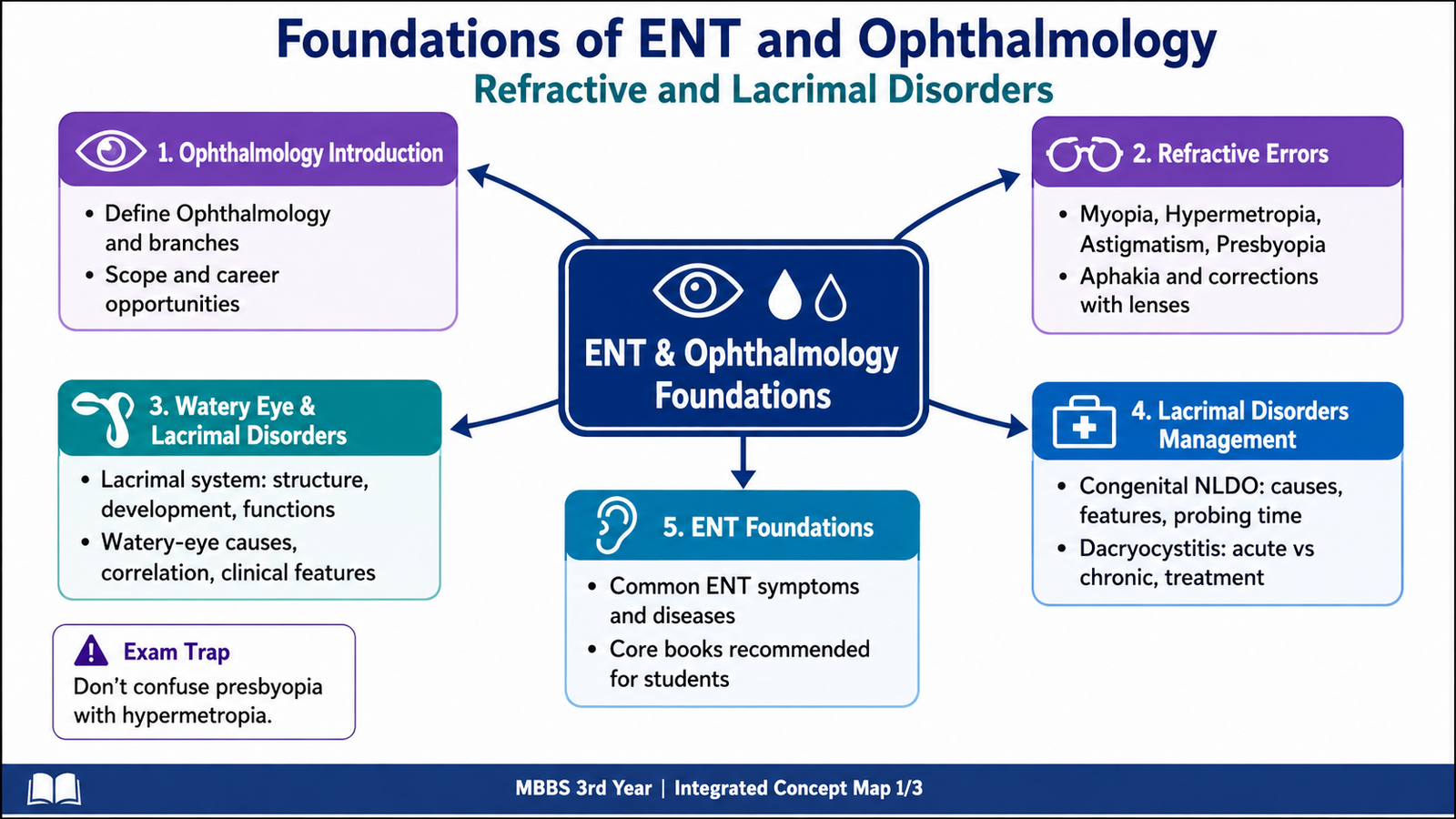
This chapter follows the supplied KMU learning outcomes and builds the topic in a simple sequence. First understand how normal vision, refractive errors and the lacrimal system work; then revise the high-yield distinguishing features at the end.
Topic 9 — Foundations of ENT and Ophthalmology: Refractive and Lacrimal Disorders
Foundation Module
A structured introduction to ophthalmology and ENT, with particular emphasis on refractive errors, their optical correction, watery-eye presentations and important lacrimal disorders.
Topic Introduction
Ophthalmology is the branch of medicine concerned with the structure, function and diseases of the eye and visual system. A major part of basic ophthalmology is understanding how light is focused on the retina and what happens when this focusing mechanism is imperfect. These problems are called refractive errors and include myopia, hypermetropia, astigmatism and presbyopia. Aphakia produces another important optical problem because the natural crystalline lens is absent. The lacrimal system is equally important because normal tear production and drainage are essential for a healthy ocular surface. Disorders of tear drainage commonly present with watering of the eye. This chapter also introduces the basic scope of ENT, common ENT symptoms and commonly encountered disease groups.
A. Foundations and Scope of Ophthalmology
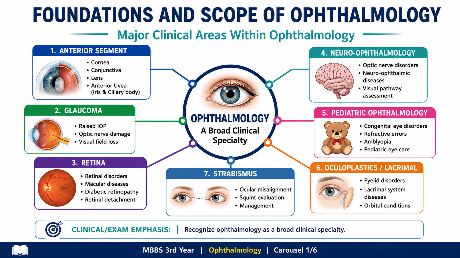
Ophthalmology deals with the normal structure and function of the eye as well as diseases that interfere with vision or ocular health. Because vision depends on several structures working together, ophthalmology includes problems involving the eyelids, lacrimal system, conjunctiva, cornea, lens, uveal tract, retina, optic nerve, ocular movements and visual pathways.
What ophthalmology includes
The field can be understood through several major areas. These divisions are useful for organizing diseases rather than treating the eye as a single structure.
- Anterior-segment ophthalmology: disorders involving structures such as the cornea, anterior chamber and lens.
- Glaucoma: disorders associated with damage to the optic nerve, often in relation to intraocular pressure.
- Retina and vitreous: diseases affecting the sensory retina and vitreous cavity.
- Neuro-ophthalmology: visual problems related to the optic nerve and neurological pathways.
- Pediatric ophthalmology: eye disorders occurring in children.
- Oculoplastics and lacrimal disorders: diseases of the eyelids, orbit and tear-drainage system.
- Strabismus: disorders of ocular alignment and coordinated eye movements.
Scope of ophthalmology as a career
Ophthalmology combines medical diagnosis with clinical examination, optical principles and procedural or surgical care. An ophthalmologist may manage problems ranging from refractive and surface disorders to cataract, glaucoma, retinal disease, ocular trauma and conditions requiring surgery. The specialty therefore requires both careful clinical observation and precise practical skills.
B. Refraction, Refractive Error and Myopia
Clear vision requires incoming light rays to be focused accurately on the retina. The cornea provides most of the eye’s refractive power, while the crystalline lens allows additional adjustment. When the optical power of the eye and its axial length are properly matched, parallel rays from a distant object focus on the retina without accommodation. This state is called emmetropia.
A refractive error exists when light is not focused correctly on the retina while accommodation is relaxed. The resulting retinal image becomes blurred. Refractive errors may occur because of excessive or insufficient optical power, abnormal axial length or unequal refraction in different meridians.
Optical power and axial length properly matched → image focuses on retina → clear distant vision.
Optical power and axial length mismatched → image focuses away from the retinal plane → blurred vision.
Myopia
Myopia, or short-sightedness, is a refractive error in which parallel rays from a distant object are focused in front of the retina when accommodation is relaxed. The patient therefore sees distant objects less clearly, while near vision may remain comparatively good.
Why myopia occurs
Myopia develops when the eye has too much refractive power for its axial length or when the eyeball is longer than the optical system can appropriately focus for.
- Axial myopia: the anteroposterior length of the eye is increased.
- Refractive myopia: the refractive power of the cornea or lens is relatively excessive.
In either situation, light is brought to a focus before it reaches the retinal plane. By the time the rays reach the retina, they have begun to diverge again, producing a blurred retinal image.
Correction of myopia
Myopia is corrected with a concave or minus lens. A concave lens diverges incoming light before it enters the eye. This reduces the effective converging power of the optical system so that the final focus moves backward from in front of the retina toward the retina.
Myopic eye focuses too early → concave lens diverges incoming rays → effective optical power is reduced → focus shifts backward onto the retina.

C. Hypermetropia, Astigmatism, Presbyopia and Aphakia
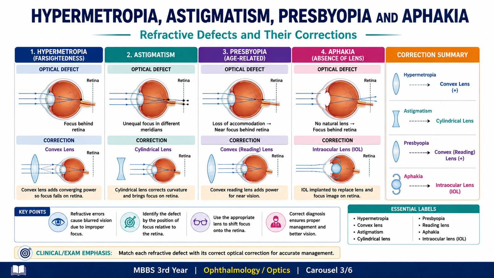
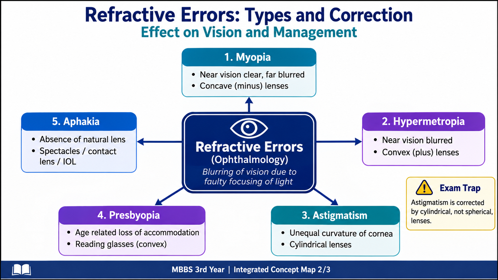
Several refractive conditions disturb vision in different ways. Hypermetropia results from insufficient optical power relative to the length of the eye. Astigmatism occurs when refractive power varies between meridians. Presbyopia reflects the age-related loss of effective accommodation, while aphakia causes major refractive change because the natural crystalline lens is absent.
Hypermetropia
Hypermetropia, also called hyperopia or long-sightedness, is a refractive error in which parallel rays would come to a focus behind the retina when accommodation is relaxed. The eye therefore has insufficient refractive power for its axial length.
The problem may occur because the eyeball is relatively short or because the refractive power of the eye is insufficient. A young patient may compensate partly by accommodation, because increasing the curvature of the lens increases its refractive power. This compensatory effort can contribute to eye strain, especially during near work.
Correction
A convex or plus lens converges incoming rays before they enter the eye. This increases effective optical power and moves the focus forward onto the retina.
Astigmatism
Astigmatism occurs when the refractive power of the eye is not the same in different meridians. Instead of bringing light from a point object to one precise point focus, the optical system produces different focal positions. This causes blurred or distorted vision.
The cornea is commonly responsible because its curvature may differ between meridians. The appropriate optical correction therefore needs to provide power in the required meridian rather than uniformly in all directions.
Cylindrical lenses
A cylindrical lens has refractive power in one principal meridian and essentially no power along the perpendicular axis. It can therefore compensate for unequal refractive power in different meridians of an astigmatic eye.
Presbyopia
Presbyopia is the progressive age-related reduction in the ability to focus clearly on near objects. Near vision requires accommodation. During accommodation, the crystalline lens must become more convex so that its refractive power increases.
With increasing age, the lens gradually becomes less flexible and the accommodative system becomes less effective. The near point therefore moves farther away, and the person may need to hold reading material at a greater distance.
Presbyopia is corrected by providing additional plus power for near vision. Depending on the person’s distance refractive status and visual needs, this may be supplied by reading spectacles or combined optical corrections.
Aphakia
Aphakia means absence of the natural crystalline lens from the eye. Because the lens contributes substantial refractive power, its absence leaves the eye markedly hypermetropic and also removes the normal ability to accommodate.
Methods of optical correction
- Strong convex spectacles: provide the missing positive refractive power externally.
- Contact lenses: provide optical correction closer to the surface of the eye.
- Intraocular lens: an artificial lens placed inside the eye can replace much of the refractive function of the natural lens.
| Condition | Main optical problem | Effect on focus | Basic correction |
|---|---|---|---|
| Myopia | Excess power or excessive axial length |
D. Lacrimal System: Structure, Development, Function and Watery Eye
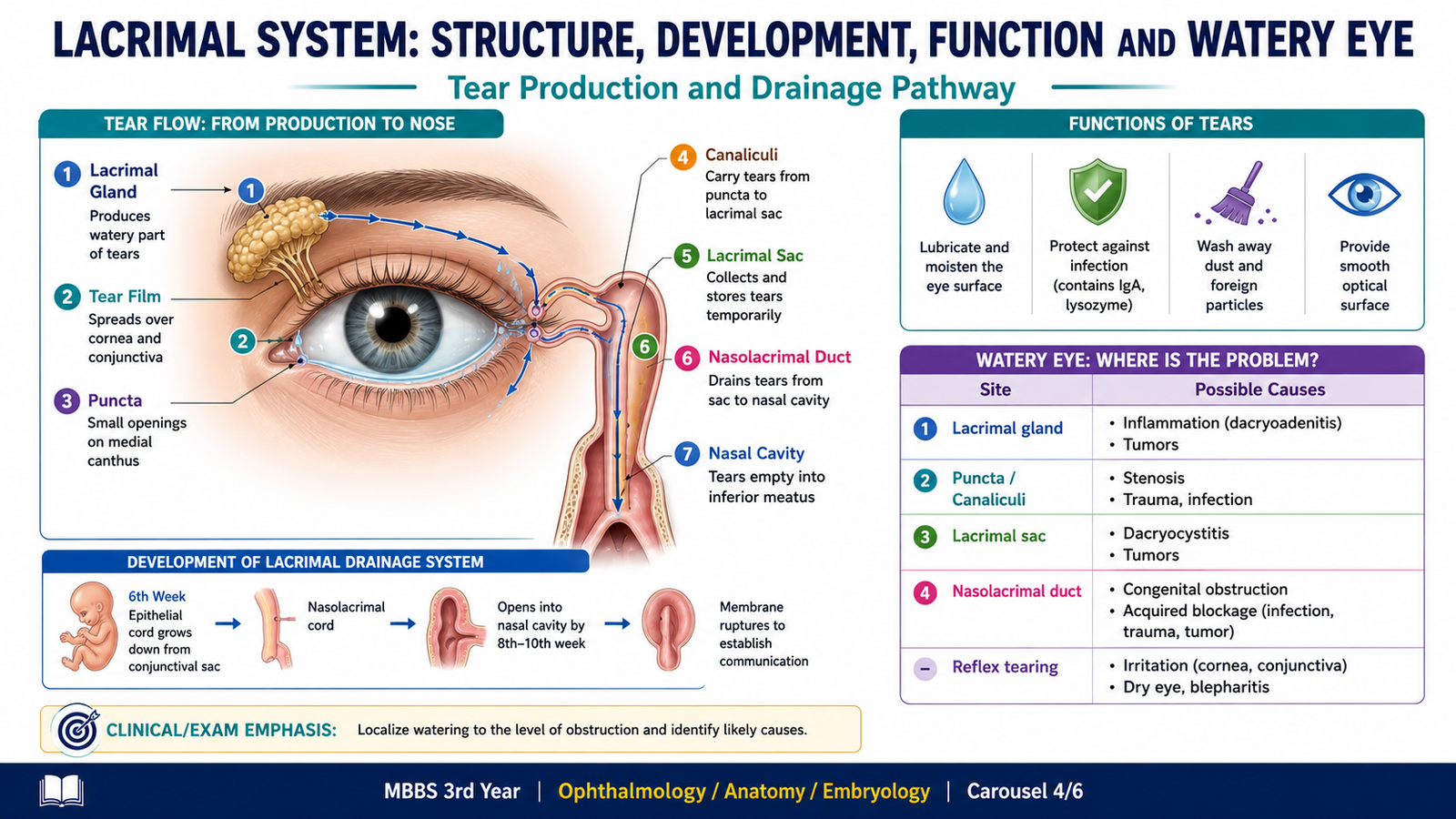
The lacrimal apparatus has two major components: a tear-producing system and a tear-drainage system. Tears must first be produced, spread across the ocular surface and then drain normally into the nose. Disturbance at any point can produce symptoms, particularly watering of the eye.
Lacrimal gland and tear production
The main lacrimal gland lies in the superolateral part of the orbit and secretes the aqueous component of tears. Accessory glands also contribute to normal tear production. Blinking distributes the tear film across the cornea and conjunctiva.
Tear-drainage pathway
After spreading across the ocular surface, tears move toward the medial side of the eyelids. They enter the small lacrimal openings called puncta, pass through the canaliculi, enter the lacrimal sac and then travel through the nasolacrimal duct into the nasal cavity.
Lacrimal secretion → tear film spreads across ocular surface → medial movement with blinking → puncta → canaliculi → lacrimal sac → nasolacrimal duct → nasal cavity.
Development of the lacrimal system
The lacrimal gland develops from surface ectodermal epithelial buds in the region of the developing conjunctiva. The drainage pathway develops from an ectodermal epithelial cord located along the line between the developing lateral nasal and maxillary facial processes. Canalization of this epithelial pathway produces the lacrimal sac and nasolacrimal duct.
This developmental concept is clinically important because incomplete opening of the lower end of the nasolacrimal duct can lead to congenital tear-drainage obstruction.
Functions of the lacrimal system
The lacrimal system maintains a stable ocular surface. Tears keep the cornea and conjunctiva moist, contribute to optical smoothness of the corneal surface, wash away small particles and participate in ocular-surface protection.
Why a patient develops a watery eye
Watering is often called epiphora when tears overflow onto the cheek because drainage is inadequate. A patient may also experience excessive tearing because the ocular surface is irritated and reflex tear production increases.
Therefore, a watery eye should be related anatomically to the tear pathway:
- Excessive tear production may follow ocular-surface irritation.
- Abnormal punctal entry can reduce drainage.
- Canalicular obstruction can interrupt tear movement toward the sac.
- Lacrimal-sac or nasolacrimal-duct obstruction can cause stagnation and overflow.
E. Congenital Nasolacrimal Duct Obstruction and Dacryocystitis
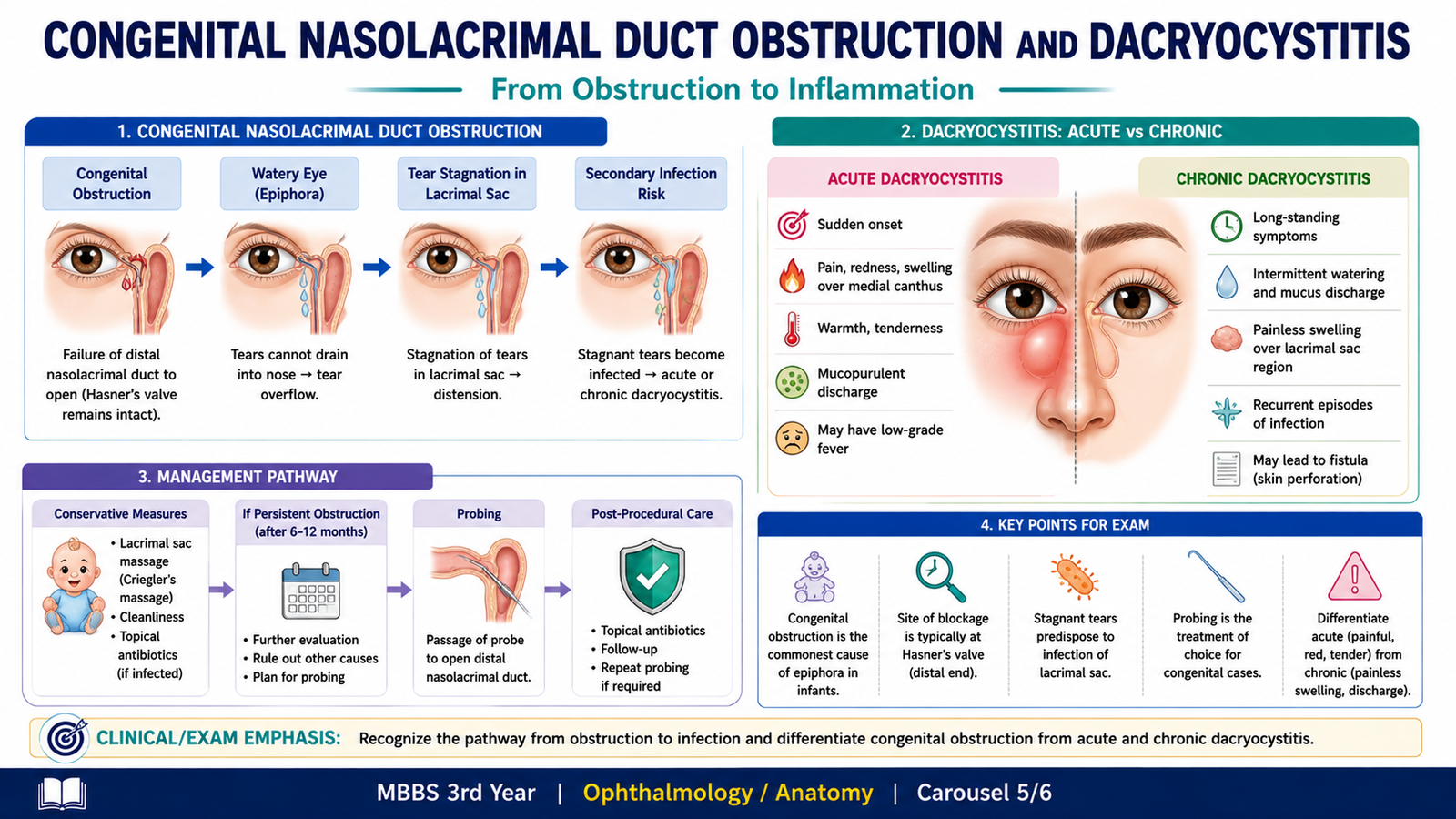
Obstruction of the lacrimal drainage pathway allows tears to remain within the system instead of flowing freely into the nose. Stagnation produces persistent watering and may also encourage accumulation of mucous material or infection. Two important clinical problems at undergraduate level are congenital nasolacrimal duct obstruction and inflammation of the lacrimal sac, known as dacryocystitis.
Congenital nasolacrimal duct obstruction
Congenital nasolacrimal duct obstruction occurs when the tear-drainage pathway is not fully patent at or soon after birth. A common anatomical basis is persistence of a membranous obstruction at the distal end of the nasolacrimal duct.
Clinical features
Because tears cannot drain normally, the infant develops persistent watering. Retained tear fluid and mucus may produce discharge, and pressure over the lacrimal sac region may sometimes cause reflux of material through the puncta.
- Watering beginning in infancy
- Tear overflow
- Mucous or mucopurulent discharge
- Recurrent matting of the eyelashes
- Possible reflux from the lacrimal sac with pressure
Basic treatment principle
Initial management is generally conservative because many congenital obstructions improve as the drainage pathway matures and opens. Appropriate lacrimal-sac massage may help movement of retained contents and promote opening of the distal obstruction. Ocular discharge or infection requires clinical assessment and appropriate treatment.
When is probing considered?
Probing mechanically passes an instrument through the lacrimal drainage pathway to overcome persistent obstruction. The important principle is that probing is considered when obstruction persists despite an appropriate period of conservative management or when the clinical situation indicates that spontaneous resolution is unlikely. The supplied learning outcomes require assessment of appropriate timing, but do not provide a specific age cut-off; therefore, this chapter does not invent one.
Dacryocystitis
Dacryocystitis is inflammation, usually associated with infection, of the lacrimal sac. Obstruction of the nasolacrimal drainage pathway promotes tear stagnation within the sac. Stagnant contents provide a setting in which infection can develop.
Acute dacryocystitis
Acute dacryocystitis presents as a painful inflammatory process in the region of the lacrimal sac, usually near the medial canthus. Because active inflammation develops rapidly, the area becomes tender, red and swollen.
- Acute pain near the medial canthus
- Redness and tenderness
- Swelling over the lacrimal sac region
- Associated watering
- Possible systemic features when infection is significant
Management is directed first toward controlling the acute infection and inflammation. Persistent underlying drainage obstruction is subsequently assessed because failure to restore adequate drainage can allow recurrent disease.
Chronic dacryocystitis
Chronic dacryocystitis develops when longstanding obstruction causes continued stagnation and low-grade inflammation of the lacrimal sac. The presentation is usually less painful than the acute form.
- Persistent or recurrent watering
- Chronic discharge
- Reflux of mucoid or mucopurulent material through the puncta may occur
- Marked acute tenderness and erythema are usually less prominent than in acute disease
Definitive management depends on the site and nature of the drainage abnormality. The basic principle is to control active infection when present and establish an adequate pathway for tear drainage when persistent obstruction requires intervention.
| Feature | Acute dacryocystitis | Chronic dacryocystitis |
|---|---|---|
| Course | Rapid inflammatory presentation | Longstanding or recurrent |
| Pain |
F. Foundations of ENT: Symptoms, Common Diseases and Learning Scope
ENT, also called otorhinolaryngology, deals with diseases of the ear, nose, paranasal sinuses, pharynx, larynx and related structures of the head and neck. At foundation level, the important goal is to recognize the common symptoms through which ENT disease presents and connect each symptom to its likely anatomical region.
Common ear-related symptoms
Ear disease frequently presents through changes in hearing, pain or abnormal sensations. Recognizing the symptom helps direct examination toward the external ear, middle ear, inner ear or associated neurological structures.
- Otalgia: ear pain.
- Hearing loss: reduced ability to hear; its anatomical basis may involve sound conduction or the sensorineural pathway.
- Otorrhea: discharge from the ear.
- Tinnitus: perception of sound without a corresponding external sound source.
- Vertigo: an abnormal sensation of movement, often associated with the vestibular system.
Common nose and sinus symptoms
- Nasal obstruction: difficulty moving air through one or both nasal cavities.
- Rhinorrhea: nasal discharge.
- Epistaxis: bleeding from the nose.
- Reduced smell: impaired olfactory function.
- Facial pressure or discomfort: may accompany disease involving the nasal cavity or paranasal sinuses.
Common throat and laryngeal symptoms
- Sore throat: discomfort arising from the pharyngeal region.
- Dysphagia: difficulty swallowing.
- Odynophagia: painful swallowing.
- Hoarseness: alteration of voice quality, often pointing toward the larynx or vocal cords.
- Stridor: a harsh respiratory sound suggesting significant upper-airway narrowing.
Common ENT disease groups
At introductory level, students should recognize the broad groups of conditions encountered in ENT rather than learn advanced management protocols.
- External and middle-ear infections
- Hearing disorders
- Vestibular disorders presenting with vertigo
- Rhinitis and nasal obstruction
- Sinus disease
- Epistaxis
- Pharyngeal and tonsillar disorders
- Laryngeal disorders presenting with hoarseness or airway symptoms
Recommended core books
The supplied curriculum specifically requires students to identify the recommended core books for ENT, but the supplied curriculum excerpt does not provide the titles of those books. Students should therefore use the officially prescribed ENT core-book list issued by their institution or KMU course documentation rather than relying on an invented title in this chapter.

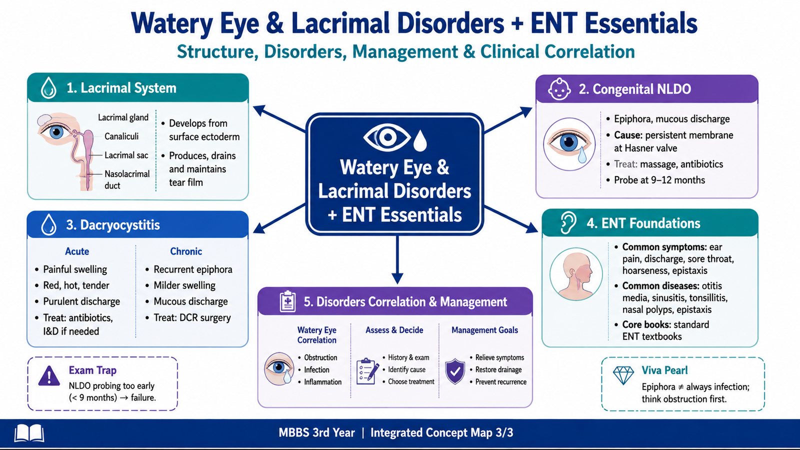
Integrated Mechanism Flow
⭐ AIM High-Yield Review
- Emmetropia means parallel distant rays are focused on the retina with accommodation relaxed.
- Myopia: focus lies in front of the retina; correction is with a concave or minus lens.
- Hypermetropia: focus would lie behind the retina; correction is with a convex or plus lens.
- Astigmatism results from unequal refractive power in different meridians and is corrected with cylindrical power.
- Presbyopia is age-related loss of effective accommodation and mainly causes difficulty with near vision.
- Aphakia means absence of the natural crystalline lens and produces marked loss of refractive power and accommodation.
- The normal tear pathway is puncta → canaliculi → lacrimal sac → nasolacrimal duct → nasal cavity.
- A watery eye may result from excessive tear production or impaired tear drainage.
- Congenital nasolacrimal duct obstruction commonly presents with watering and discharge beginning in infancy.
- Persistent congenital obstruction despite appropriate conservative management may require probing; an exact age threshold was not supplied in the curriculum excerpt.
- Acute dacryocystitis is characterized by painful, red and tender inflammation over the lacrimal sac region.
- Chronic dacryocystitis more commonly produces longstanding watering, discharge and possible reflux from the lacrimal sac.
- Common ENT symptoms include otalgia, hearing loss, otorrhea, tinnitus, vertigo, nasal obstruction, rhinorrhea, epistaxis, dysphagia and hoarseness.
- Stridor is an important sign of upper-airway narrowing and requires particular attention.
🎥 AIM Video Learning — Refractive Errors
Review myopia, hypermetropia, astigmatism, presbyopia and the basic principles of optical correction.
🎥 AIM Video Learning — Lacrimal System
Review the lacrimal apparatus, tear-drainage pathway and important lacrimal disorders including dacryocystitis.