
Study Tip
This chapter follows the supplied KMU learning outcomes in a logical sequence. First understand how and why cell injury develops, then use the high-yield review at the end to revise the most important examination concepts.
Cellular Injury: Causes, Mechanisms and Subcellular Responses
Foundation Module • Pathology
Understand why cells become injured, how reversible injury progresses to irreversible damage, and how cells respond to damaged or unwanted intracellular material.
Topic Introduction
Cell injury develops when a harmful stress exceeds the ability of a cell to maintain normal structure and function. Mild or short-lasting injury may produce reversible changes, allowing the cell to recover when the stress is removed. More severe or persistent injury can damage essential cellular systems beyond recovery and produce irreversible injury and cell death. In this chapter, you will first learn the basic language of pathology, then examine the major causes and biochemical mechanisms of cell injury. Special attention is given to hypoxic and ischemic injury, free radicals, ischemia-reperfusion injury, chemical injury, reversible versus irreversible cellular changes, and important subcellular responses such as lysosomal digestion and autophagy.
A. Foundations of Pathology and Cell Injury
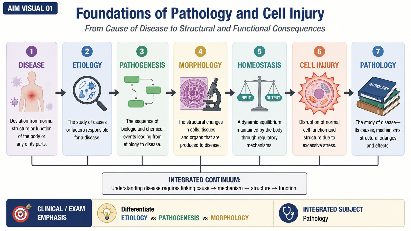
Pathology is the study of disease. It explains what causes disease, how the disease develops, and what structural and functional changes occur in affected cells, tissues and organs. Understanding a few basic terms makes the later mechanisms of cell injury much easier to follow.
Important Definitions
Disease is an abnormal condition in which normal structure or function is disturbed. Disease develops when cells, tissues or organs can no longer maintain their normal state in response to harmful influences.
Etiology means the cause of a disease. The cause may be genetic, acquired or a combination of several factors.
Pathogenesis describes the sequence of events through which the cause of a disease produces cellular, tissue and organ abnormalities.
Morphology refers to structural changes produced by disease. These changes may be visible with the naked eye as gross changes or may require microscopic examination.
Homeostasis is the ability of cells and tissues to maintain a relatively stable internal environment despite changes around them. Normal cells continuously adjust their metabolism and activity to remain within this stable range.
Cell injury occurs when a stress or harmful stimulus exceeds the cell’s adaptive capacity. The cell can no longer maintain normal homeostasis, so biochemical, functional and eventually structural abnormalities develop.
Classes of Pathology
Pathology can be considered broadly according to whether disease processes are studied at a general level or within particular organs and systems.
- General pathology: studies basic reactions of cells and tissues to abnormal stimuli, such as cell injury, inflammation and healing.
- Systemic pathology: studies diseases as they affect particular organs and organ systems.
- Anatomic pathology: studies structural changes in cells, tissues and organs, including examination of surgical specimens, biopsies and cytology.
- Clinical pathology: studies disease through laboratory investigation of blood and other body fluids.
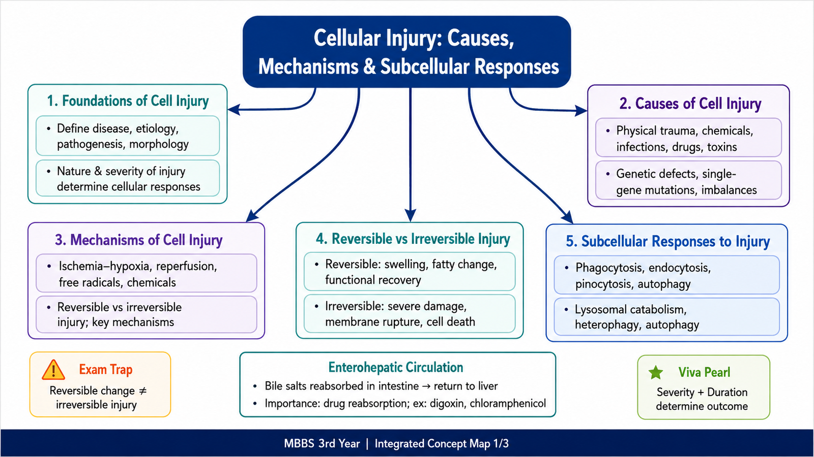
B. Causes of Cell Injury and the Cellular Response
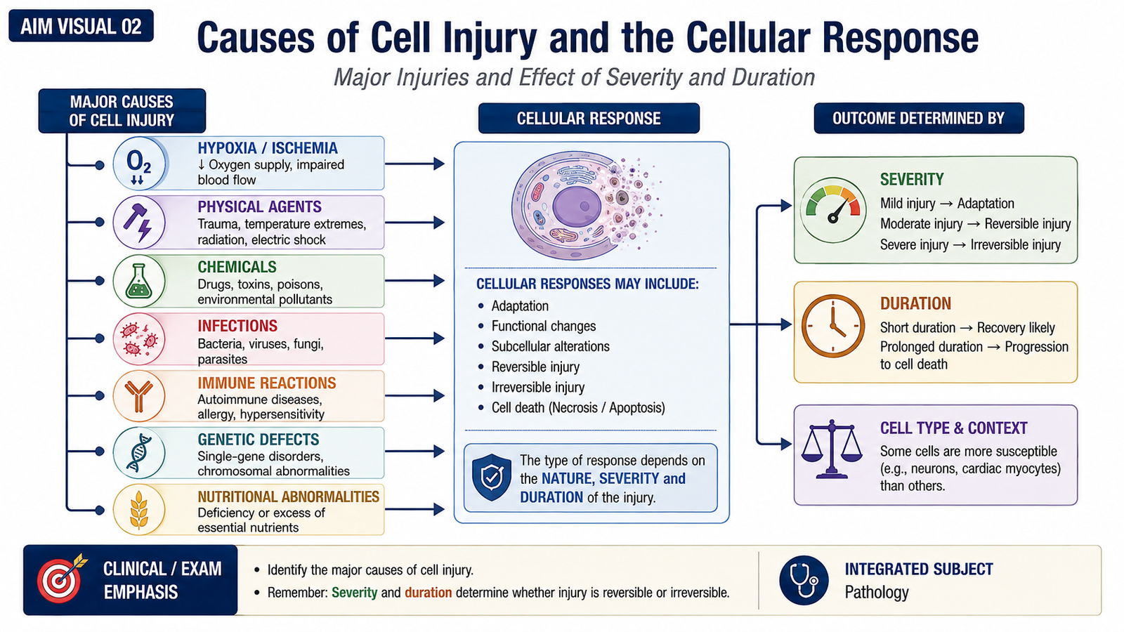
Cells may be injured by many different harmful influences. The final effect does not depend only on the type of injury. It also depends on its intensity, duration and the characteristics of the affected cell. A brief disturbance may therefore be completely reversible, while a severe or prolonged disturbance may cause irreversible injury.
Major Causes of Cell Injury
- Oxygen deprivation: hypoxia and ischemia reduce the oxygen available for cellular metabolism.
- Physical agents: mechanical trauma, extremes of temperature, radiation, electric injury and major changes in environmental pressure can damage cells.
- Chemical agents and drugs: harmful chemicals may directly damage cellular components or may become toxic after metabolic conversion.
- Infectious agents: microorganisms can injure cells directly or through host inflammatory and immune responses.
- Immunological reactions: inappropriate or excessive immune responses may damage normal cells and tissues.
- Genetic abnormalities: defects ranging from chromosomal abnormalities to single-gene defects may alter proteins, enzymes or cellular structures.
- Nutritional abnormalities: deficiencies and excesses of nutrients can disturb normal cellular metabolism.
Why the Same Injury Does Not Affect Every Cell Equally
The cellular response depends on several factors. The nature of the injury determines which cellular components are targeted. The severity and duration determine whether the cell can recover. The type and metabolic state of the cell also matter because different cells have different oxygen requirements, energy reserves and capacities for adaptation.
C. General Biochemical Mechanisms of Cell Injury
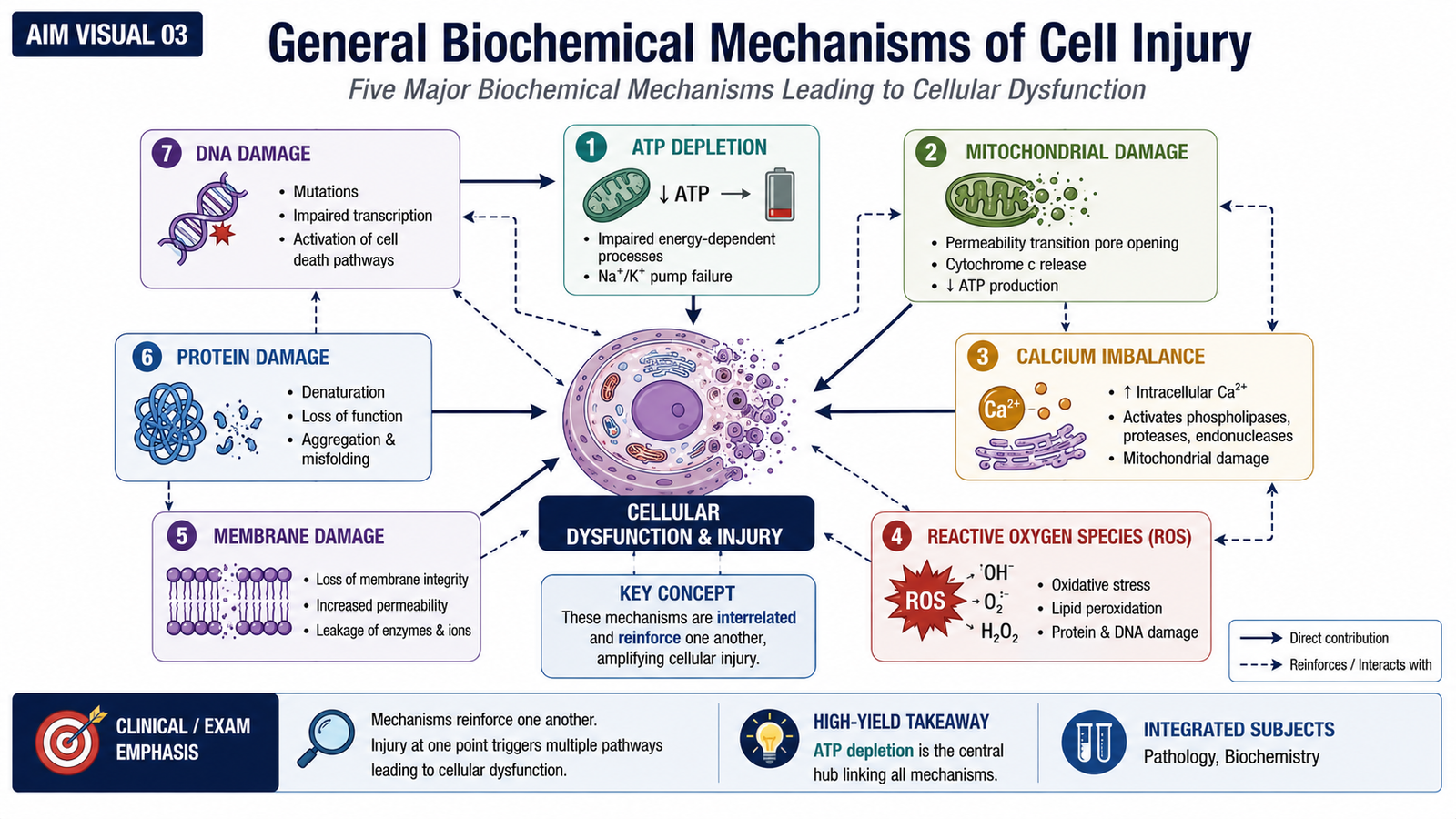
Although many different agents can injure cells, they often damage a limited number of critical cellular systems. These disturbances are interconnected. For example, mitochondrial injury reduces ATP production, while loss of calcium control and formation of reactive oxygen species further damage mitochondria and membranes. Therefore, several mechanisms commonly operate together rather than in isolation.
1. ATP Depletion
ATP provides energy for membrane pumps, protein synthesis and many metabolic processes. ATP depletion is especially important during hypoxia and ischemia because oxidative phosphorylation falls when oxygen delivery is inadequate.
As ATP falls, the sodium-potassium ATPase pump becomes less effective. Sodium accumulates inside the cell and water follows, producing cellular swelling. Calcium pumping also becomes impaired. The cell increasingly depends on anaerobic glycolysis, which consumes glycogen and produces lactic acid. The resulting reduction in intracellular pH affects cellular proteins and nuclear chromatin.
2. Mitochondrial Damage
Mitochondria are central to cellular energy production. Injury to mitochondria decreases ATP generation and can increase the formation of reactive oxygen species. Severe mitochondrial dysfunction is therefore an important step toward irreversible cell injury.
3. Loss of Calcium Homeostasis
Normally, cytosolic calcium concentration is kept much lower than calcium concentrations outside the cell and within intracellular stores. Cellular injury can increase cytosolic calcium. Excess calcium activates enzymes that damage important cellular structures, including membranes, proteins and nucleic acids.
4. Membrane Damage
Damage to cellular membranes interferes with normal compartmentalization. Plasma membrane damage allows leakage of cellular contents and disturbed ion movement. Mitochondrial membrane damage further reduces energy production, while lysosomal membrane damage can release hydrolytic enzymes into the cytoplasm.
5. Damage to Proteins and DNA
Cells depend on correctly folded proteins and intact genetic material. Significant damage to proteins and DNA activates cellular stress responses. If the damage is too severe to repair, the cell may progress toward death.
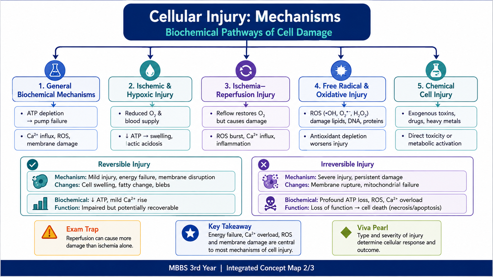
D. Hypoxic, Ischemic, Reperfusion, Free-Radical and Chemical Injury
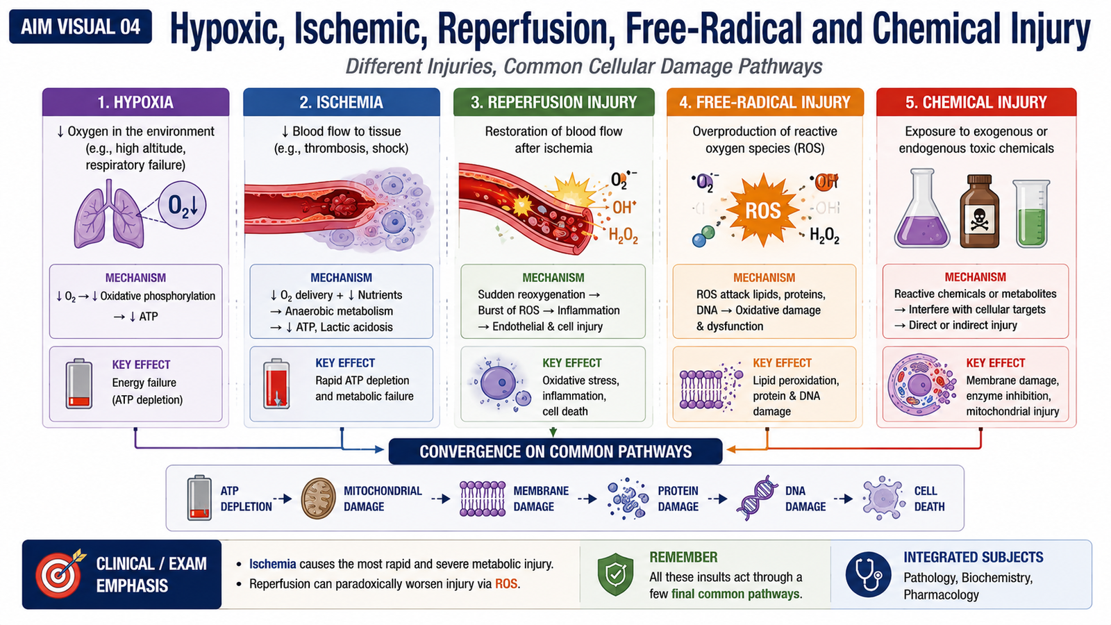
Several major forms of cell injury are best understood by applying the biochemical mechanisms already described. Hypoxia and ischemia mainly interfere with oxygen-dependent energy production, while free radicals and chemicals may directly damage membranes, proteins, DNA and mitochondria.
Hypoxic and Ischemic Cell Injury
Hypoxia means reduced oxygen availability to tissues. Ischemia means reduced blood flow. Ischemia is generally more damaging than isolated hypoxia because blood flow supplies not only oxygen but also metabolic substrates and removes waste products.
↓ oxygen delivery
↓ oxidative phosphorylation
↓ ATP production
failure of energy-dependent membrane pumps
sodium and water enter the cell + calcium balance is disturbed
cellular swelling and metabolic dysfunction
prolonged injury → membrane and mitochondrial failure → irreversible injury
Initially, reduced ATP causes reversible functional and structural abnormalities. If oxygen deprivation continues, mitochondrial dysfunction and membrane damage become severe enough that the cell cannot regain normal function even if oxygen delivery is restored.
Ischemia-Reperfusion Injury
Restoration of blood flow to ischemic tissue is essential for survival of cells that are still viable. However, in some cells, reperfusion can paradoxically produce additional damage. This is known as ischemia-reperfusion injury.
The renewed supply of oxygen can increase the formation of reactive oxygen species. Damaged cells may also have abnormal calcium handling, and inflammatory processes may contribute further injury. Thus, tissue damage after reperfusion may be greater than that produced by ischemia alone in some settings.
Free Radical-Induced Cell Injury
A free radical is a chemical species containing an unpaired electron, making it highly reactive. Reactive oxygen species are important mediators of cellular injury when their production exceeds the cell’s antioxidant defenses.
Reactive oxygen species can produce:
- Lipid peroxidation: damage to membrane lipids, reducing membrane stability.
- Protein modification: altered structure and function of cellular proteins and enzymes.
- DNA damage: changes that interfere with normal genetic function and cellular survival.
Cells normally possess protective antioxidant systems. Injury develops when production of reactive species becomes excessive or protective mechanisms are overwhelmed.
Chemical Cell Injury
Chemicals may injure cells in two main ways. Some act directly by binding to and damaging important cellular components. Others are initially less harmful but are converted by cellular metabolism into reactive toxic metabolites.
Once formed, toxic compounds may damage membranes, proteins, mitochondria or DNA. Chemical injury therefore commonly operates through mechanisms such as oxidative stress, membrane damage and disturbed cellular metabolism.
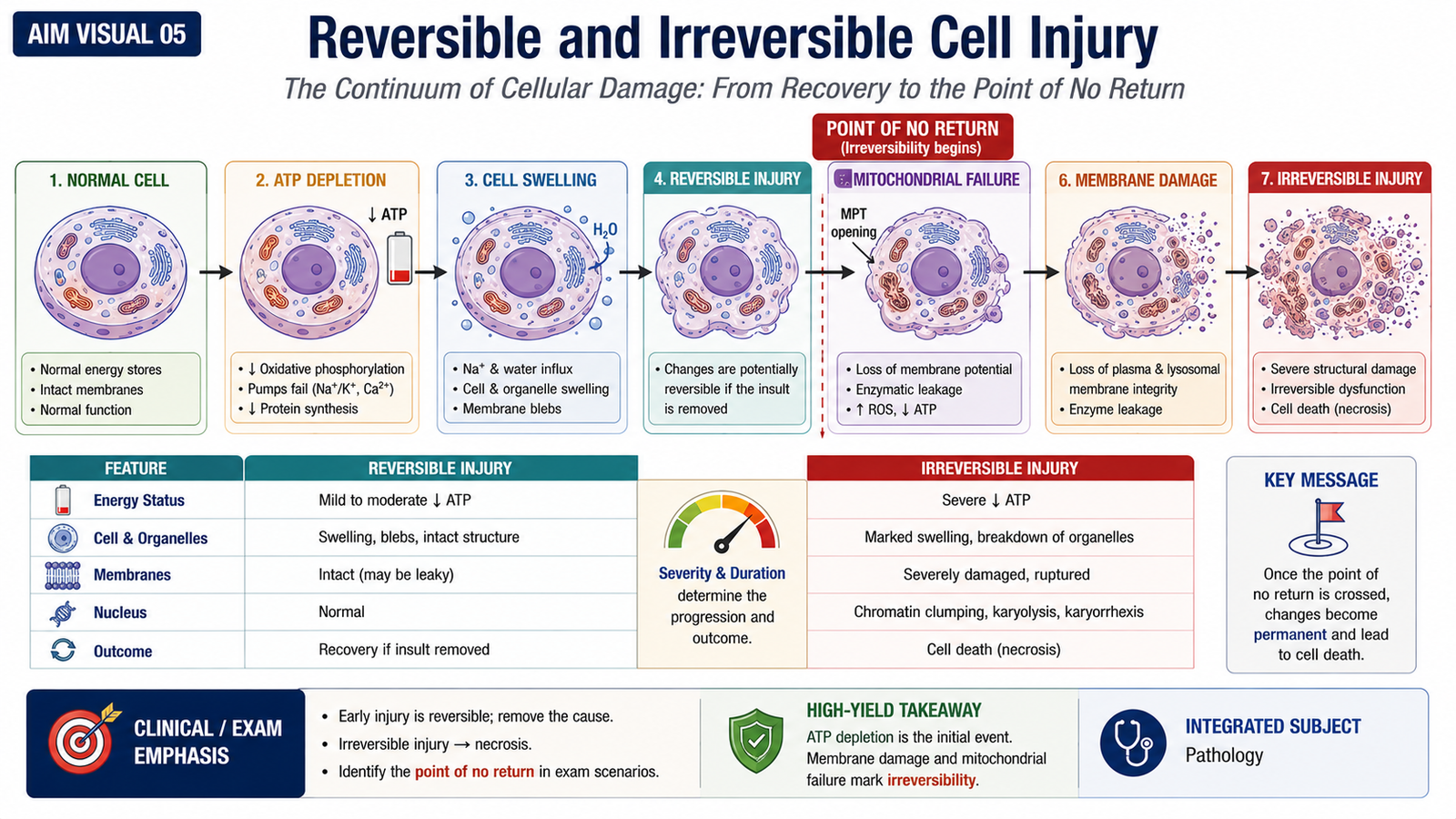
E. Reversible and Irreversible Cell Injury
The distinction between reversible and irreversible injury is fundamental in pathology. A reversibly injured cell is abnormal but can return to normal if the harmful stimulus is removed. An irreversibly injured cell has crossed a critical point beyond which normal structure and function cannot be restored.
Biochemical and functional changes include:
- reduced ATP production;
- increased anaerobic glycolysis;
- glycogen depletion;
- increased lactic acid;
- reduced intracellular pH;
- reduced protein synthesis;
- temporary impairment of specialized cellular function.
The key feature is that these abnormalities can improve if the injury is removed before critical structures are permanently damaged.
Irreversible Cell Injury
Irreversible injury develops when cellular damage becomes severe enough that recovery is no longer possible. Two particularly important features are the inability to restore normal mitochondrial function and severe damage to cellular membranes.
Major consequences include:
- profound failure of energy production;
- severe plasma membrane dysfunction;
- loss of cellular contents;
- major calcium disturbance;
- lysosomal membrane damage with release of degradative enzymes;
- progressive structural breakdown of the cell.
As membrane integrity is lost and cellular components are degraded, the cell progresses toward death.
| Feature | Reversible Injury | Irreversible Injury |
|---|---|---|
| Recovery | Possible after removal of stimulus | Not possible |
| ATP | Reduced | Severely impaired with persistent mitochondrial dysfunction |
| Cell swelling | Common | May progress to major structural breakdown |
| Membranes | Altered but potentially recoverable | Severely damaged |
| Cell contents | Largely retained | May leak from damaged membranes |
| Outcome | Return toward normal | Cell death |
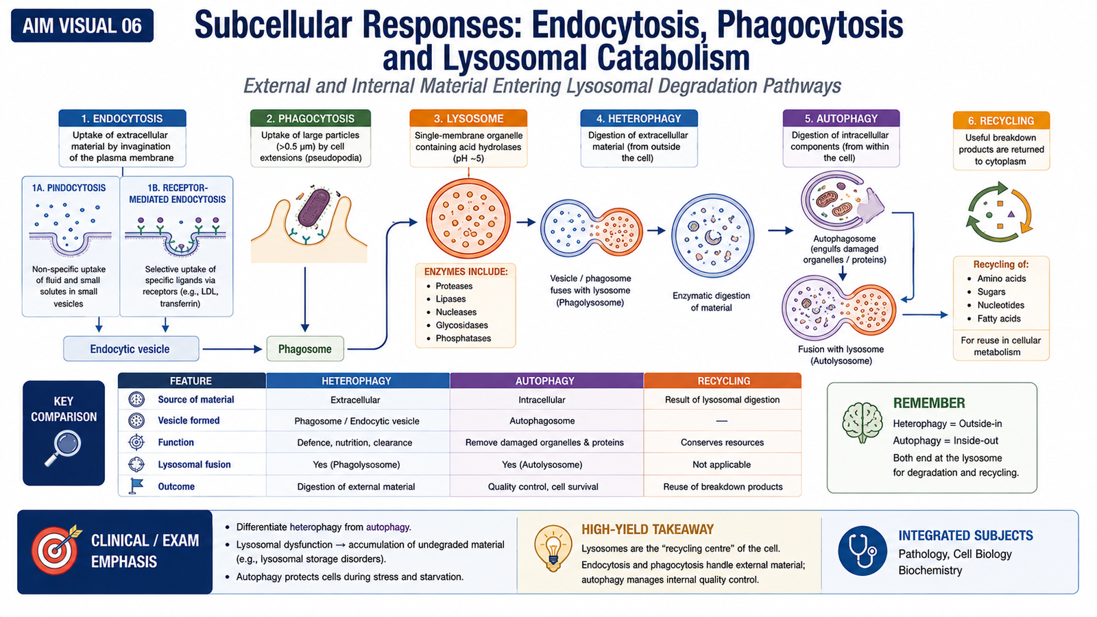
F. Subcellular Responses: Endocytosis, Phagocytosis and Lysosomal Catabolism
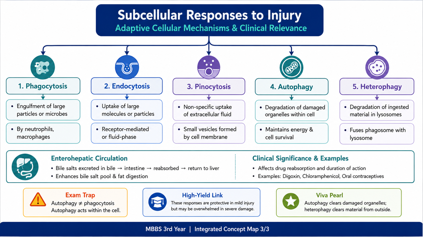
Cells must continuously handle material from outside the cell and remove damaged or unnecessary material from within the cell. Several related processes bring substances into intracellular compartments or deliver them to lysosomes for degradation. These mechanisms are especially important when cells respond to injury and cellular stress.
Endocytosis
Endocytosis is the general process by which a cell takes material from its surroundings into membrane-bound vesicles. The cell membrane surrounds the material and brings it into the cytoplasm.
Pinocytosis
Pinocytosis is a form of endocytosis in which small amounts of extracellular fluid and dissolved substances are taken into the cell in small vesicles. It may be understood as cellular uptake of fluid-phase material.
Phagocytosis
Phagocytosis is the uptake of relatively large particles, such as microorganisms or cellular debris. The particle is surrounded by the cell membrane and enclosed within a phagosome. The phagosome can then fuse with lysosomes, allowing lysosomal enzymes to digest the engulfed material.
Lysosomal Catabolism
Lysosomes contain hydrolytic enzymes capable of degrading proteins, lipids, carbohydrates and other cellular material. Lysosomal catabolism may process substances taken into the cell from outside or damaged material originating inside the cell.
This leads to two important concepts:
- Heterophagy: lysosomal digestion of material that originated outside the cell and entered through processes such as endocytosis or phagocytosis.
- Autophagy: lysosomal digestion of the cell’s own intracellular components, particularly damaged or unnecessary organelles and cytoplasmic material.
Autophagy as a Cellular Response to Stress
During cellular stress, autophagy allows the cell to remove damaged components and recycle their constituents. Portions of cytoplasm or organelles are enclosed within membrane-bound structures and ultimately delivered to lysosomes for degradation. The resulting breakdown products can then be reused by the cell.
Autophagy: damaged intracellular component → sequestration → lysosomal digestion → recycling
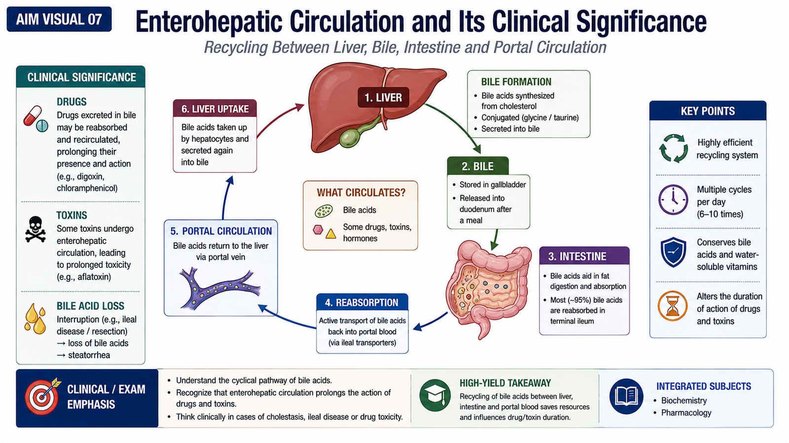
G. Enterohepatic Circulation and Its Clinical Significance
Enterohepatic circulation is the recycling of certain substances between the liver and intestine. A substance is secreted by the liver into bile, enters the intestine, and may then be reabsorbed from the intestine into the portal circulation. It returns to the liver, where it may again be processed or secreted.
The basic sequence is:
↓ secretion into bile
intestine
↓ intestinal reabsorption
portal blood
↓
return to liver
This recycling can prolong the time that a substance remains in the body because it is not immediately eliminated after biliary secretion.
Clinical Significance
Enterohepatic circulation is relevant to endogenous substances as well as some drugs. Bile acids are physiologically recycled in this way. Some drug compounds or their metabolites may also enter bile and later be reabsorbed, which can prolong their presence and biological effect in the body.
For example, substances conjugated in the liver may be secreted into bile. In the intestine, these conjugates may sometimes be modified, allowing the original substance to be reabsorbed and returned to the liver.
Integrated Mechanism Flow
↓
ATP depletion, mitochondrial dysfunction, calcium disturbance and/or reactive oxygen species
↓
metabolic dysfunction and cellular swelling
↓
continuing injury causes increasing membrane and organelle damage
↓
reversible injury crosses into irreversible injury
↓
severe mitochondrial and membrane dysfunction prevents recovery
↓
cell death
Important Comparison: Hypoxia and Ischemia
| Feature | Hypoxia | Ischemia |
|---|---|---|
| Main disturbance | Reduced oxygen availability | Reduced blood flow |
| Oxygen delivery | Reduced | Reduced |
| Nutrient delivery | May continue | Reduced because blood flow is reduced |
| Waste removal | May continue | Impaired |
| Typical cellular severity | Depends on degree and duration | Often more rapidly damaging |
⭐ AIM High-Yield Review
- Etiology is the cause of disease; pathogenesis is the sequence through which that cause produces disease.
- Morphology refers to structural abnormalities produced by disease.
- Cell injury occurs when harmful stress exceeds the cell’s ability to maintain homeostasis.
- Important causes include oxygen deprivation, physical and chemical agents, infections, immune reactions, genetic abnormalities and nutritional disturbances.
- ⭐ The response to injury depends on its nature, severity and duration and on the type of cell affected.
- ATP depletion produces ion-pump failure, sodium and water accumulation and cellular swelling.
- Major mechanisms of cell injury include ATP depletion, mitochondrial damage, calcium disturbance, oxidative stress and membrane injury.
- ⭐ Ischemia is usually more damaging than isolated hypoxia because blood flow, nutrient delivery and waste removal are all impaired.
- Reperfusion may sometimes increase tissue injury through reactive oxygen species, calcium disturbances and inflammatory mechanisms.
- Reactive oxygen species damage cells through lipid peroxidation, protein modification and DNA damage.
- ⭐ Reversible injury can recover after removal of the stimulus; irreversible injury cannot.
- Severe mitochondrial dysfunction and major membrane damage are important features of irreversible injury.
- Heterophagy digests material originating outside the cell; autophagy digests the cell’s own damaged or unnecessary components.
- Phagocytosis involves uptake of relatively large particles, whereas pinocytosis involves uptake of extracellular fluid and dissolved substances.
- ⭐ Enterohepatic circulation recycles substances between the liver and intestine and may prolong their presence in the body.
Cell Injury — Reversible vs Irreversible Cell Injury
Watch this video after completing the learning material to reinforce the major mechanisms and morphological changes of reversible and irreversible cell injury.
causes of cell injury, ATP depletion, cellular swelling, membrane damage, and the transition from reversible to irreversible injury.