
This chapter follows the supplied KMU learning outcomes for cellular adaptation, necrosis, apoptosis and cellular ageing. First understand why cells adapt or die, then use the final high-yield review to revise the most important distinctions.
Cellular Adaptation, Necrosis, Apoptosis and Cellular Ageing
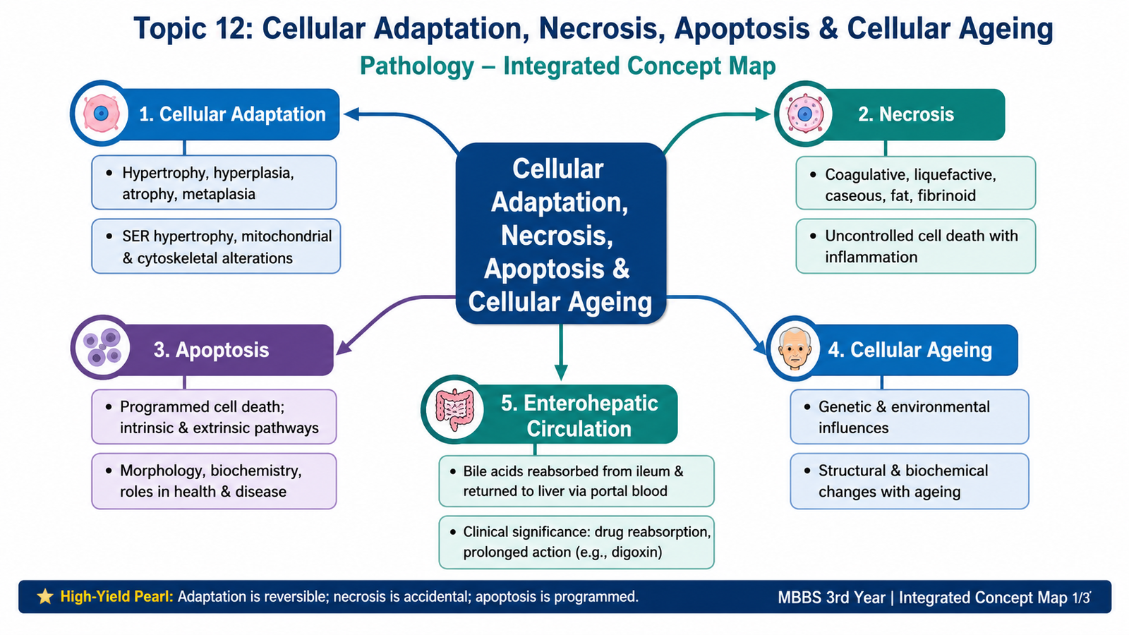
Cells constantly respond to changes in their environment. Some responses allow survival through adaptation, while severe or persistent injury may produce cell death by necrosis or apoptosis. With advancing age, accumulated molecular and cellular damage gradually reduces cellular function and regenerative capacity.
Topic Introduction
Cells normally maintain a stable internal state called homeostasis. When the environment changes, a cell may alter its size, number, phenotype or metabolic activity so that it can continue functioning. These reversible changes are called cellular adaptations. If stress becomes excessive or damaging, the cell may die. Necrosis usually results from severe injury and is associated with membrane damage and inflammation, whereas apoptosis is a regulated process in which individual cells are removed with minimal inflammation. Cells also undergo progressive changes with ageing because of accumulated molecular damage and declining repair mechanisms. Understanding these processes explains many common pathological changes seen in tissues.
A. Cellular Adaptation: General Principles and Types
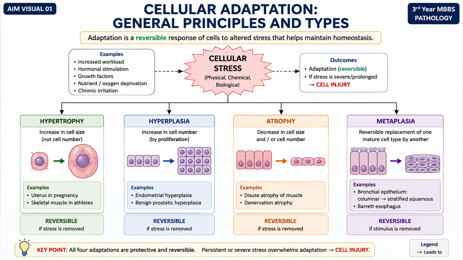
Cellular adaptation is a reversible change in cell size, cell number, phenotype, metabolic activity or function that occurs when a cell faces altered physiological demands or pathological stress. The purpose of adaptation is survival. Instead of immediately becoming injured, the cell establishes a new steady state that is better suited to the changed environment.
Whether adaptation is successful depends on the nature of the stimulus, its severity, its duration and the ability of the affected cell to respond. If the adaptive capacity is exceeded, cell injury develops and may eventually become irreversible.
Major Types of Cellular Adaptation
- Hypertrophy: increase in the size of individual cells.
- Hyperplasia: increase in the number of cells.
- Atrophy: decrease in cell size and often in the size of the affected organ.
- Metaplasia: reversible replacement of one mature differentiated cell type by another mature cell type better able to tolerate the stress.
Physiological versus Pathological Adaptation
Physiological adaptations occur in response to normal stimuli, such as hormones or increased functional demand. For example, skeletal muscles enlarge with exercise because individual muscle fibers undergo hypertrophy.
Pathological adaptations occur because of disease-related stress. They may initially be protective but can eventually impair function or predispose to further pathological change. For example, persistent pressure overload can produce cardiac hypertrophy.
B. Hypertrophy and Hyperplasia
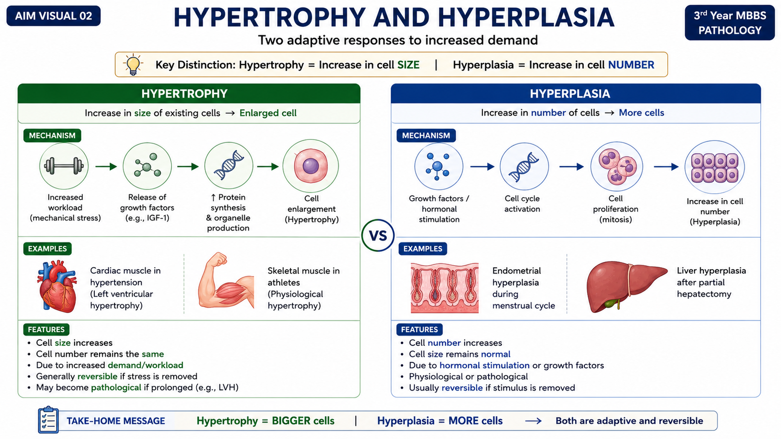
Hypertrophy and hyperplasia both cause enlargement of a tissue or organ, but they do so by different mechanisms. In hypertrophy, cells become larger. In hyperplasia, the number of cells increases. Both processes may occur together when a tissue contains cells capable of division.
Hypertrophy
Hypertrophy is an increase in cell size caused by increased synthesis of structural proteins and cellular components. It is especially important in tissues composed of cells with limited ability to divide, such as cardiac muscle.
Hypertrophy develops when cells are exposed to increased mechanical workload or trophic stimulation by hormones and growth factors. These stimuli activate intracellular signaling pathways that alter gene expression and increase protein synthesis. As more structural proteins and organelles are produced, the cell becomes larger.
Increased workload or hormonal stimulation → activation of cellular signaling → altered gene expression → increased synthesis of proteins and organelles → increased cell size.
Physiological examples include enlargement of skeletal muscle fibers during repeated exercise and enlargement of uterine smooth muscle during pregnancy.
Pathological hypertrophy can occur when an organ works against increased resistance. For example, prolonged pressure overload increases the workload of cardiac muscle cells, producing myocardial hypertrophy. Initially this helps the heart generate greater force, but excessive hypertrophy may eventually impair function.
Hyperplasia
Hyperplasia is an increase in the number of cells within a tissue or organ. It occurs only in populations that are capable of cell division. The process is driven mainly by growth factors and hormonal stimulation that increase proliferation of mature cells or, in some tissues, stem cells.
Physiological hyperplasia may be:
- Hormonal: proliferation of glandular tissue in the female breast during puberty and pregnancy.
- Compensatory: proliferation of remaining cells after loss or removal of part of a tissue.
Pathological hyperplasia results from excessive hormonal or growth-factor stimulation. Although the process remains controlled and is different from neoplasia, persistent excessive proliferation may create conditions in which further genetic abnormalities can develop.
| Feature | Hypertrophy | Hyperplasia |
|---|---|---|
| Main change | Increased cell size | Increased cell number |
| Requirement for cell division | No | Yes |
| Major stimulus | Workload, hormones, growth factors | Hormones and growth factors |
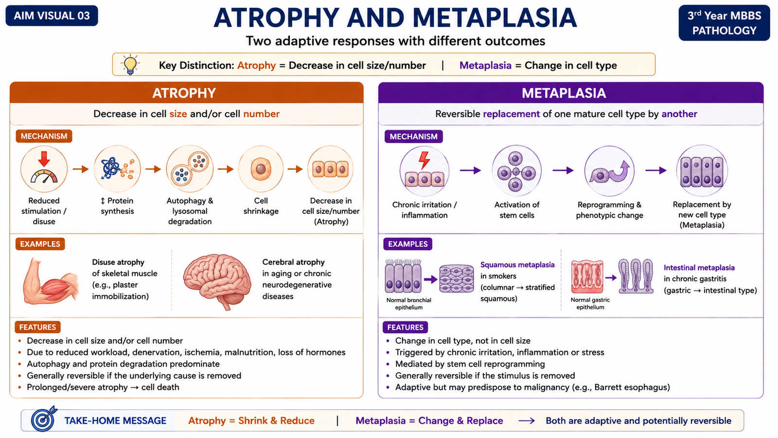
C. Atrophy and Metaplasia
Atrophy
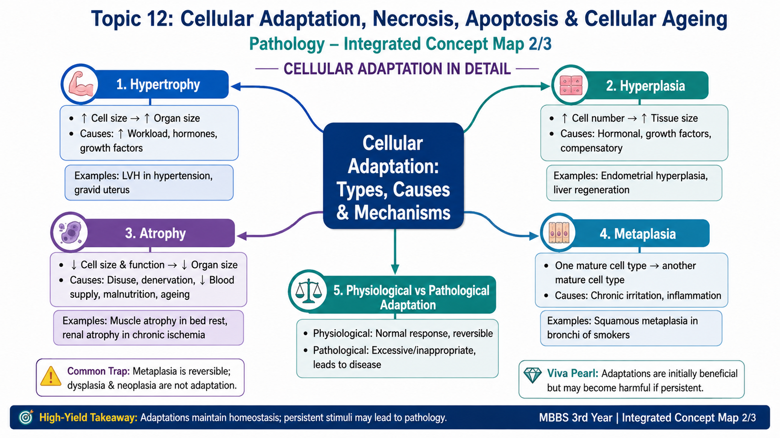
Atrophy is a reduction in the size of cells caused by loss of cellular substance. When enough cells are affected, the entire tissue or organ becomes smaller. Atrophy does not mean that the cell is dead. The cell remains alive but reduces its structural components and metabolic activity to match a lower level of demand or nutrition.
Important causes include:
- Decreased workload, also called disuse.
- Loss of nerve supply.
- Reduced blood supply.
- Inadequate nutrition.
- Loss of endocrine stimulation.
- Pressure on a tissue.
- Ageing.
Atrophy develops through two major cellular processes. First, protein synthesis decreases. Second, degradation of cellular proteins increases, particularly through the ubiquitin-proteasome pathway. Cells may also digest some of their own components through autophagy, allowing them to survive with fewer resources.
Metaplasia
Metaplasia is a reversible change in which one mature cell type is replaced by another mature cell type that is better able to tolerate persistent stress. The original mature cells do not directly transform into another mature type. Instead, tissue stem cells or undifferentiated precursor cells are reprogrammed by cytokines, growth factors and signals from the altered environment.
A classic example is squamous metaplasia of respiratory epithelium in response to chronic irritation. The normal columnar epithelial cells are replaced by stratified squamous cells that are more resistant to injury.
This adaptation provides better mechanical protection, but it has a functional cost. The normal respiratory epithelium contains specialized cells that contribute to mucus production and mucociliary clearance. Replacing it with squamous epithelium reduces these specialized functions.
Persistent stimuli may also make metaplastic tissue more susceptible to progression toward dysplasia and malignant transformation. Metaplasia itself, however, is an adaptive and potentially reversible process.
D. Subcellular Adaptations and Cytoskeletal Abnormalities
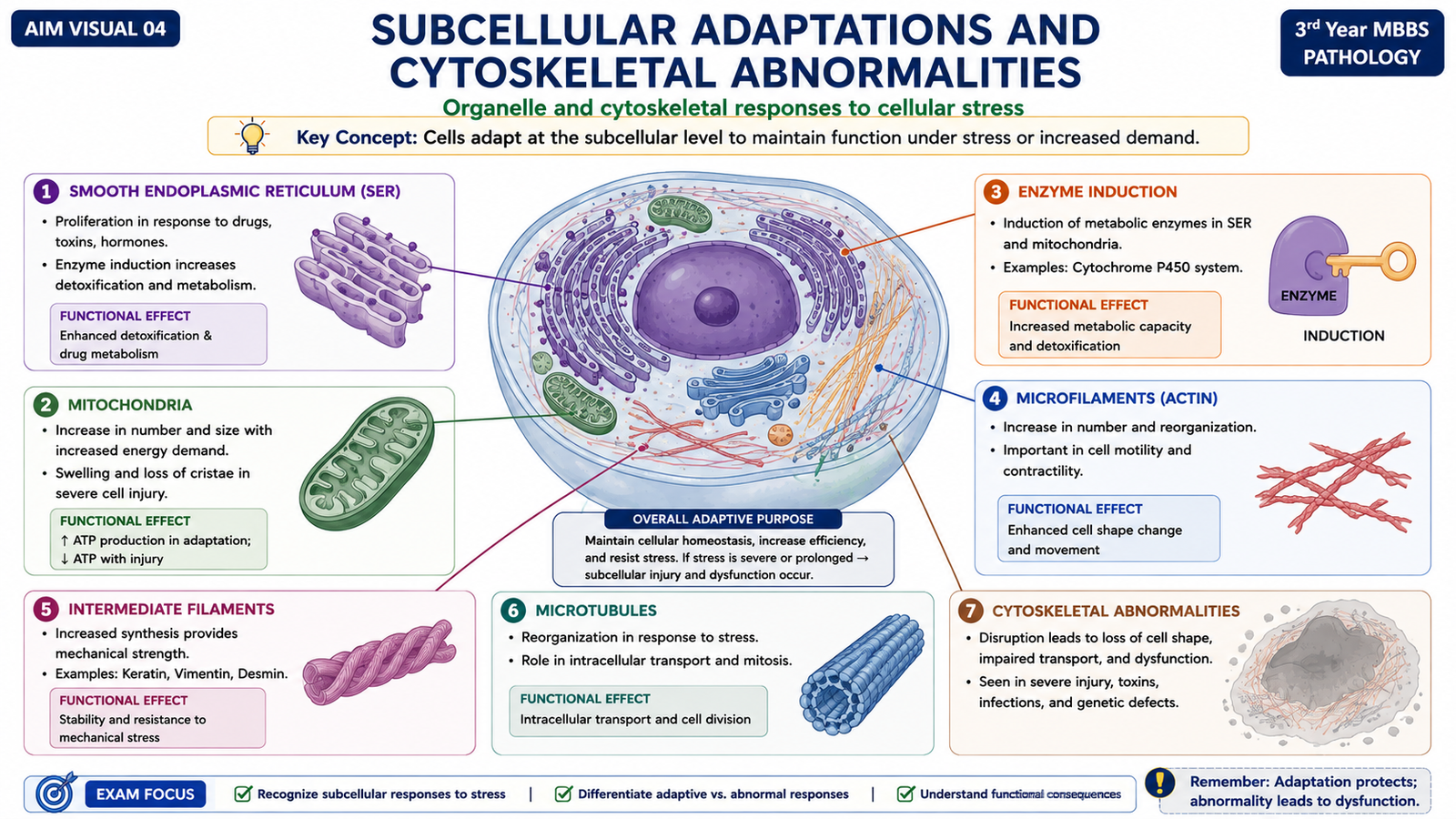
Adaptation does not always involve the whole cell becoming larger, smaller or changing phenotype. Cells may also alter individual organelles and structural proteins in response to changing functional demands. These subcellular adaptations can be useful, but excessive or abnormal changes may contribute to disease.
Smooth Endoplasmic Reticulum Hypertrophy
The smooth endoplasmic reticulum contains enzymes involved in the metabolism of several substances, including drugs. Repeated exposure to compounds that require metabolism can stimulate cells to increase the amount of smooth endoplasmic reticulum and its associated enzymes.
This explains why repeated exposure to some drugs may increase the ability of hepatocytes to metabolize those substances. The adaptation may also influence metabolism of other compounds handled by the same enzymatic systems.
Mitochondrial Alterations
Mitochondria respond to changes in cellular energy requirements. Cells with increased energy demands may increase mitochondrial number or size. In contrast, mitochondrial injury can produce abnormal swelling, altered internal structure and impaired ATP production. Severe mitochondrial dysfunction is especially important because failure of energy production can contribute to irreversible cell injury and death.
Cytoskeletal Abnormalities
The cytoskeleton maintains cell shape, intracellular organization, movement and transport. It consists mainly of microfilaments, intermediate filaments and microtubules. Abnormalities involving these structures can alter cell function and may produce characteristic pathological changes.
Examples include:
- Intermediate filament accumulation: abnormal collections of cytoskeletal proteins may form within injured cells.
- Microtubule dysfunction: may interfere with intracellular transport and movement of cellular structures.
- Defective structural proteins: can reduce mechanical stability and alter tissue function.
The important principle is that cytoskeletal abnormalities may result from either inherited defects in structural proteins or acquired cell injury, and the functional effect depends on which cellular system is disturbed.
E. Necrosis: Mechanism, Morphology and Major Patterns
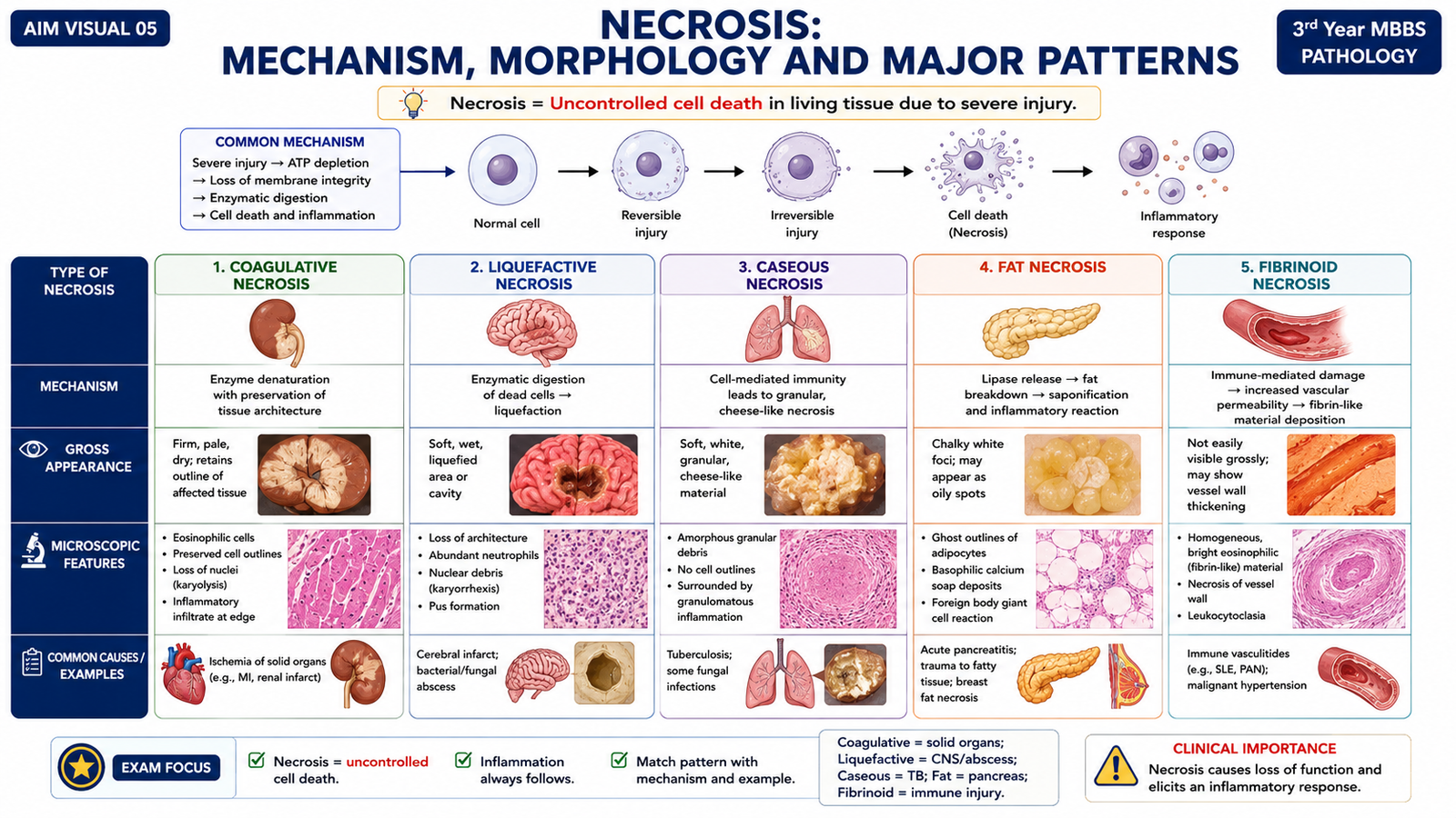
Necrosis is cell death that occurs after severe irreversible injury in living tissue. The cell loses membrane integrity, intracellular contents leak into the surrounding tissue and an inflammatory response commonly develops. Necrosis therefore affects not only the dead cell but also the surrounding tissue environment.
Mechanism of Necrosis
Severe injury causes failure of essential cellular systems. ATP depletion, mitochondrial dysfunction, loss of calcium homeostasis, membrane damage and degradation of cellular proteins and nucleic acids progressively destroy the cell. Once the plasma membrane is severely damaged, cellular enzymes and other intracellular contents escape into the extracellular space and provoke inflammation.
Morphological Changes
Necrotic cells usually become more eosinophilic on routine histological staining because of protein denaturation and loss of normal cytoplasmic RNA. Their nuclei undergo characteristic progressive changes:
- Pyknosis: the nucleus becomes small, dark and shrunken.
- Karyorrhexis: the pyknotic nucleus fragments.
- Karyolysis: nuclear material fades because DNA is degraded.
Major Patterns of Necrosis
Different tissues and mechanisms of injury produce recognizable morphological patterns of necrosis.
- Coagulative necrosis: tissue architecture is temporarily preserved because structural proteins and enzymes are denatured. It commonly follows ischemic injury in solid organs other than the brain.
- Liquefactive necrosis: dead tissue is digested into a liquid or viscous mass. It occurs characteristically in bacterial infections and in ischemic injury of the brain.
- Caseous necrosis: produces soft, friable, cheese-like necrotic material. It is classically associated with tuberculosis and is often surrounded by granulomatous inflammation.
- Fat necrosis: refers to focal destruction of adipose tissue. Released fatty acids may combine with calcium to produce chalky deposits through saponification.
- Fibrinoid necrosis: occurs mainly in vessel walls, where immune-mediated vascular injury produces deposition of intensely eosinophilic material resembling fibrin.
- Gangrenous necrosis: is a clinical term rather than a distinct microscopic pattern. Ischemic coagulative necrosis of a limb is commonly called dry gangrene; superimposed infection and liquefaction produce wet gangrene.
| Type | Key Feature | Typical Example |
|---|---|---|
| Coagulative | Architecture initially preserved | Ischemic injury of solid organs |
| Liquefactive | Enzymatic digestion produces liquid material | Brain infarction or bacterial infection |
| Caseous | Friable cheese-like material | Tuberculosis |
| Fat | Fat destruction with possible saponification | Enzymatic injury to fat |
| Fibrinoid | Bright eosinophilic vessel-wall material | Immune-mediated vascular injury |
F. Apoptosis: Programmed Cell Death in Health and Disease
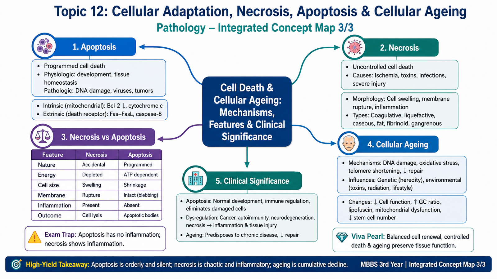
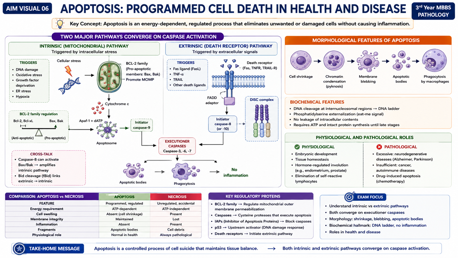
Apoptosis is a regulated form of cell death in which a cell activates internal mechanisms that lead to its controlled removal. Unlike necrosis, the plasma membrane generally remains intact long enough to prevent leakage of intracellular material, so apoptosis usually produces little or no surrounding inflammation.
Physiological Causes
Apoptosis is essential for normal development and tissue homeostasis. It removes cells that are no longer required or that could be harmful.
- Removal of cells during embryonic development.
- Reduction of hormone-dependent tissues when hormonal stimulation falls.
- Deletion of excess cells in continuously proliferating tissues.
- Elimination of potentially harmful self-reactive lymphocytes.
- Removal of immune cells after an immune response has finished.
Pathological Causes
Apoptosis also removes cells that are damaged beyond repair while limiting injury to surrounding tissue. Important examples include cells with severe DNA damage, cells containing misfolded proteins and certain infected cells targeted by cytotoxic lymphocytes.
Morphological Features
An apoptotic cell typically shrinks. Chromatin becomes condensed, the nucleus fragments and the cell forms membrane-bound fragments called apoptotic bodies. These bodies are rapidly phagocytosed by neighboring cells or macrophages.
Because the membrane-bound fragments are removed before intracellular contents spill into the tissue, significant inflammation is usually absent.
Biochemical Basis: Caspases
The central enzymes of apoptosis are caspases. They are proteolytic enzymes produced as inactive precursors and activated in a controlled sequence. Initiator caspases activate executioner caspases, which then degrade cytoskeletal and nuclear proteins and activate enzymes that break down DNA.
Intrinsic or Mitochondrial Pathway
The intrinsic pathway is triggered by internal cellular stress such as severe DNA damage, growth-factor withdrawal or accumulation of misfolded proteins. Its key control point is the mitochondrial membrane.
Members of the BCL-2 family regulate mitochondrial permeability. Some family members promote cell survival, whereas others promote apoptosis. When pro-apoptotic signals dominate, mitochondrial permeability increases and cytochrome c is released into the cytoplasm. Cytochrome c participates in activation of initiator caspases, which then activate executioner caspases.
Extrinsic or Death-Receptor Pathway
The extrinsic pathway begins when specific extracellular signals bind to death receptors on the cell surface. These receptors belong to the tumor necrosis factor receptor family and contain intracellular death domains. Receptor activation recruits adaptor proteins and activates initiator caspases, which then activate executioner caspases.
Role in Health and Disease
Normal apoptosis maintains tissue balance by matching cell production with cell removal. Too little apoptosis can permit survival of abnormal cells, whereas excessive apoptosis may contribute to tissue loss. Therefore, precise regulation of apoptosis is important for both normal physiology and disease prevention.
G. Cellular Ageing: Mechanisms and Structural Changes
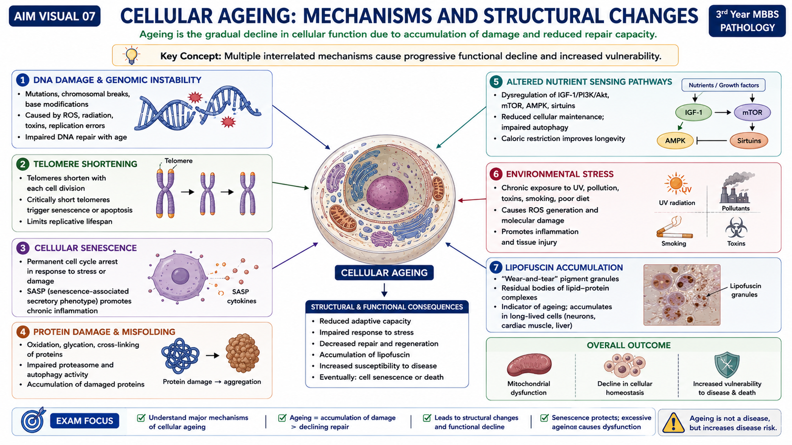
Cellular ageing is the progressive decline in cellular function and the ability to respond to stress. It develops because molecular damage accumulates over time while repair and regenerative mechanisms become less effective. Ageing therefore reflects the combined effects of genetic programming, environmental exposure and repeated cellular injury.
Accumulation of DNA Damage
DNA is continuously exposed to damaging influences generated during normal metabolism and from environmental sources. Cells possess repair mechanisms, but damage that is not completely repaired accumulates over time. Persistent DNA injury can alter gene function, reduce cellular performance and promote cellular senescence or apoptosis.
Replicative Senescence and Telomeres
At the ends of chromosomes are specialized DNA sequences called telomeres. Telomeres shorten progressively during repeated cell division in many somatic cells. When they become critically short, the cell recognizes the chromosome ends as damaged DNA and may stop dividing. This permanent growth arrest is an important contributor to replicative senescence.
Defective Protein Homeostasis
Normal cells continuously fold, repair and remove proteins. With ageing, mechanisms responsible for maintaining normal proteins become less efficient. Damaged or abnormally folded proteins may therefore accumulate and interfere with normal cellular function.
Altered Nutrient-Sensing and Cellular Maintenance
Cells contain signaling pathways that adjust metabolism, growth and repair according to nutrient availability. Alterations in these pathways influence the balance between cellular growth and maintenance. Mechanisms that support repair, stress resistance and removal of damaged components contribute to cellular longevity.
Genetic and Environmental Influences
The rate of ageing is influenced by inherited differences in genes controlling DNA repair, metabolism, stress responses and protein maintenance. Environmental factors add further cellular stress. Repeated exposure to damaging agents can accelerate accumulation of molecular injury.
Structural and Biochemical Changes
Ageing cells may become less efficient at protein synthesis, energy production, degradation of damaged components and response to growth signals. Some long-lived cells accumulate intracellular material such as lipofuscin, a yellow-brown pigment containing products of lipid and protein damage. Its accumulation is a morphological marker of previous free-radical injury and lipid peroxidation rather than a direct cause of cell death.
Overall, these changes reduce the functional reserve of tissues. Ageing cells are therefore less able to adapt to physiological stress and may recover more slowly after injury.
Integrated Mechanism Flow
↓
Successful reversible response → hypertrophy, hyperplasia, atrophy or metaplasia
↓
If damaging stress develops → cellular injury
↓
Severe uncontrolled injury → membrane damage → necrosis → inflammation
OR
Regulated death signal → caspase activation → apoptosis → controlled cell removal
↓
Repeated molecular damage and declining repair over time → cellular ageing and reduced functional reserve
Important Comparison — Necrosis versus Apoptosis
| Feature | Necrosis | Apoptosis |
|---|---|---|
| Usual setting | Severe pathological injury | Physiological or pathological |
| Cell size | Usually swelling | Shrinkage |
| Plasma membrane | Disrupted | Remains intact around apoptotic fragments |
| Cell contents | Leak out | Remain contained in apoptotic bodies |
| Nuclear change | Pyknosis, karyorrhexis, karyolysis | Chromatin condensation and fragmentation |
| Mechanism | Uncontrolled consequences of severe injury | Regulated, caspase-dependent process |
| Inflammation | Common | Usually minimal or absent |
⭐ AIM High-Yield Review
- Hypertrophy means increased cell size; hyperplasia means increased cell number.
- Hypertrophy is particularly important in cells with limited ability to divide, such as cardiac muscle cells.
- Atrophy involves decreased cell size through reduced protein synthesis and increased degradation of cellular components.
- Metaplasia is reversible replacement of one mature cell phenotype by another through reprogramming of stem or precursor cells.
- Persistent metaplastic stress can predispose tissue to further abnormal growth, but metaplasia itself is not malignant transformation.
- Smooth-ER hypertrophy may occur when repeated chemical exposure increases the demand for metabolic enzymes.
- Necrosis involves loss of membrane integrity, leakage of intracellular contents and inflammation.
- The major nuclear changes in necrosis are pyknosis → karyorrhexis → karyolysis.
- Coagulative necrosis is typical of ischemia in most solid organs; brain ischemia characteristically produces liquefactive necrosis.
- Caseous necrosis is classically associated with tuberculosis.
- Apoptosis produces cell shrinkage, chromatin condensation and apoptotic bodies with little inflammation.
- Both intrinsic and extrinsic apoptotic pathways ultimately activate caspases.
- The intrinsic pathway is regulated at the mitochondria and involves the BCL-2 family and cytochrome c.
- Cellular ageing is associated with accumulated DNA damage, telomere shortening, defective protein homeostasis and altered cellular maintenance.
- Lipofuscin accumulation is a morphological marker of previous oxidative and lipid damage in long-lived cells.