
This chapter follows the KMU learning outcomes for cataract in a simple sequence. First understand how the lens becomes opaque and how the major cataract types differ; then revise the complications and basic management principles from the high-yield section.
Topic 15 — Cataract: Pathogenesis, Types, Complications and Management
A clear guide to what cataract is, how lens opacity develops, the important types, complications of advanced cataract and the principles of treatment.
Topic Introduction
A cataract is an opacity of the crystalline lens of the eye that reduces its normal transparency. The lens must remain transparent so that light can pass through it and form a clear image on the retina. When lens proteins, lens fibers or lens hydration change abnormally, transparency is lost and vision becomes progressively impaired. Cataracts may develop with ageing or may occur because of congenital abnormalities, trauma, systemic disease, ocular disease or certain drugs. In this chapter, you will understand the definition and major types of cataract, the basic mechanisms by which lens opacity develops, the important complications of advanced cataract and the main principles of cataract management.
A. Definition and Core Concept of Cataract
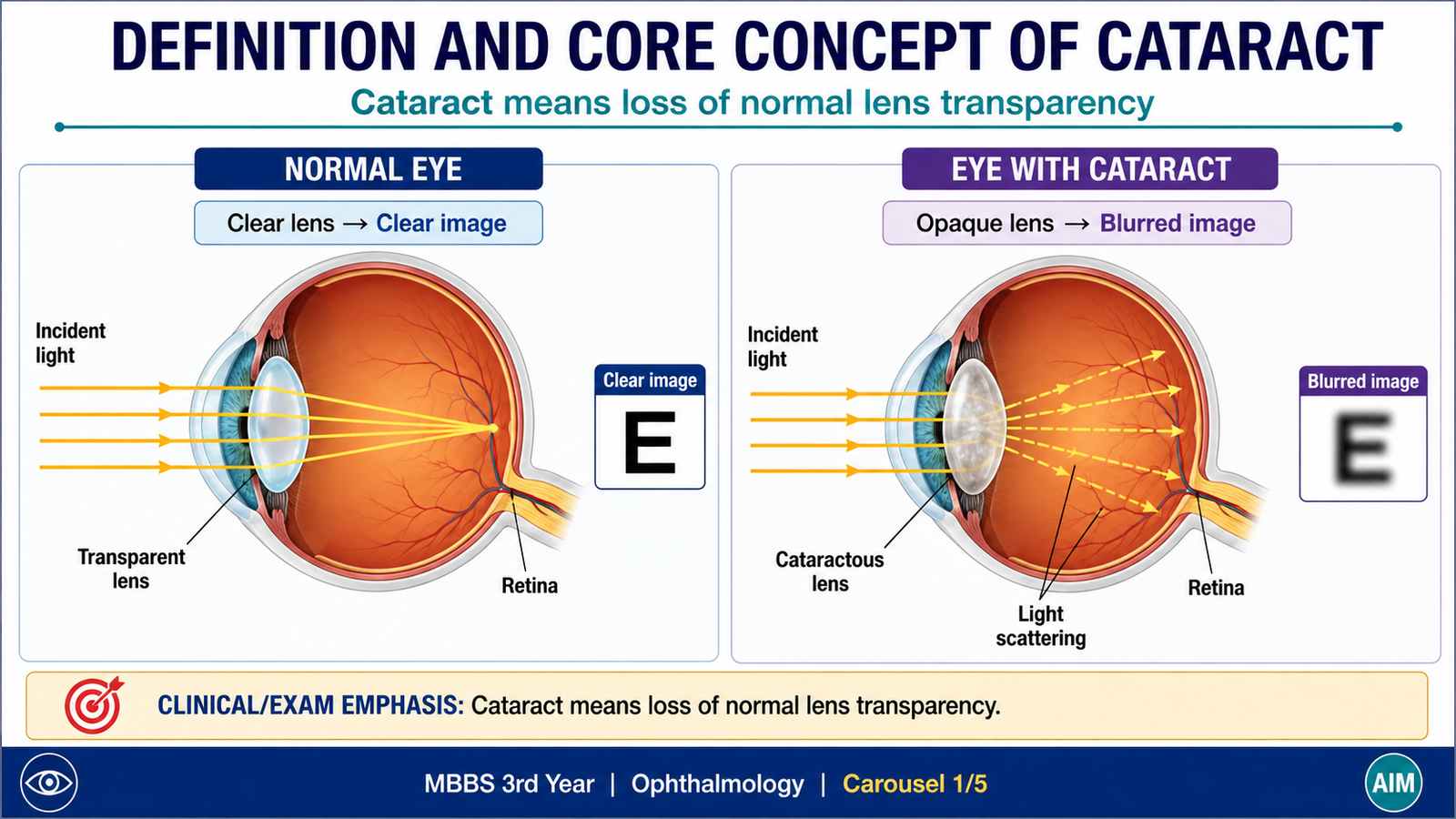
A cataract is any opacity of the normally transparent crystalline lens or its capsule that interferes with the passage of light. The lens is transparent because its fibers are arranged in an orderly manner, contain highly organized soluble proteins called crystallins, and normally have no blood vessels. This organization allows light to pass through the lens with minimal scattering. When the arrangement or chemical state of lens proteins is disturbed, light is scattered instead of being transmitted clearly. The degree of visual disturbance therefore depends on the site, density and extent of the opacity. A small peripheral opacity may cause little visual difficulty, whereas an opacity close to the visual axis may significantly affect vision.
The word cataract describes the lens opacity itself. It does not specify its cause. Cataracts therefore form a group of conditions with different causes and patterns but a common final result: loss of lens transparency.
B. Types of Cataract
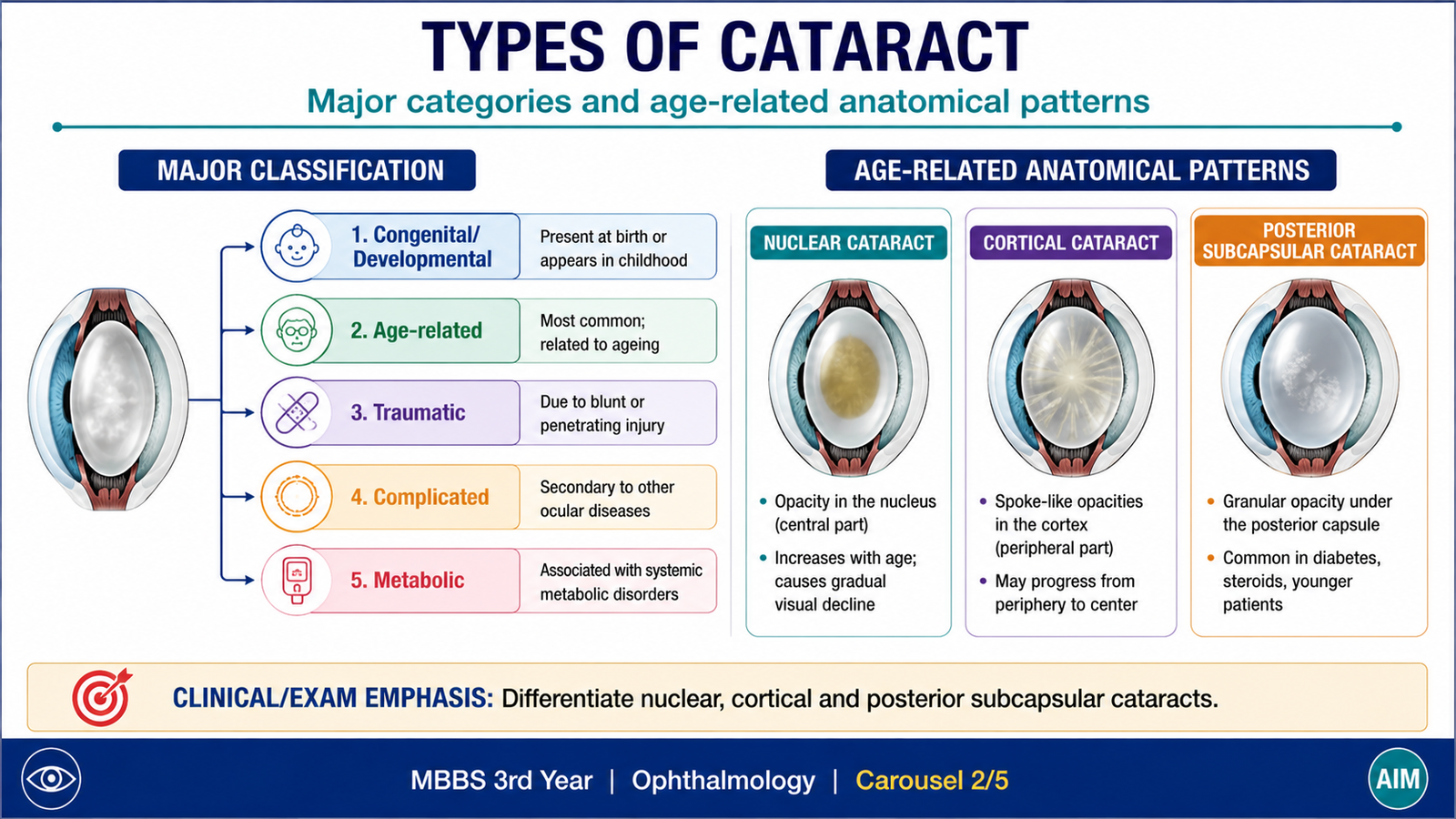
Cataracts can be classified according to their age of onset, cause, anatomical location and stage of development. For undergraduate learning, it is useful first to distinguish congenital or developmental cataracts from acquired cataracts, and then recognize the common morphological patterns of age-related cataract.
Congenital and developmental cataracts
These cataracts are present at birth or develop during childhood because normal lens development or lens transparency has been disturbed. They may be unilateral or bilateral. Their importance is greater in children because a dense cataract that blocks clear retinal images during visual development can interfere with normal maturation of vision.
Acquired cataracts
Acquired cataracts develop after the lens has initially formed normally. Important categories include:
- Age-related cataract: develops as part of progressive degenerative changes in the ageing lens and is the most familiar acquired form.
- Traumatic cataract: develops following mechanical, penetrating, blunt, electrical or other significant injury to the lens.
- Complicated cataract: occurs secondary to another ocular disorder that disturbs normal lens metabolism.
- Metabolic cataract: develops when systemic metabolic abnormalities alter the biochemical or osmotic environment of the lens.
- Drug-related cataract: may occur following prolonged exposure to certain drugs, particularly corticosteroids.
Major patterns of age-related cataract
| Type | Main site | Basic change | Important point |
|---|---|---|---|
| Nuclear cataract | Central lens nucleus | Progressive nuclear sclerosis and discoloration | Central lens becomes increasingly dense |
| Cortical cataract | Lens cortex | Hydration and disruption of cortical lens fibers | Opacities may form spoke-like patterns |
| Posterior subcapsular cataract | Posterior lens just beneath the capsule | Granular or plaque-like opacity | A relatively small central opacity may interfere markedly with vision |
Stages of age-related cataract
As a cataract progresses, the lens may pass through stages commonly described as immature, mature and hypermature. In an immature cataract, some transparent lens material remains. In a mature cataract, the lens becomes completely opaque. In a hypermature cataract, further degeneration occurs and lens proteins may liquefy or leak from the lens.
C. Pathogenesis of Cataract
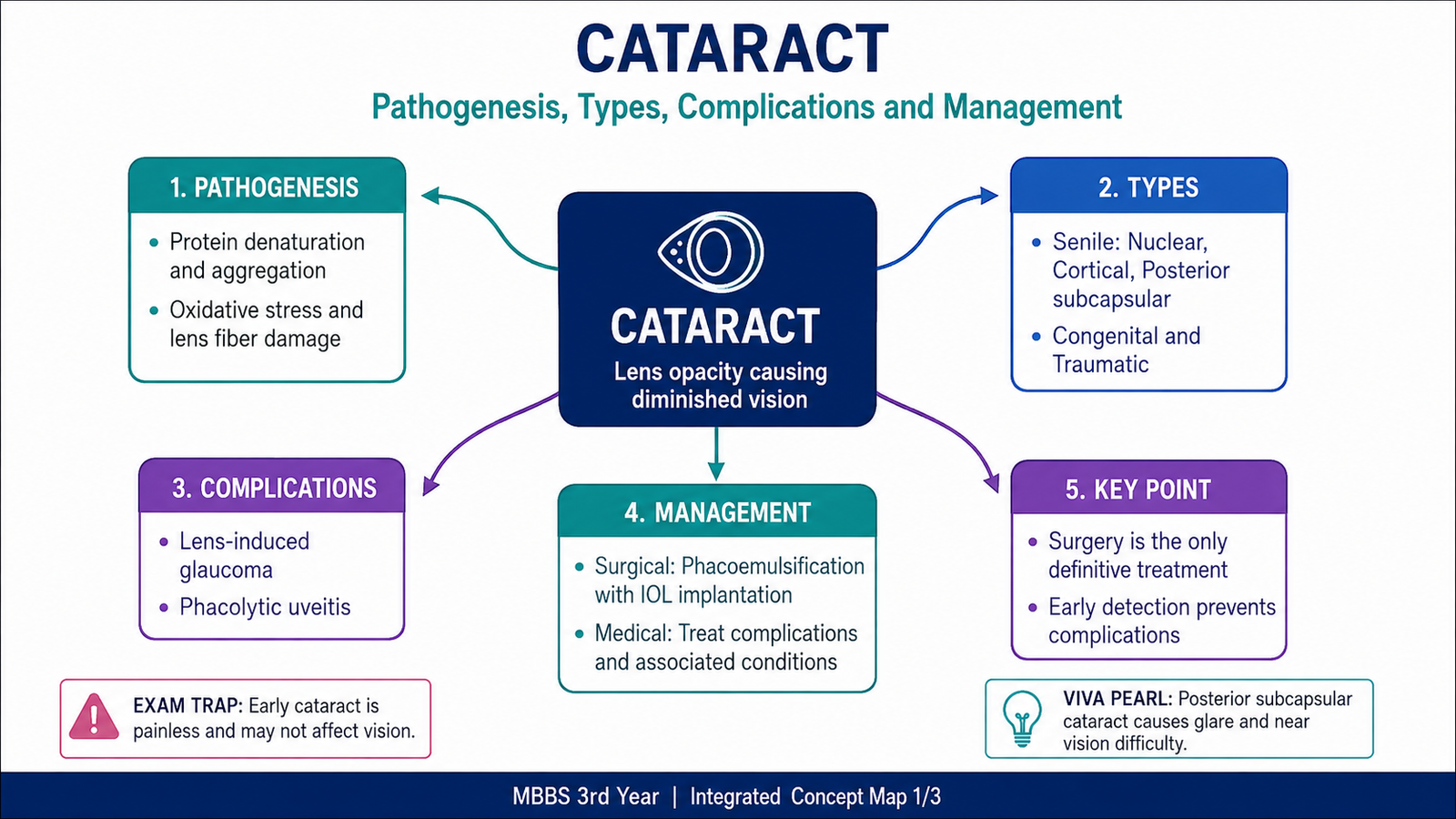
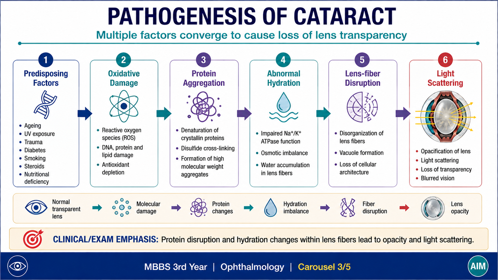
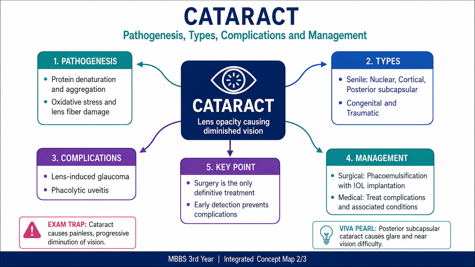
Although different cataracts have different initiating causes, the central pathogenic event is a disturbance of the highly organized lens fibers and proteins that normally maintain transparency. Lens cells have limited ability to replace damaged proteins. Therefore, biochemical and structural damage can accumulate gradually until the lens begins to scatter light.
Age-related changes
With ageing, lens proteins undergo progressive chemical modification and aggregation. Oxidative damage contributes to alteration of crystallin proteins, while lens fibers become increasingly compact and less flexible. Insoluble protein aggregates scatter light, producing opacity.
Role of lens hydration
The transparency of the lens also depends on carefully controlled water and electrolyte balance. Abnormal movement of water into lens fibers causes swelling and separation of the normally ordered fibers. This change is especially important in cortical cataract formation because disrupted cortical fibers lose their normal transparency.
Metabolic cataract formation
Systemic metabolic disturbances can change the chemical environment of the lens. In diabetes mellitus, excess glucose enters the lens and can be converted to sorbitol. Sorbitol tends to remain within the lens and contributes to osmotic stress and water accumulation. At the same time, chronic metabolic and oxidative stress can damage lens proteins. These changes promote loss of lens transparency.
Traumatic cataract formation
Trauma can directly damage the lens capsule or lens fibers. Once the orderly structure of the lens is disturbed, local hydration, degeneration and protein changes occur. The resulting opacity may remain localized or progress to involve a larger portion of the lens.
Drug-related cataract formation
Long-term corticosteroid exposure is associated particularly with posterior subcapsular cataract. The important undergraduate concept is that prolonged drug exposure can disturb normal lens-cell metabolism and organization, producing opacity in a characteristic anatomical region.
D. Complications of Cataract
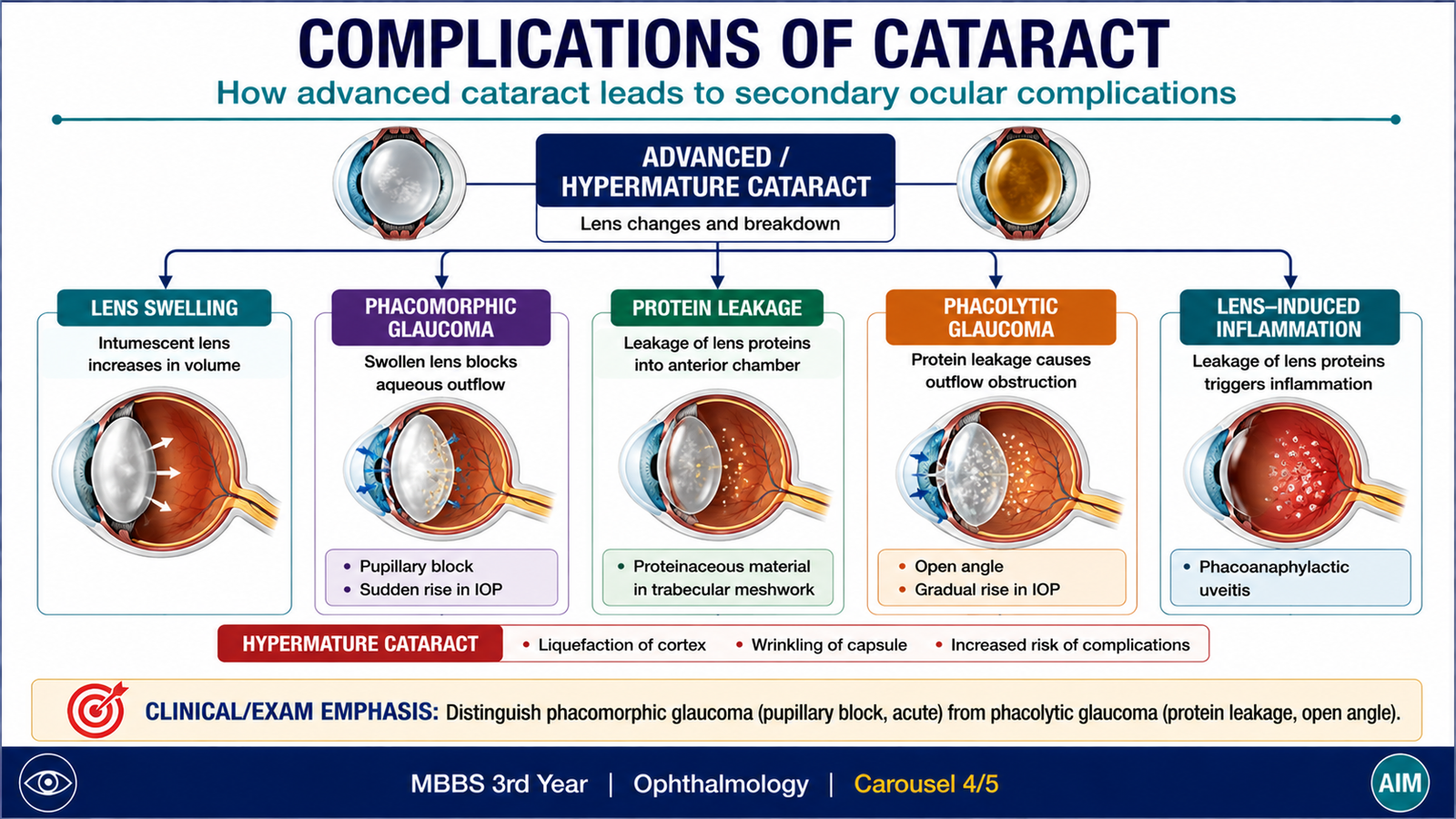
A cataract may initially be limited to loss of lens transparency, but an advanced or hypermature cataract can produce secondary problems within the eye. These complications arise because the cataractous lens may enlarge, degenerate or release altered lens proteins. Understanding the mechanism helps distinguish the complications from the cataract itself.
Progressive visual impairment
The most direct consequence is progressive reduction in vision. As the lens becomes more opaque, progressively more light is scattered or blocked before reaching the retina. A dense cataract may therefore cause severe visual limitation even when the retina and optic nerve are otherwise healthy.
Phacomorphic glaucoma
An intumescent or swollen cataractous lens may become enlarged. The enlarged lens can crowd the anterior segment and interfere with normal aqueous humour drainage, leading to a secondary rise in intraocular pressure.
Phacolytic glaucoma
In a hypermature cataract, degraded high-molecular-weight lens proteins may escape through the lens capsule. Macrophages ingest these proteins, and protein-rich material together with macrophages can obstruct the trabecular meshwork. Aqueous outflow is reduced and intraocular pressure rises.
Lens-induced inflammation
Lens proteins are normally enclosed within the lens capsule. When altered lens proteins become exposed or leak into surrounding ocular tissues, they can provoke an inflammatory response. This can produce lens-induced uveitis.
Changes in a hypermature cataract
Continued degeneration may cause liquefaction of the lens cortex. In some cases, the dense nucleus becomes mobile within liquefied cortical material. Such advanced changes increase the risk of secondary lens-related complications and make the cataract more than a simple optical opacity.
E. Management of Cataract
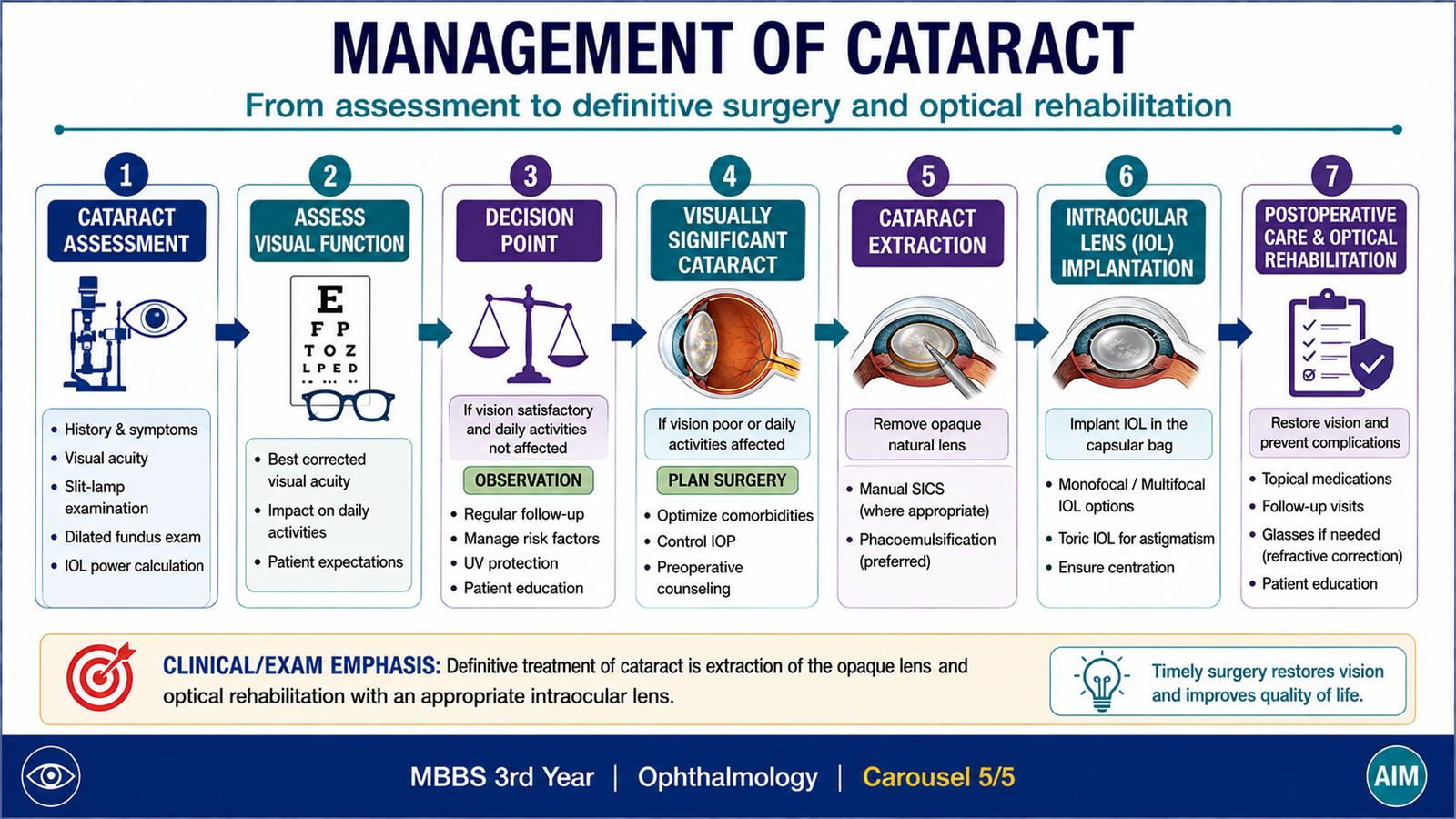
Management depends on how much the cataract affects useful vision and whether it is producing complications. The presence of a lens opacity alone does not automatically mean that immediate surgery is necessary. The central principle is to assess the effect of the cataract on vision and daily function, exclude other causes of visual loss and treat the cataract definitively when its impact becomes significant or complications develop.
Initial assessment
The eye should be evaluated to confirm that the reduction in vision is compatible with cataract and to identify other ocular disease that could limit the visual result after treatment. Visual acuity and examination of the lens are therefore basic parts of assessment.
Conservative measures
In early cataract, when useful vision remains adequate, simple measures may temporarily improve function. These do not remove the cataract because established lens opacity cannot be reversed by spectacles or routine medication.
- Correction of associated refractive error may improve useful vision.
- Improved lighting may help some patients perform daily activities more comfortably.
- Modifiable contributing factors, where present, should be addressed appropriately.
- The cataract should be reviewed for progression when clinically required.
Definitive treatment
The definitive treatment of a visually significant cataract is surgical removal of the opaque lens with optical rehabilitation, usually by implantation of an intraocular lens (IOL). Surgery removes the structure responsible for light scattering and replaces its refractive function.
Main surgical approaches
Phacoemulsification is a commonly used technique in which the lens nucleus is fragmented using ultrasonic energy and removed through a small incision. An intraocular lens is then implanted. The key concept is small-incision removal of the cataractous lens followed by IOL implantation. Extracapsular cataract extraction (ECCE) removes the lens nucleus and cortex while leaving the posterior part of the lens capsule to support an intraocular lens. It requires a larger incision than standard phacoemulsification. In selected situations, other surgical techniques may be used, but detailed operative protocols are beyond the required undergraduate learning outcome.
When surgery becomes important
Surgery is considered when the cataract causes sufficient visual impairment to interfere with the patient’s useful visual function or when the cataract itself is producing an ocular complication. The decision is therefore based on the functional and clinical effect of the cataract rather than simply on the presence of opacity.
Integrated Mechanism Flow
Important Comparison — Major Age-Related Cataract Patterns
| Feature | Nuclear | Cortical | Posterior Subcapsular |
|---|---|---|---|
| Main location | Lens nucleus | Lens cortex | Posterior lens beneath capsule |
| Basic process | Nuclear sclerosis and protein alteration | Cortical hydration and fiber disruption | Posterior granular/plaque-like opacity |
| Characteristic idea | Central lens becomes dense | Spoke-like cortical opacities may occur | Small central opacity may have marked visual effect |
⭐ AIM High-Yield Review
- Cataract is opacity of the crystalline lens or its capsule causing loss of normal lens transparency.
- Lens transparency depends on the orderly arrangement of lens fibers and soluble transparent crystallin proteins.
- ⭐ The common acquired categories include age-related, traumatic, complicated, metabolic and drug-related cataracts.
- The major age-related morphological patterns are nuclear, cortical and posterior subcapsular cataracts.
- Nuclear cataract mainly affects the central lens nucleus; cortical cataract primarily affects cortical lens fibers.
- Posterior subcapsular cataract lies close to the visual axis, so even a relatively small opacity may significantly interfere with vision.
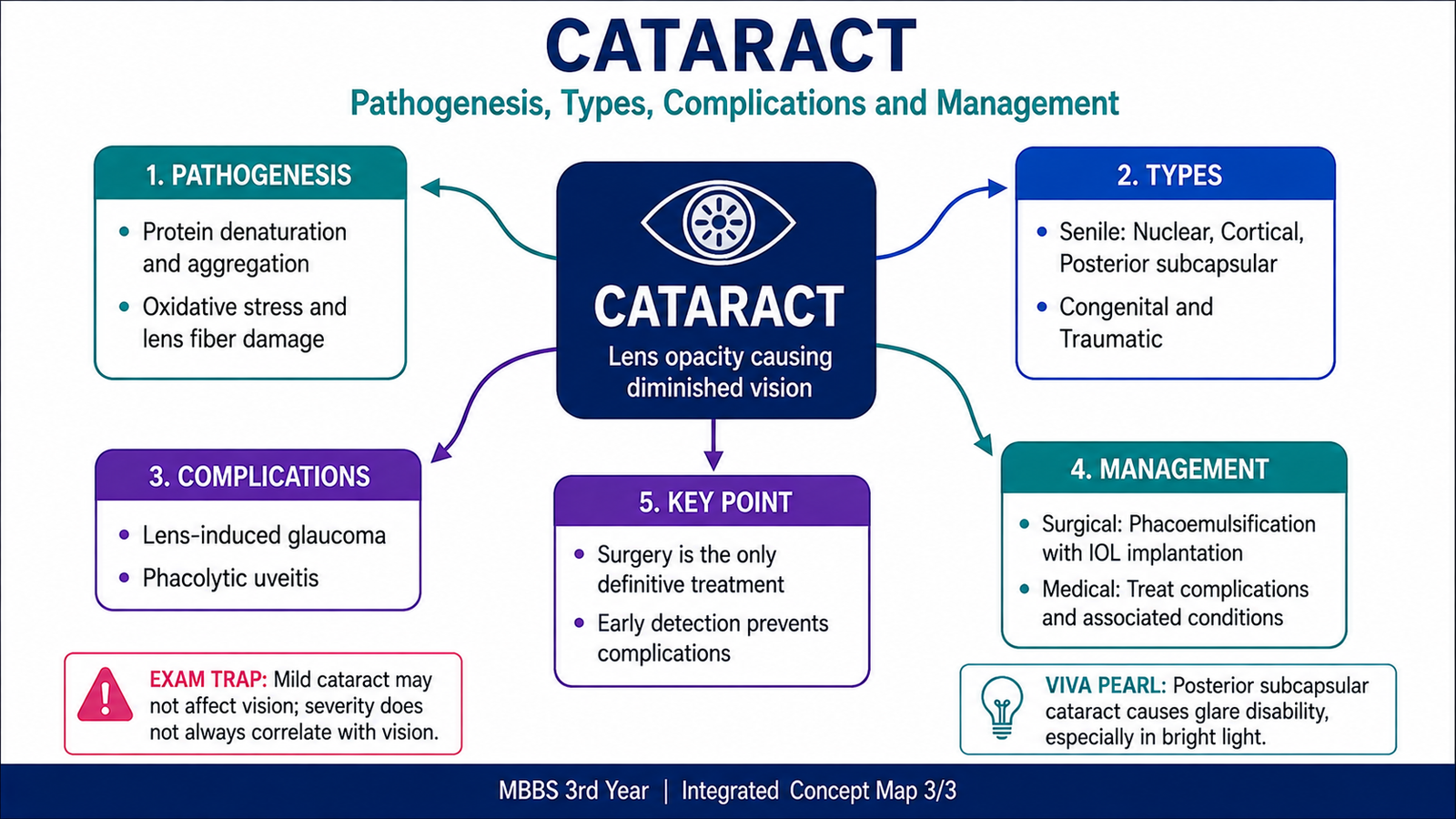
- Core pathogenesis: lens protein damage and/or abnormal hydration → loss of organized lens structure → light scattering → opacity.
- In diabetes, abnormal glucose metabolism within the lens can increase sorbitol and osmotic stress, contributing to lens damage.
- ⭐ Phacomorphic glaucoma is related to enlargement of a swollen cataractous lens and secondary interference with aqueous drainage.
- ⭐ Phacolytic glaucoma occurs when proteins leak from an advanced cataract and obstruct aqueous outflow.
- Leakage or exposure of lens proteins can also provoke lens-induced ocular inflammation.
- A mature cataract is completely opaque; a hypermature cataract shows further degeneration and may produce secondary complications.
- Established cataract opacity is not removed by spectacles or routine medication.
- ⭐ Definitive management of a visually significant cataract is surgical removal of the opaque lens with optical rehabilitation, usually an intraocular lens.
- The decision for surgery is based mainly on the functional effect of cataract and the presence of complications, not merely on the existence of a lens opacity.
🎥 AIM Video Learning — Intracellular Accumulations
Use this video to reinforce intracellular accumulations, including steatosis, protein, glycogen and pigments. :contentReference[oaicite:0]{index=0}
AIM Focus: While watching, focus on the mechanisms of intracellular accumulation, fatty change, protein and glycogen accumulation, and endogenous versus exogenous pigments.