
This chapter follows the KMU learning outcomes in a logical sequence. First understand why substances accumulate inside cells and how cells respond, then use the final high-yield review to revise the important examination points.
Intracellular Accumulations, Steatosis, Pigments and Pathologic Calcification
Foundation Module • Pathology
Understand why abnormal substances collect within cells, how fatty change develops, how proteins and glycogen accumulate, how pigments are classified, and why calcium may be deposited abnormally in tissues.
Topic Introduction
Cells continuously synthesize, process, store and remove many substances. When these normal handling mechanisms are disturbed, substances may accumulate abnormally inside the cytoplasm or within organelles. These substances may be normal cellular constituents present in excessive amounts, abnormal endogenous products, or materials that the cell cannot break down. Important examples include lipids, proteins, glycogen and pigments. Steatosis is a particularly important form of lipid accumulation, especially in the liver. In addition, abnormal deposits may occur outside or within cells, as seen in pathologic calcification. Understanding the cause, mechanism and morphology of these changes helps explain how apparently simple deposits can reflect significant cellular injury or metabolic disturbance.
A. Intracellular Accumulations: Basic Mechanisms
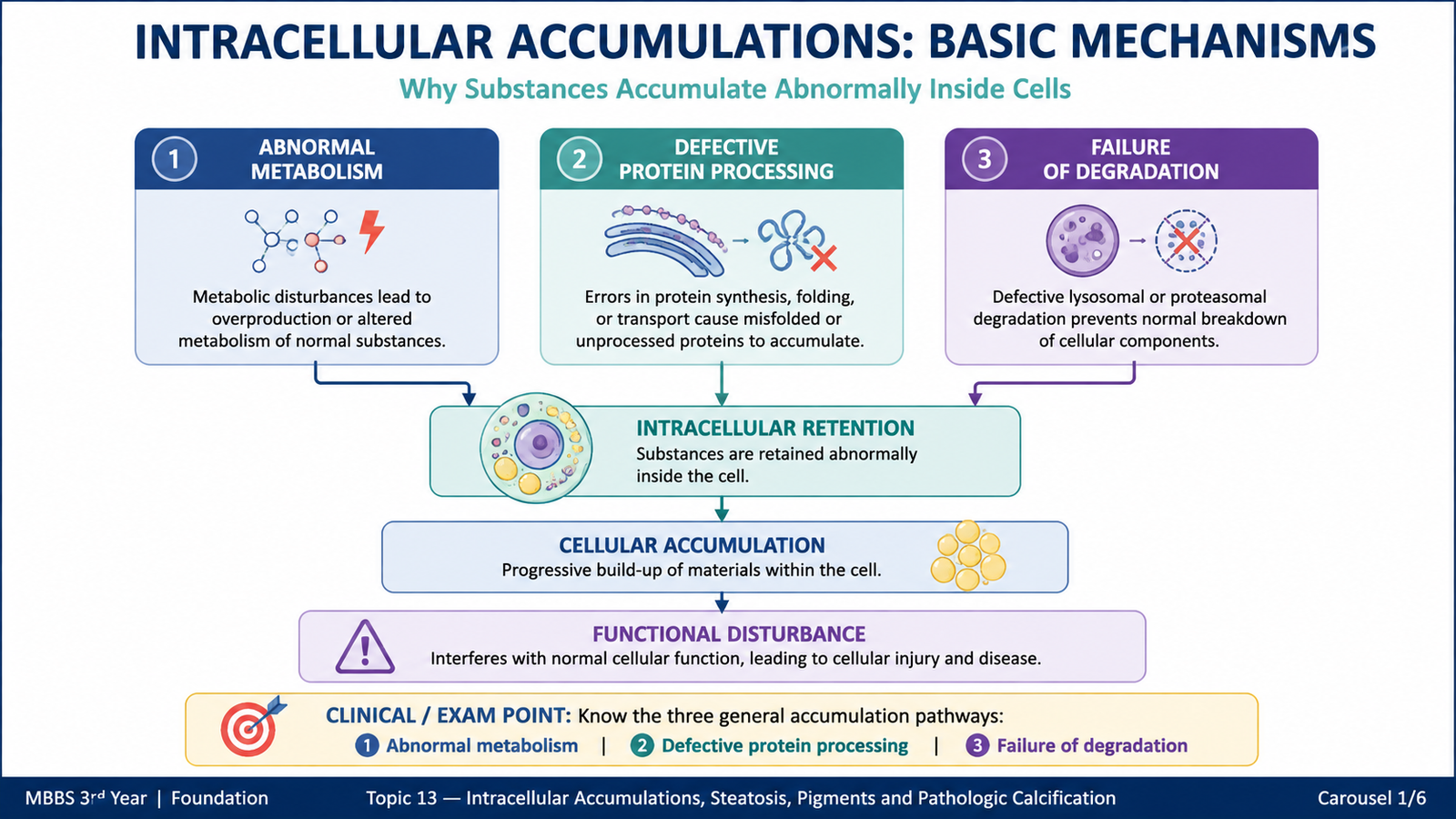
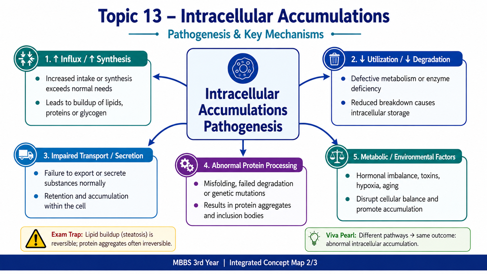
Intracellular accumulation means abnormal collection of a substance within a cell. The material may be located in the cytoplasm, inside organelles such as lysosomes, or occasionally within the nucleus. An accumulation develops when the rate at which a substance enters or is produced within a cell becomes greater than the cell’s ability to metabolize, transport or remove it.
The accumulated substance may be a normal cellular component such as triglyceride, protein or glycogen, or it may be an abnormal substance that the cell cannot process efficiently.
Three general pathways
Most abnormal intracellular accumulations can be understood through three general mechanisms.
- Abnormal metabolism of a normal endogenous substance: a normal substance is produced at a normal or increased rate but cannot be adequately metabolized or removed. It therefore accumulates. Fat accumulation in hepatocytes is an important example.
- Defects in protein folding, transport or secretion: an abnormal or altered endogenous protein is produced, but the cell cannot transport or secrete it normally. The protein remains within the cell and accumulates.
- Failure to degrade a substance: cells may take up an endogenous or exogenous material but lack the enzymes required to break it down. The material then persists, commonly within lysosomes.
These pathways share the same basic principle: the amount of material delivered to or produced within the cell exceeds the cell’s capacity to process and eliminate it.

B. Lipid Accumulation and Steatosis
Lipids may accumulate inside cells when their uptake, synthesis, utilization or export becomes disturbed. The most important form is steatosis, also called fatty change, in which triglycerides accumulate abnormally within parenchymal cells. The liver is the major organ affected because it plays a central role in fatty-acid metabolism, although fatty change may also occur in other tissues.
Definition of steatosis
Steatosis is the abnormal accumulation of triglycerides within parenchymal cells. It most commonly affects hepatocytes.
Causes
Fatty change develops when normal lipid handling by the cell is disturbed. Important causes include:
- Excessive alcohol exposure.
- Obesity and excessive delivery of fatty acids to the liver.
- Diabetes mellitus and other metabolic disturbances.
- Protein malnutrition, which can interfere with lipoprotein formation.
- Hypoxia, which reduces energy-dependent lipid metabolism.
- Certain toxins and drugs that interfere with lipid metabolism.
Mechanisms of hepatic steatosis
The liver normally receives fatty acids from the blood, synthesizes triglycerides, oxidizes fatty acids for energy and packages triglycerides into lipoproteins for export. Disturbance at any of these steps can cause triglycerides to accumulate.
Increased fatty-acid delivery or synthesis
→ increased triglyceride formation
→ reduced fatty-acid oxidation and/or impaired lipoprotein synthesis or secretion
→ triglycerides remain within hepatocytes
→ fatty liver.
Several mechanisms may operate together in the same patient. For example, a toxic or metabolic disturbance may simultaneously increase fatty-acid delivery and interfere with their oxidation or export.
Morphology of fatty liver
Early fatty change is usually reversible if the underlying cause is removed. As triglyceride accumulates, the appearance of both the organ and individual hepatocytes changes.
Gross morphology:
- The liver becomes enlarged.
- It is soft and yellow.
- Its cut surface has a greasy appearance because of the accumulated lipid.
Microscopic morphology:
- Small lipid droplets may initially appear within the cytoplasm of hepatocytes.
- The droplets may enlarge and merge into larger vacuoles.
- In marked steatosis, a large fat vacuole may occupy much of the cytoplasm and displace the nucleus toward the edge of the cell.
Consequences
Mild steatosis may have little immediate effect on cellular function and can disappear after removal of the cause. Severe or persistent fatty accumulation can impair hepatocyte function and may accompany more significant forms of liver injury. Therefore, the importance of steatosis depends mainly on its severity, duration and underlying cause.

C. Protein Accumulation
Proteins may accumulate within cells when they are taken up in excessive amounts, synthesized excessively, incorrectly folded, or inadequately transported and secreted. The appearance of the accumulated material depends on the protein involved and the affected cell.
Major mechanisms
1. Excessive protein reabsorption
Renal tubular epithelial cells normally reabsorb small amounts of protein filtered through the glomerulus. When unusually large amounts of protein enter the filtrate, tubular cells reabsorb more protein than usual. Protein-containing droplets may therefore become visible within their cytoplasm.
2. Excessive synthesis of secretory proteins
Cells that actively synthesize immunoglobulins or other secretory proteins may accumulate these products if production becomes greater than the ability to secrete them.
3. Defective folding and intracellular transport
Some abnormal proteins cannot fold into their normal three-dimensional shape. Because cellular quality-control systems prevent these proteins from being transported normally, they are retained inside the cell, particularly within the endoplasmic reticulum.
4. Accumulation of cytoskeletal proteins
Abnormal aggregation of cellular structural proteins may produce intracellular inclusions. The important principle is that altered structural proteins are not processed normally and therefore accumulate.
Morphology and consequences
Protein accumulations frequently appear as rounded eosinophilic droplets, aggregates or inclusions within the cytoplasm. Whether they cause significant dysfunction depends on the amount accumulated and the biological importance of the affected cell. Some protein accumulations are largely markers of cellular stress, whereas others interfere with normal cellular function.

D. Glycogen Accumulation
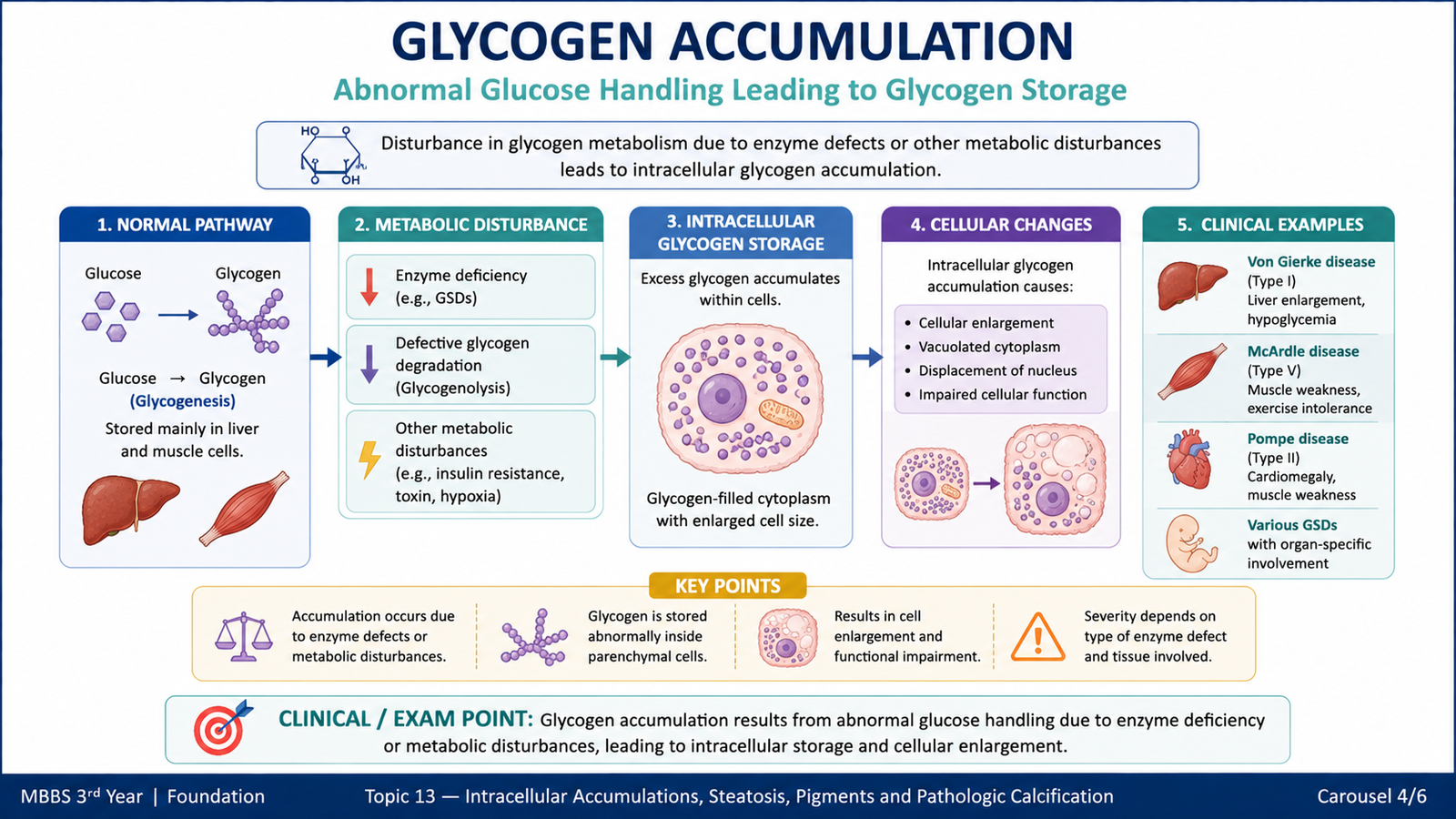
Glycogen is the normal storage form of glucose. It is mainly stored in the liver and skeletal muscle. Excessive intracellular glycogen becomes visible when glucose or glycogen metabolism is disturbed. The accumulation may therefore occur because cells receive excessive amounts of glucose or because inherited enzyme abnormalities prevent normal glycogen breakdown.
Important causes and mechanisms
Diabetes mellitus: Abnormal glucose metabolism can lead to excessive glycogen accumulation in particular cells. The basic problem is altered regulation and handling of glucose, causing increased intracellular storage in some tissues.
Glycogen storage diseases: These inherited disorders result from deficiency of enzymes involved in glycogen synthesis or degradation. Because glycogen cannot be processed normally, it accumulates within cells, especially in organs in which glycogen metabolism is important.
Defect in glucose or glycogen metabolism
→ impaired utilization or degradation of glycogen
→ intracellular glycogen retention
→ cellular enlargement and possible organ dysfunction.
Morphology
Excess glycogen produces clear cytoplasmic vacuoles in routine tissue sections because glycogen may be dissolved during tissue processing. Special histochemical methods can demonstrate glycogen within cells.
Consequences
The functional effect depends on the amount of glycogen and the organ involved. In inherited glycogen storage disorders, extensive accumulation may distort cells and interfere with normal organ function.
E. Pathological Pigments
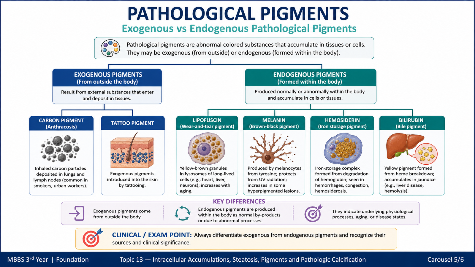
Pigments are colored substances that may normally occur in cells or may accumulate as a result of environmental exposure, metabolic activity or tissue breakdown. They are broadly classified into exogenous pigments, which enter the body from outside, and endogenous pigments, which are formed within the body.
Exogenous pigments
Carbon: Carbon particles are inhaled from polluted air and smoke. Alveolar macrophages ingest these particles and transport them through lymphatic channels. Carbon may therefore accumulate in the lungs and regional lymph nodes, producing a dark discoloration known as anthracotic pigmentation.
Tattoo pigment: Pigments introduced into the dermis are taken up by macrophages. Because many of these particles are resistant to degradation, they may remain in the dermis for many years.
Endogenous pigments
Lipofuscin: Lipofuscin is a yellow-brown intracellular pigment composed largely of material derived from lipid peroxidation and cellular membrane breakdown. It tends to accumulate slowly with ageing and chronic cellular stress, particularly in long-lived cells such as cardiac muscle cells and hepatocytes. It is generally considered a marker of previous free-radical injury rather than a major cause of cellular dysfunction.
Melanin: Melanin is a brown-black pigment produced by melanocytes. It normally contributes to skin, hair and eye pigmentation and helps protect tissues against ultraviolet radiation. Abnormal amounts or distribution of melanin can alter tissue pigmentation.
Hemosiderin: Hemosiderin is a golden-yellow to brown iron-storage pigment derived mainly from hemoglobin breakdown. Small amounts can occur normally, but excessive accumulation indicates increased local or systemic iron deposition.
After local hemorrhage, macrophages break down hemoglobin from extravasated red cells and store some of the released iron as hemosiderin. When excessive iron accumulates throughout the body, hemosiderin may be deposited in several organs.
Bilirubin: Bilirubin is a yellow-green pigment produced during heme breakdown. Excess bilirubin in tissues produces yellow discoloration. Its accumulation reflects disturbance of bilirubin production, metabolism or excretion rather than a primary storage process within a single cell type.
| Pigment | Origin | Typical appearance/significance |
|---|---|---|
| Carbon | Exogenous | Black pigment in lungs and lymph nodes |
| Tattoo pigment | Exogenous | Persistent pigment within dermal macrophages |
| Lipofuscin | Endogenous | Yellow-brown ageing and wear-and-tear pigment |
| Melanin | Endogenous | Brown-black pigment produced by melanocytes |
| Hemosiderin | Endogenous | Golden-brown iron-storage pigment |
| Bilirubin | Endogenous | Yellow-green pigment derived from heme breakdown |
F. Pathologic Calcification
Pathologic calcification is abnormal deposition of calcium salts in tissues. It occurs in two major forms: dystrophic calcification and metastatic calcification. The distinction is important because the two processes occur in different settings and have different mechanisms.
Dystrophic calcification
Dystrophic calcification occurs in dead or damaged tissues despite a normal concentration of calcium in the blood. Cellular injury creates sites that favor deposition and crystallization of calcium salts.
Important settings include:
- Areas of necrosis.
- Atherosclerotic plaques.
- Damaged or ageing heart valves.
- Old areas of tissue injury.
The central idea is that local tissue damage, rather than increased blood calcium, initiates the deposition.
→ local binding and concentration of calcium and phosphate
→ crystal formation
→ visible calcium deposits
→ possible tissue stiffness or impaired function.
Metastatic calcification
Metastatic calcification occurs in otherwise viable tissues because calcium-phosphate balance in the body is disturbed, most importantly in settings associated with increased serum calcium. Calcium salts may then be deposited widely in different tissues.
Important causes of hypercalcemia that can promote metastatic calcification include increased parathyroid hormone activity, destruction of bone, disorders that increase vitamin D activity, and reduced renal phosphate excretion in chronic renal failure.
Metastatic deposits tend to occur particularly in tissues that have conditions favorable for calcium precipitation, including the kidneys, lungs, gastric mucosa and systemic arteries.
Morphology of calcification
Grossly, calcium deposits may appear as fine white granules or firm gritty areas. Microscopically, calcium salts usually appear as basophilic, granular or clumped material. Deposits may occur within cells, outside cells, or in both locations.
Functional alterations
The effect of calcification depends on its location and extent. Small deposits may have little functional effect. Extensive calcification can make tissues rigid, interfere with movement or narrowing and obstructing luminal structures, and impair normal organ function. For example, calcification of a heart valve may limit normal valve movement.
| Feature | Dystrophic calcification | Metastatic calcification |
|---|---|---|
| Main setting | Dead or damaged tissue | Otherwise viable tissues |
| Serum calcium | Usually normal | Usually increased or calcium-phosphate balance disturbed |
| Underlying problem | Local tissue injury | Systemic mineral imbalance |
| Typical examples | Necrotic tissue, atherosclerotic plaque, damaged valve | Kidney, lung, gastric mucosa and arteries |

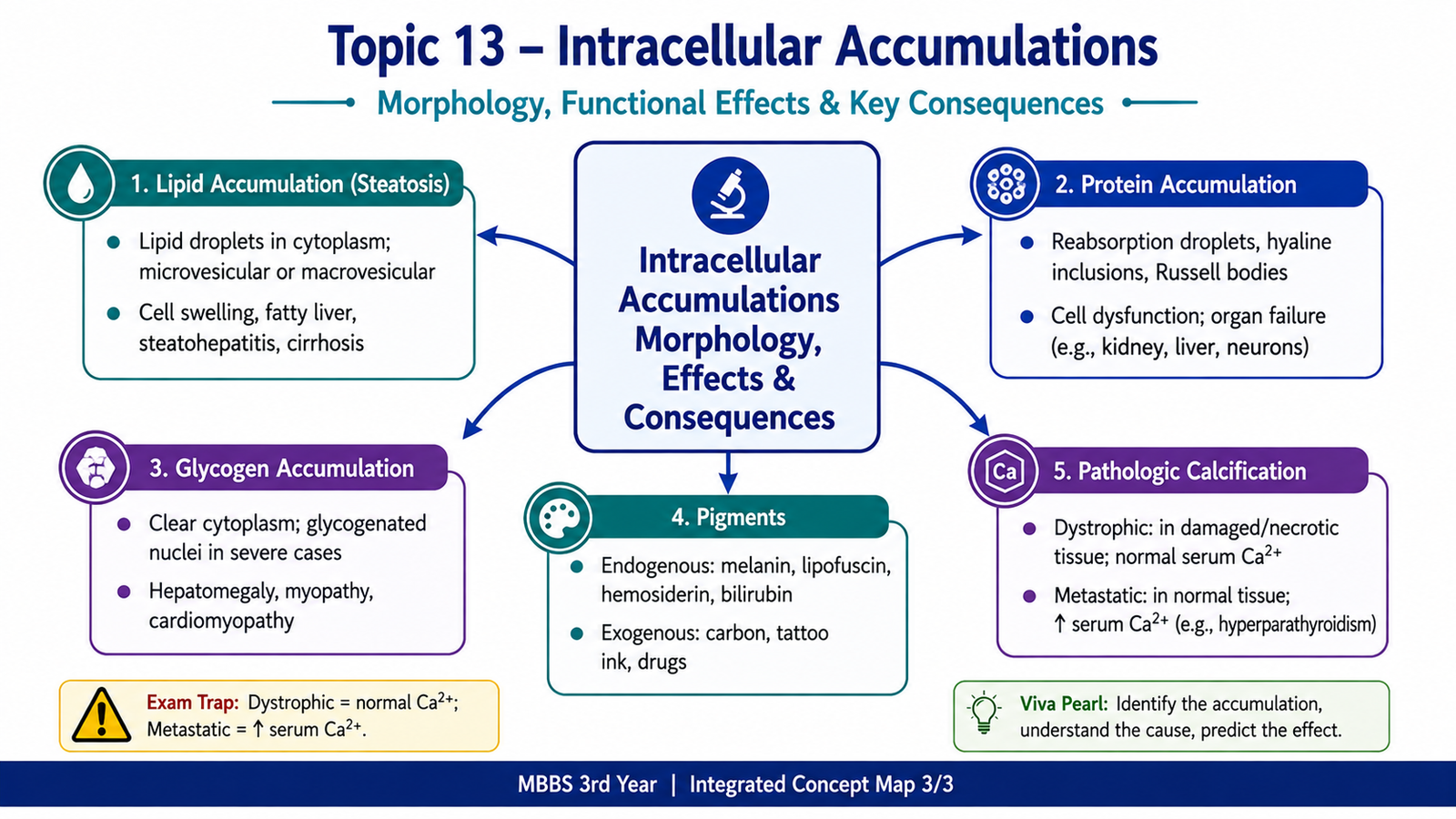
Integrated Mechanism Flow
↓
Increased production, uptake or retention of a substance
↓
Cellular processing, transport or degradation capacity is exceeded
↓
Lipid, protein, glycogen or pigment accumulates
↓
Characteristic intracellular morphological change develops
↓
Mild accumulation may be reversible, whereas persistent or severe accumulation may impair cellular function.
⭐ AIM High-Yield Review
- Intracellular accumulation occurs when production or uptake of a substance exceeds the cell’s ability to metabolize, transport or remove it.
- The three general pathways involve abnormal metabolism of normal substances, defective processing of endogenous products, and failure to degrade accumulated material.
- ⭐ Steatosis is abnormal intracellular accumulation of triglycerides and most commonly affects the liver.
- Hepatic steatosis can result from increased fatty-acid delivery or synthesis, reduced oxidation, or impaired lipoprotein formation and export.
- A markedly fatty liver is typically enlarged, yellow, soft and greasy.
- Protein may accumulate because of excessive uptake or synthesis, defective folding or impaired intracellular transport.
- Glycogen accumulates when glucose metabolism is abnormal or when enzymes required for glycogen metabolism are deficient.
- Exogenous pigments include carbon and tattoo pigments.
- Lipofuscin is a yellow-brown pigment associated with ageing and previous free-radical injury.
- Hemosiderin is an iron-containing golden-brown pigment derived largely from hemoglobin breakdown.
- ⭐ Dystrophic calcification occurs in dead or damaged tissue while serum calcium is usually normal.
- ⭐ Metastatic calcification occurs because of systemic disturbance of calcium-phosphate metabolism and affects otherwise viable tissues.
- Microscopic calcium deposits are typically basophilic and granular or clumped.
- Extensive pathologic calcification can cause tissue stiffness and functional impairment.