
Pituitary and Growth Disorders: Pathology, Clinical Evaluation, Pharmacotherapy and Surgical Management
An integrated study of pituitary structure and regulation, pituitary adenomas, growth-hormone disorders, hyperprolactinemia, hypopituitarism, pediatric growth assessment and the medical and surgical principles used to manage these disorders.
1. Topic Introduction
The pituitary gland coordinates several endocrine systems that control growth, reproduction, thyroid function, adrenal function and lactation. Pituitary disease may therefore present in two broad ways: a hormone may be produced in excess, or normal pituitary hormones may become deficient. Pituitary tumors can also produce symptoms simply by enlarging and compressing nearby structures, especially the optic chiasm. In this chapter, you will first understand normal pituitary structure and hormonal control. You will then study pituitary adenomas, growth-hormone excess, hyperprolactinemia and hypopituitarism, followed by pediatric assessment of short stature and the principles of pituitary surgery. Pharmacological treatment is explained at the point where each drug becomes clinically relevant.
2. Core Learning Material
A. Pituitary Structure, Hormones and Hypothalamic Regulation
The pituitary is a small endocrine gland located within the sella turcica of the sphenoid bone. It is connected to the hypothalamus by the pituitary stalk and lies immediately below the optic chiasm. These anatomical relations are clinically important because a pituitary mass can extend upward and compress the optic chiasm.
Gross Structure
The gland is divided into two functionally different components:
- Anterior pituitary or adenohypophysis: glandular endocrine tissue that synthesizes and secretes several trophic hormones.
- Posterior pituitary or neurohypophysis: neural tissue that stores and releases hormones synthesized in the hypothalamus.
The anterior pituitary includes the pars distalis, which forms most of the gland, together with the pars tuberalis and a small pars intermedia. The posterior pituitary consists mainly of the pars nervosa and the infundibular connection with the hypothalamus.
Microscopic Structure
The anterior pituitary is composed of endocrine cells arranged in cords and nests around a rich network of fenestrated capillaries. The major functional cell types are identified by the hormones they produce:
- Somatotrophs: growth hormone (GH).
- Lactotrophs: prolactin.
- Corticotrophs: adrenocorticotropic hormone (ACTH).
- Thyrotrophs: thyroid-stimulating hormone (TSH).
- Gonadotrophs: follicle-stimulating hormone (FSH) and luteinizing hormone (LH).
The posterior pituitary contains unmyelinated axons arising mainly from neurons in the supraoptic and paraventricular nuclei of the hypothalamus. Antidiuretic hormone and oxytocin travel down these axons and are stored in terminal swellings before release. Supporting glial-like cells called pituicytes are also present.
Hormonal Control
The hypothalamus controls the anterior pituitary through releasing and inhibitory hormones carried in the hypothalamic-hypophyseal portal circulation.
- GHRH stimulates GH; somatostatin inhibits GH.
- TRH stimulates TSH.
- CRH stimulates ACTH.
- GnRH stimulates FSH and LH.
- Dopamine inhibits prolactin secretion.
Most endocrine axes operate through negative feedback. For example, GH stimulates production of IGF-1, mainly in the liver. Increasing GH and IGF-1 then reduce further GH secretion through effects at the hypothalamus and pituitary. This arrangement explains why pituitary disease may produce both hormone excess and hormone deficiency. It also explains the stalk effect: interruption of dopamine delivery through the pituitary stalk removes normal inhibition of prolactin secretion and can raise serum prolactin.

B. Hyperpituitarism and Pituitary Adenomas
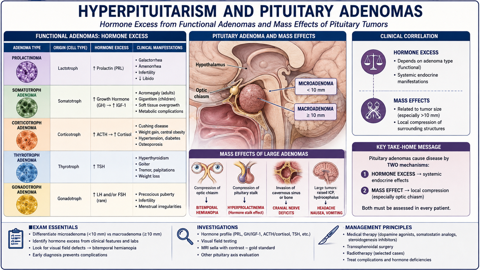
Hyperpituitarism means excessive secretion of one or more pituitary hormones. The most important cause is a functioning pituitary adenoma. Less commonly, pituitary cell hyperplasia may occur when a cell population receives persistent stimulatory signals. The effects of hyperpituitarism depend on both the hormone produced and the size of the pituitary lesion.
Pituitary Adenomas
Pituitary adenomas are usually benign neoplasms arising from anterior pituitary cells. They can be classified by size and by functional activity.
- Microadenoma: less than 10 mm.
- Macroadenoma: 10 mm or larger.
- Functioning adenoma: produces clinically important hormone excess.
- Non-functioning adenoma: does not produce a clinically recognizable hormone-excess syndrome.
Morphology
Grossly, an adenoma is commonly a soft, well-circumscribed lesion within the sella. A small tumor may remain confined to the gland, whereas a macroadenoma can enlarge the sella and extend upward into the suprasellar region. Microscopically, pituitary adenomas usually contain relatively uniform endocrine cells arranged in sheets, cords or nests. Compared with normal pituitary tissue, the normal reticulin framework is disrupted. Immunohistochemistry can demonstrate hormone production and help identify the tumor cell lineage.
Major Functional Adenomas
- Prolactinoma: prolactin excess causes hypogonadism, menstrual disturbance, infertility and sometimes galactorrhea.
- Somatotroph adenoma: GH excess causes gigantism in children or acromegaly in adults.
- Corticotroph adenoma: ACTH excess stimulates adrenal cortisol production and causes Cushing disease.
- Thyrotroph adenoma: TSH excess produces inappropriate thyroid stimulation and hyperthyroidism.
Mass Effect
A large adenoma may produce symptoms independently of its hormonal activity. Upward growth can compress the optic chiasm and damage crossing nasal retinal fibers. The result is classically bitemporal hemianopia. Compression of the remaining normal pituitary gland can also cause hypopituitarism. Core relationship: pituitary adenoma → hormone excess and/or tumor enlargement → endocrine syndrome, visual-field disturbance or loss of normal pituitary function.
C. Growth Hormone Pharmacology and Control of GH Excess
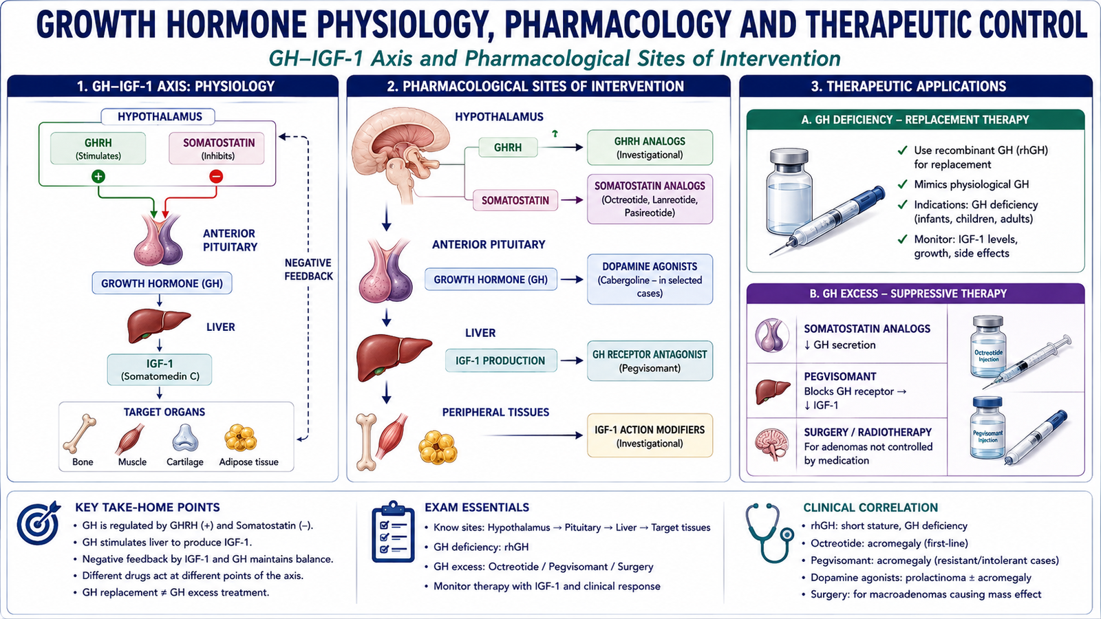
Growth hormone is a peptide hormone released by anterior-pituitary somatotrophs. Its secretion is stimulated by GHRH and inhibited by somatostatin. Many of its growth-promoting effects occur through IGF-1, produced mainly by the liver. Pharmacological treatment can therefore either replace deficient GH, reduce excessive GH secretion or block the action of GH at its receptor.
Pharmacodynamic Principle
GH binds to growth-hormone receptors and activates intracellular signaling that increases IGF-1 production and promotes growth of bone and soft tissues. GH also increases protein synthesis, promotes lipolysis and reduces insulin sensitivity.
GH → GH receptor activation → increased IGF-1 → skeletal and soft-tissue growth
Old and New Sources of Growth Hormone
Historically, therapeutic GH was extracted from human cadaveric pituitary glands. This practice was discontinued because pituitary-derived preparations could transmit Creutzfeldt-Jakob disease. Modern treatment uses recombinant human growth hormone, usually somatropin, produced through recombinant-DNA technology. Recombinant GH avoids the infection risk associated with cadaveric pituitary extracts and provides a consistent pharmaceutical product.
Relevant Pharmacokinetics
GH and somatostatin-related medicines are peptides, so oral administration is ineffective because digestive enzymes would degrade them. Recombinant GH is therefore usually given subcutaneously. Natural somatostatin has a very short duration of action, which is why longer-acting analogues such as octreotide are useful clinically.
Clinical Use and Adverse Effects of Recombinant GH
The main use in this topic is replacement therapy for confirmed growth-hormone deficiency. Treatment aims to restore appropriate growth in children rather than simply make every short child taller. Important adverse effects include:
- fluid retention and edema;
- arthralgia and muscle discomfort;
- insulin resistance and impaired glucose tolerance;
- intracranial hypertension in susceptible patients;
- slipped upper femoral epiphysis in susceptible growing children.
Drugs Used Against GH Excess
A true GH-receptor antagonist is pegvisomant. It binds to the GH receptor without producing normal receptor activation, thereby reducing IGF-1 production. Other medicines control GH excess by reducing its secretion rather than directly antagonizing the GH receptor.
- Somatostatin analogues: octreotide and lanreotide suppress GH secretion.
- GH-receptor antagonist: pegvisomant blocks peripheral GH action.
- Dopamine agonists: can reduce GH secretion in selected patients, although their effect is generally weaker.
Octreotide in Acromegaly and Gigantism
Octreotide is a synthetic somatostatin analogue with a much longer duration of action than natural somatostatin. It activates somatostatin receptors on pituitary somatotrophs, particularly SSTR2, and suppresses GH release.
Octreotide → somatostatin-receptor activation → reduced GH secretion → reduced IGF-1 → improvement in GH-excess manifestations
Immediate-release octreotide may be started at approximately 50 micrograms subcutaneously three times daily and adjusted according to biochemical and clinical response. Doses in the range of approximately 100–200 micrograms every 8 hours may be required in some patients. Long-acting octreotide formulations can be administered by deep intramuscular injection approximately every four weeks after tolerability has been established. Important adverse effects include:
- abdominal discomfort, nausea and diarrhea;
- reduced gallbladder contraction and gallstone formation;
- altered glucose regulation;
- bradycardia in susceptible patients;
- injection-site discomfort.
D. Acromegaly and Gigantism: Clinical Evaluation and Management
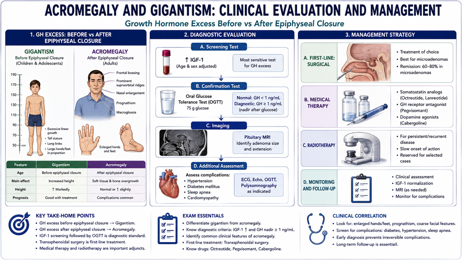
Acromegaly and gigantism are caused by persistent growth-hormone excess, usually from a GH-secreting pituitary adenoma. The major difference depends on whether GH excess begins before or after closure of the epiphyseal growth plates. Before epiphyseal closure, long bones can continue to lengthen, producing gigantism. After epiphyseal closure, linear growth is no longer possible, so GH and IGF-1 mainly cause enlargement of bones, soft tissues and internal organs, producing acromegaly.
Clinical Features of Acromegaly
Changes are usually gradual. The patient may notice increasing shoe or ring size before recognizing the typical facial changes.
- Enlarged hands and feet due to bone and soft-tissue growth.
- Coarse facial features from enlargement of facial bones and soft tissues.
- Prognathism from mandibular growth.
- Increased spacing of teeth as the jaw enlarges.
- Macroglossia from soft-tissue overgrowth.
- Thick, sweaty skin because of soft-tissue and sweat-gland effects.
- Arthropathy due to excessive cartilage and bone growth.
- Carpal tunnel syndrome from soft-tissue expansion around the median nerve.
GH antagonizes insulin action, so chronic excess may cause insulin resistance and diabetes mellitus. Cardiovascular effects include hypertension and cardiomyopathy. Upper-airway soft-tissue enlargement can contribute to obstructive sleep apnea. Headache and visual-field loss result from the pituitary tumor rather than directly from GH. A macroadenoma compressing the optic chiasm can cause bitemporal hemianopia.
Clinical Features of Gigantism
The hallmark is excessive linear growth before epiphyseal closure. Children may become unusually tall for age with accelerated height velocity. Other manifestations of GH excess, including soft-tissue enlargement, metabolic effects and features of the pituitary mass, may also occur.
Investigations
A random GH concentration is unreliable because normal GH secretion is pulsatile. Serum IGF-1, interpreted for age, provides a more stable estimate of overall GH activity and is an important initial biochemical test. Biochemical confirmation is obtained by demonstrating that GH fails to suppress appropriately after an oral glucose load. In healthy individuals, glucose suppresses GH secretion; this suppression is lost in acromegaly. After biochemical confirmation, pituitary MRI is used to identify the adenoma, determine its size and assess its relation to the optic chiasm and surrounding structures. Visual-field testing is particularly important when a macroadenoma approaches the optic chiasm.
Treatment
Treatment aims to control GH and IGF-1, remove or reduce the pituitary tumor, relieve mass effect and reduce long-term complications.
- Transsphenoidal surgery: important first-line treatment for many resectable GH-secreting adenomas, particularly when tumor mass effect is present.
- Somatostatin analogues: octreotide or related agents suppress GH secretion.
- Pegvisomant: blocks the GH receptor and lowers IGF-1.
- Dopamine agonists: may be useful in selected patients.
- Radiotherapy: may be considered in persistent disease that remains inadequately controlled by surgery and medical treatment.
Complications
Untreated GH excess increases morbidity because it affects multiple organs. Important complications include hypertension, cardiomyopathy, glucose intolerance or diabetes, obstructive sleep apnea, degenerative joint disease and consequences of tumor mass effect.
E. Hyperprolactinemia and Dopamine-Agonist Therapy
Hyperprolactinemia is an increase in circulating prolactin. A prolactinoma is an important cause, but prolactin may also rise because normal hypothalamic dopamine inhibition is reduced or because another physiological, pharmacological or endocrine stimulus is present.
Why Dopamine Is Important
Dopamine released from the hypothalamus normally activates D2 receptors on lactotrophs and continuously suppresses prolactin release.
Dopamine → D2 receptors on lactotrophs → inhibition of prolactin secretion
This is why stalk compression can increase prolactin: less dopamine reaches the anterior pituitary, so inhibition is removed.
Important Causes
- pregnancy and lactation;
- prolactinoma;
- pituitary-stalk compression;
- drugs that block dopamine action;
- primary hypothyroidism, in which increased TRH can stimulate prolactin release;
- selected systemic disorders affecting prolactin metabolism or clearance.
Clinical Features
High prolactin suppresses hypothalamic GnRH, reducing FSH and LH secretion. Many clinical manifestations therefore result from hypogonadism. Women may develop oligomenorrhea or amenorrhea, infertility, reduced libido and galactorrhea. Men may develop reduced libido, erectile dysfunction and infertility. A large pituitary tumor may additionally produce headache, visual-field loss and deficiency of other pituitary hormones.
Investigations
The first step is to confirm persistent hyperprolactinemia and look for an alternative explanation. Clinical assessment should therefore consider pregnancy, medication use and symptoms of hypothyroidism or pituitary mass effect. Relevant investigations include serum prolactin, pregnancy testing when appropriate and thyroid-function testing. If persistent hyperprolactinemia remains unexplained or a pituitary lesion is suspected, pituitary MRI is performed.
Treatment
The underlying reversible cause should be corrected when possible. A prolactinoma is usually treated initially with a dopamine agonist, because these drugs can reduce both prolactin secretion and tumor size. Cabergoline is commonly preferred in clinical practice because of its effectiveness and tolerability, while bromocriptine remains an important prototype drug.
Bromocriptine
Bromocriptine is a dopamine D2-receptor agonist.
Bromocriptine → D2-receptor stimulation → reduced prolactin synthesis and release → improved gonadal function and reduction in prolactinoma activity
Its major use in this topic is treatment of hyperprolactinemia and prolactinoma. It can also suppress GH secretion in some patients with acromegaly. Important adverse effects include:
- nausea and vomiting;
- postural hypotension and dizziness;
- headache;
- nasal congestion;
- neuropsychiatric effects in susceptible patients.
Surgery is considered when a prolactinoma cannot be adequately controlled with appropriate medical therapy, when medication is not tolerated or when a compelling compressive indication exists.

F. Hypopituitarism and Sheehan Syndrome
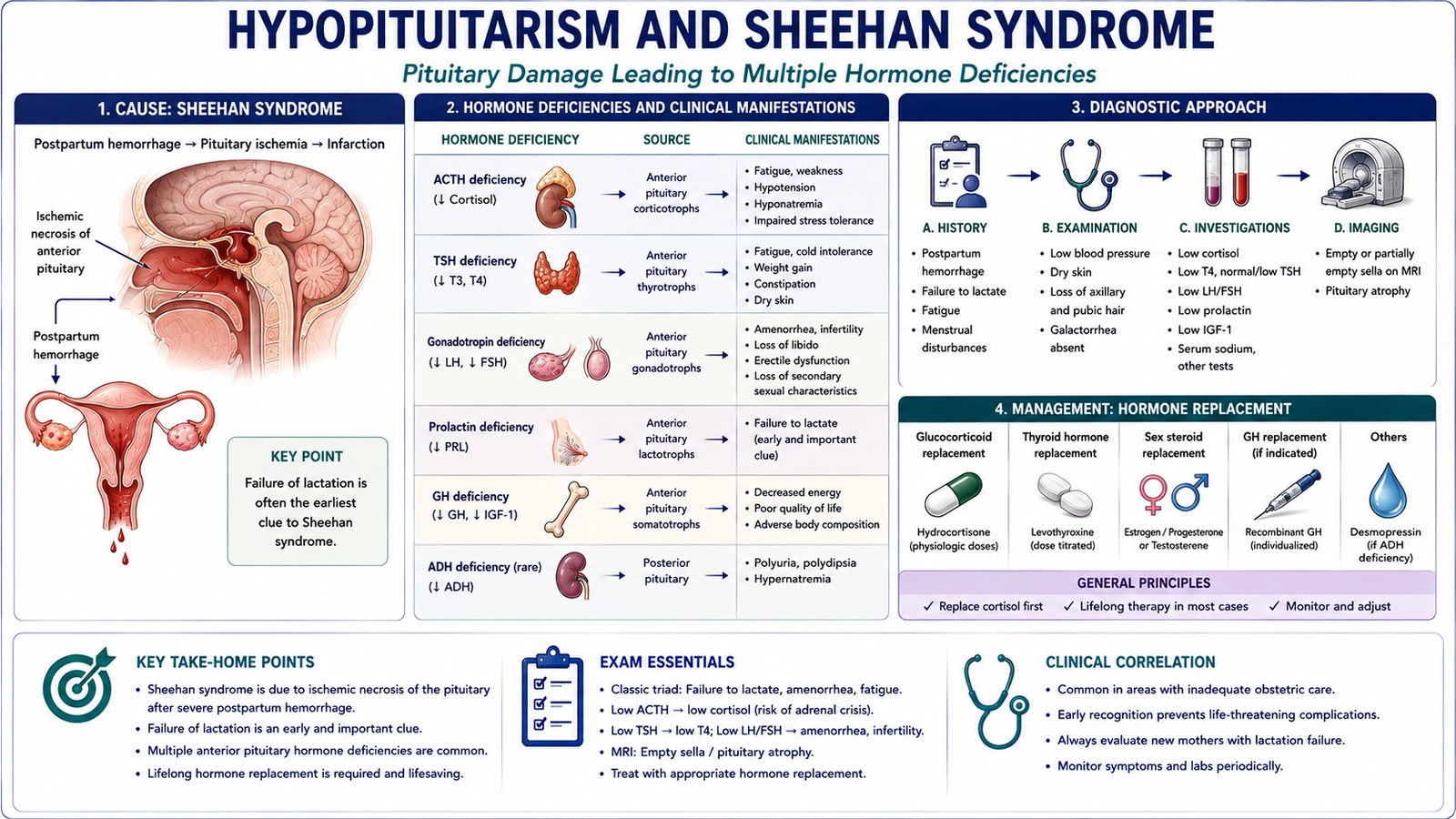
Hypopituitarism is deficiency of one or more pituitary hormones. Clinical manifestations depend on which hormone-producing cells are affected, the extent of damage and whether hormone loss develops suddenly or gradually.
Etiology
Pituitary function can be lost through destruction, compression or vascular injury. Important causes include:
- pituitary macroadenoma compressing normal pituitary tissue;
- pituitary surgery or radiotherapy;
- pituitary apoplexy;
- postpartum pituitary ischemic necrosis;
- inflammatory or infiltrative pituitary disease;
- structural or traumatic damage involving the hypothalamic-pituitary region.
Clinical Manifestations
The easiest way to understand hypopituitarism is to connect each missing pituitary hormone with loss of stimulation of its target tissue.
- ACTH deficiency → secondary adrenal insufficiency: fatigue, weakness, hypotension and impaired response to physiological stress.
- TSH deficiency → secondary hypothyroidism: fatigue, cold intolerance and other manifestations of reduced thyroid-hormone action.
- FSH/LH deficiency → hypogonadism: menstrual disturbance, infertility and loss of libido in women; reduced libido, erectile dysfunction and infertility in men.
- GH deficiency: reduced linear growth in children.
- Prolactin deficiency: failure of lactation after childbirth.
Because ACTH is low in pituitary disease, hyperpigmentation is not expected in secondary adrenal insufficiency. This contrasts with primary adrenal failure, where ACTH rises.
Sheehan Syndrome
During pregnancy, the anterior pituitary enlarges, mainly because lactotrophs increase in size and number. The enlarged gland becomes particularly vulnerable to a major reduction in blood flow.
Severe postpartum hemorrhage or hypotension → reduced pituitary perfusion → ischemic necrosis of anterior pituitary → pituitary hormone deficiencies
An early clue may be failure to lactate. Later manifestations can include amenorrhea, infertility, fatigue, symptoms of hypothyroidism and features of secondary adrenal insufficiency.
Investigations
Both the pituitary hormone and its target-gland hormone must be interpreted together. In central endocrine failure, the target hormone is low while the corresponding pituitary hormone is low or inappropriately normal. Depending on the clinical presentation, assessment may include cortisol and ACTH, free thyroid hormone and TSH, gonadal hormones with FSH/LH, prolactin and evaluation of the GH–IGF-1 axis. Pituitary MRI is appropriate when a structural lesion is suspected.
Management
Management includes treatment of the underlying cause where possible and replacement of deficient hormones. When both ACTH and TSH deficiency are suspected, glucocorticoid deficiency must be corrected before thyroid-hormone replacement, because increasing metabolic demand in a cortisol-deficient patient can precipitate adrenal crisis. Other deficiencies are replaced according to clinical need, including sex hormones and GH in appropriately selected patients. Long-term endocrine follow-up is important when multiple axes are affected.
G. Pediatric Short Stature and Growth-Hormone Deficiency
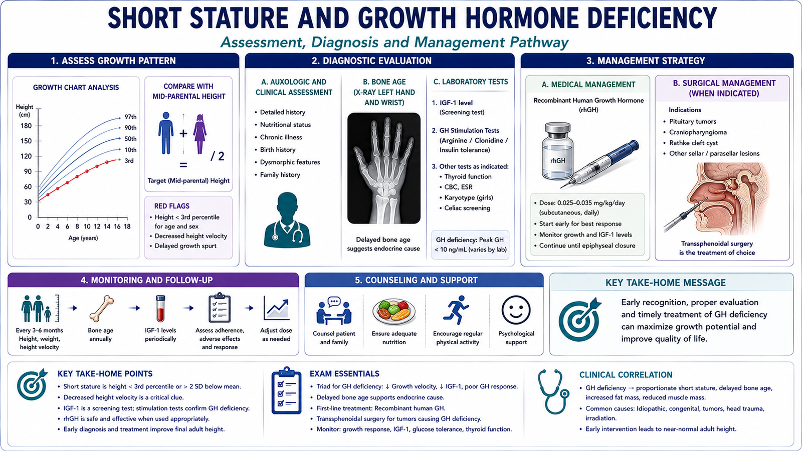
Short stature in a child cannot be interpreted from one height measurement alone. The key question is whether the child is growing normally over time and whether the growth pattern is appropriate for the child’s genetic potential. For this reason, height velocity, parental height, bone age and overall health are considered together.
Height Velocity
Height velocity is the rate at which a child gains height over time. Serial measurements plotted on a growth chart are therefore more useful than a single measurement. A child with familial short stature may remain short but continue to grow at an appropriate rate. In contrast, a child whose height velocity falls or who crosses downward through growth centiles requires evaluation for an underlying disorder.
Mid-Parental Height and Target Height
Parental height provides an estimate of the child’s genetic height potential. A commonly used calculation is:
This is an estimate rather than an exact prediction. It must be interpreted together with serial growth, pubertal stage and bone age.
Differentiating Major Causes of Growth Deficiency
Familial short stature usually shows short parents, relatively preserved growth velocity and bone age close to chronological age. Constitutional delay of growth and puberty usually shows delayed skeletal maturation and delayed puberty, but growth velocity is relatively appropriate once the child’s individual pattern is established. Growth-hormone deficiency produces reduced linear growth velocity. Bone age is commonly delayed, while body weight may be relatively preserved because GH deficiency affects linear growth more than nutritional weight gain. Chronic systemic illness or undernutrition often affects both height and weight. Poor weight gain may therefore point away from an isolated endocrine growth disorder. Abnormal body proportions or dysmorphic features should prompt consideration of skeletal or genetic causes rather than assuming an isolated GH problem.
Evaluation of Suspected GH Deficiency
Assessment begins with accurate serial height measurements, growth charts, family history, nutritional and systemic history, pubertal assessment and physical examination. Serum IGF-1 can support assessment of the GH axis. A random GH concentration is not reliable because GH secretion is pulsatile. When appropriate, specialist evaluation may include GH stimulation testing. Bone-age radiography helps assess skeletal maturation, while pituitary MRI is used when a hypothalamic-pituitary structural abnormality is suspected.
Management
Confirmed GH deficiency is treated with recombinant human growth hormone under specialist supervision. Response is assessed through improvement in height velocity and overall growth pattern. Other causes of short stature require treatment of the underlying disorder. Nutritional deficiency, chronic systemic disease and endocrine disorders such as hypothyroidism should not be treated simply by giving GH.
Communication and Multidisciplinary Care
Families should be told clearly that short stature is a finding, not a diagnosis. Explaining the child’s growth chart and growth velocity helps parents understand why repeated measurements are necessary and why a single hormone test cannot answer every question. Depending on the cause, management may involve pediatricians, pediatric endocrinologists, dietitians, genetic services and other relevant specialists. The child’s emotional wellbeing and family concerns should also be addressed respectfully.
H. Neurosurgical Management of Pituitary Adenomas
Surgery is an important component of pituitary-adenoma management when the tumor produces significant mass effect or when a functioning adenoma requires definitive tumor removal. The aim is not simply to remove tissue; the surgeon must relieve compression while preserving normal pituitary function and protecting nearby structures.
When Surgery Is Considered
Important indications include:
- optic-chiasm compression or progressive visual impairment;
- symptomatic enlarging macroadenoma;
- many GH-, ACTH- or TSH-secreting adenomas when resection is appropriate;
- selected tumors that remain inadequately controlled by medical therapy;
- selected cases of pituitary apoplexy with significant neurological or visual compromise.
Prolactinomas are an important exception. Because dopamine agonists can markedly reduce prolactin secretion and tumor size, medical treatment is generally preferred initially. Surgery is reserved for selected patients with treatment failure, intolerance or another compelling indication.
Microadenoma
A microadenoma is small and often confined to the pituitary. A functioning microadenoma may be removed selectively when surgical treatment is appropriate. Because the lesion is localized, there may be a greater possibility of preserving surrounding normal pituitary tissue.
Macroadenoma
A macroadenoma may expand beyond the sella, compress the optic chiasm or involve adjacent structures. Complete removal may therefore be more difficult. In a large compressive tumor, an important surgical objective is decompression of the optic apparatus, even when complete removal cannot safely be achieved.
Transsphenoidal Surgery
The standard route for many pituitary adenomas is transsphenoidal resection, commonly using an endoscopic endonasal approach. The pituitary is reached through the nasal cavity and sphenoid sinus. Detailed operative technique is beyond undergraduate requirements; the important principle is that this route provides access to the sella without a large open cranial exposure.
Important Complications
- Cerebrospinal-fluid leak if the skull-base barrier is breached.
- Diabetes insipidus from disturbance of the hypothalamic-posterior pituitary pathway.
- Hyponatremia from postoperative water-balance disturbance.
- New or worsened hypopituitarism from damage to functioning pituitary tissue.
- bleeding or vascular injury;
- infection;
- rare worsening of visual function.
Postoperative Monitoring
Postoperative assessment focuses on visual function, fluid balance, serum electrolytes and pituitary hormone status. Long-term follow-up is required to detect residual or recurrent tumor and to identify hormone deficiencies requiring replacement.
3. Integrated Mechanism Flow
4. Important Comparison
Acromegaly versus Gigantism
| Feature | Gigantism | Acromegaly |
|---|---|---|
| Timing of GH excess | Before epiphyseal closure | After epiphyseal closure |
| Dominant skeletal effect | Excessive linear growth | Bone and soft-tissue enlargement |
| Typical age | Childhood/adolescence | Adults |
| Common biochemical abnormality | Raised GH activity and IGF-1 | Raised GH activity and IGF-1 |
| Usual pituitary cause | GH-secreting adenoma | GH-secreting adenoma |
Pituitary Microadenoma versus Macroadenoma
| Feature | Microadenoma | Macroadenoma |
|---|---|---|
| Size | <10 mm | ≥10 mm |
| Mass effect | Usually limited | More likely |
| Optic-chiasm compression | Uncommon | Important concern with suprasellar extension |
| Hypopituitarism | Less likely from compression | More likely from compression |
| Surgical objective | Selective tumor removal when indicated | Tumor removal/debulking and decompression |