
This chapter follows the KMU learning outcomes and builds adrenal cortical hyperfunction in a logical sequence. First understand how normal adrenal hormones are produced and regulated, then connect hormone excess with clinical features, investigations and treatment. Use the final High-Yield Review only after understanding the main explanations.
Topic 10 — Adrenal Cortical Hyperfunction: Cushing Syndrome, Hyperaldosteronism and Adrenogenital Syndrome
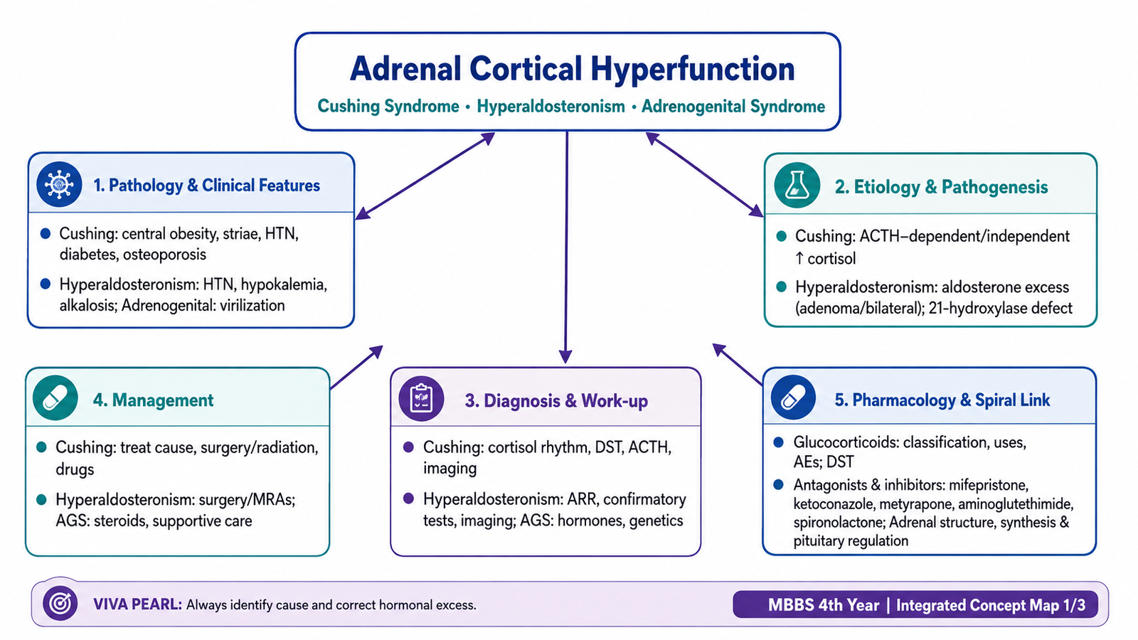
Understand how excessive adrenal cortical hormones produce characteristic clinical syndromes, how these disorders are investigated and managed, and how glucocorticoids and adrenal hormone antagonists are used therapeutically.
1. Topic Introduction
The adrenal cortex produces three major groups of steroid hormones: mineralocorticoids, glucocorticoids and adrenal androgens. Excess production of these hormones produces different clinical patterns. Excess cortisol causes Cushing syndrome, excess aldosterone causes primary hyperaldosteronism, and excessive adrenal androgen production is an important feature of the adrenogenital syndromes. Understanding these disorders becomes easier when the normal adrenal cortical zones, hormone synthesis and regulation are first understood. This chapter therefore connects normal adrenal physiology with the causes, clinical manifestations, diagnostic approach and management of adrenal cortical hyperfunction. It also explains the pharmacology of glucocorticoids, the dexamethasone suppression test, drugs that reduce cortisol synthesis or action, and the aldosterone antagonist spironolactone.
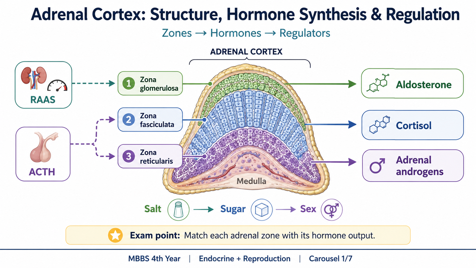
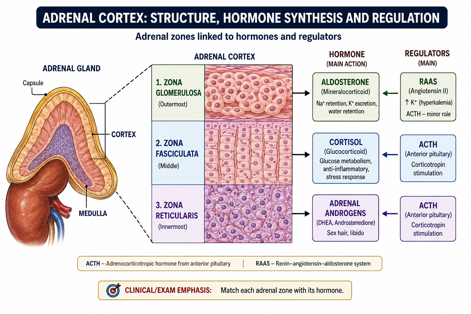
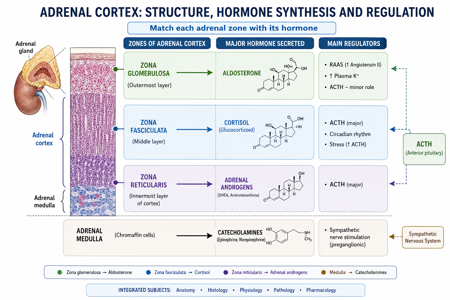
A. Adrenal Cortex: Structure, Hormone Synthesis and Regulation
The adrenal glands lie above the kidneys and consist of an outer cortex and an inner medulla. The cortical cells contain abundant lipid because cholesterol is the starting material for steroid hormone synthesis. The different cortical zones contain different steroidogenic enzymes, so each zone produces a characteristic group of hormones.
Gross and microscopic structure
The adrenal cortex forms the major outer portion of the gland and appears yellow because of its lipid-rich steroid-producing cells. Histologically, it is divided into three zones from outside inward.
| Zone | Microscopic arrangement | Main hormone | Main regulator |
|---|---|---|---|
| Zona glomerulosa | Cells arranged in rounded clusters beneath the capsule | Aldosterone | Renin–angiotensin system and plasma potassium |
| Zona fasciculata | Long cords of lipid-rich cells; the thickest cortical zone | Cortisol | ACTH |
| Zona reticularis | Smaller cells arranged in an interconnected network | Adrenal androgens | ACTH |
Synthesis of adrenal cortical hormones
All adrenal cortical hormones are steroid hormones derived from cholesterol. Cholesterol is first converted to pregnenolone. From this common starting point, different enzymes direct steroid synthesis toward aldosterone, cortisol or adrenal androgens.
Cholesterol → pregnenolone → enzyme-specific steroid pathways → aldosterone, cortisol or adrenal androgens
This enzyme-dependent pathway is clinically important. A tumor may produce too much of one hormone, while an inherited enzyme deficiency may block one pathway and divert steroid precursors into another pathway. This explains why some congenital adrenal disorders produce androgen excess even though the primary abnormality is an enzyme deficiency.
Regulation of cortisol
Cortisol is controlled mainly by the hypothalamic–pituitary–adrenal axis. The hypothalamus secretes corticotropin-releasing hormone, which stimulates the anterior pituitary to release adrenocorticotropic hormone (ACTH). ACTH acts mainly on the zona fasciculata and stimulates cortisol synthesis and secretion.
Cortisol then produces negative feedback on both the hypothalamus and anterior pituitary.
Cortisol secretion follows a daily rhythm and also rises during physiological stress. Therefore, abnormal cortisol production may disturb both its concentration and its normal daily pattern.
Regulation of aldosterone
Aldosterone is controlled differently. Its major regulator is the renin–angiotensin–aldosterone system. A fall in effective circulating volume stimulates renin release, leading to formation of angiotensin II. Angiotensin II stimulates zona glomerulosa cells to produce aldosterone. An increase in plasma potassium also directly promotes aldosterone secretion.
Aldosterone acts mainly on the distal nephron to increase sodium reabsorption and promote potassium and hydrogen-ion secretion. Therefore, excessive aldosterone tends to produce hypertension, potassium loss and metabolic alkalosis.
General pharmacodynamic principle of steroid hormones
Adrenocortical hormones are lipid-soluble. They diffuse through the cell membrane and bind to intracellular receptors. The hormone–receptor complex then interacts with DNA and changes gene transcription. Because many effects depend on synthesis of new proteins, steroid actions may develop more slowly than the effects of drugs acting directly on membrane ion channels, but they can persist after the circulating drug concentration begins to fall.
B. Cushing Syndrome: Etiology, Pathogenesis and Clinical Features
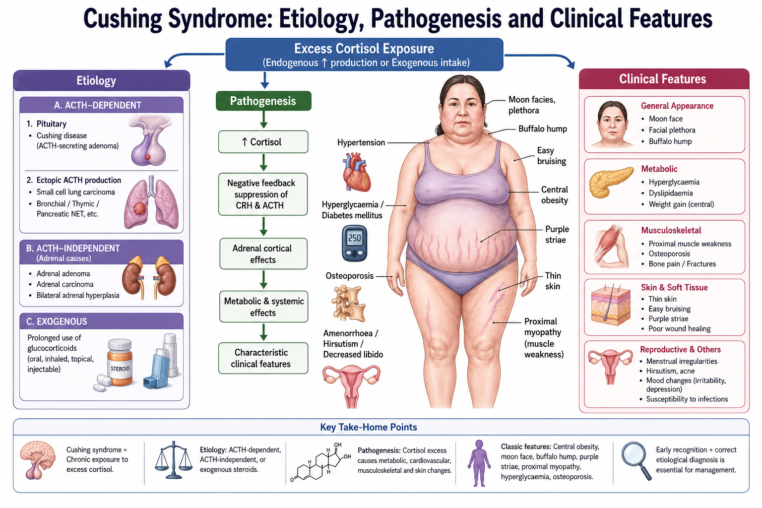
Cushing syndrome is the clinical state produced by prolonged excessive exposure to glucocorticoids, particularly cortisol. It may result from administration of glucocorticoid drugs or from excessive endogenous cortisol production. The term Cushing disease has a narrower meaning: it refers specifically to Cushing syndrome caused by an ACTH-secreting pituitary adenoma.
Etiology
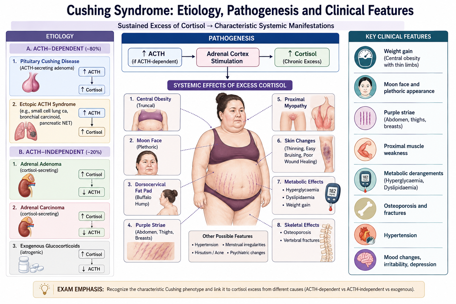
The first important distinction is between exogenous and endogenous Cushing syndrome. Prolonged therapeutic glucocorticoid exposure is an important cause of the syndrome. Endogenous Cushing syndrome is classified according to whether cortisol excess is driven by ACTH.
- Exogenous glucocorticoid exposure: prolonged administration of glucocorticoid drugs.
- ACTH-dependent endogenous disease:
- Pituitary ACTH-secreting adenoma — Cushing disease.
- Ectopic ACTH secretion from a non-pituitary tumor.
- ACTH-independent endogenous disease:
- Adrenal cortical adenoma.
- Adrenal cortical carcinoma.
- Autonomous nodular adrenal cortical disease.
How excess cortisol produces the clinical syndrome
Cortisol affects carbohydrate, protein and fat metabolism and also influences bone, skin, the immune system, blood pressure and the central nervous system. Cushing syndrome therefore affects several organ systems at the same time.
Cortisol increases hepatic glucose production and reduces effective glucose utilization in peripheral tissues. This produces insulin resistance and hyperglycaemia. At the same time, cortisol promotes protein catabolism. Loss of muscle proteins causes thin limbs and proximal muscle weakness.
Fat is redistributed rather than simply accumulating uniformly. Patients may develop central obesity, a rounded facial appearance and increased fat over the dorsocervical region while the limbs remain relatively thin.
Protein breakdown also reduces collagen and other structural proteins in the skin. The skin becomes thin, bruises easily and heals poorly. Stretching of weakened skin over expanding subcutaneous tissue produces the characteristic broad violaceous or purple striae.
Excess cortisol decreases bone formation, increases bone resorption and interferes with normal calcium balance. Over time, this contributes to osteoporosis and fractures. Its immunosuppressive action increases susceptibility to infection and may reduce the inflammatory response to infection.
In high concentrations cortisol can also produce mineralocorticoid-like effects, promoting sodium retention and potassium loss. This contributes to hypertension and, in some patients, hypokalaemia.
Clinical features
Cushing syndrome should be recognized as a pattern rather than by one isolated finding. Several common features such as obesity or hypertension are nonspecific, but the combination of characteristic fat redistribution, muscle weakness, skin changes and metabolic abnormalities strongly suggests glucocorticoid excess.
- Central weight gain with relatively thin extremities.
- Rounded facial appearance and facial fullness.
- Dorsocervical fat accumulation.
- Proximal muscle weakness due to protein catabolism.
- Thin skin, easy bruising and poor wound healing.
- Broad purple or violaceous abdominal striae.
- Hypertension.
- Glucose intolerance or diabetes mellitus.
- Osteoporosis and increased fracture risk.
- Increased susceptibility to infection.
- Menstrual disturbance, acne and hirsutism in women when androgen production is increased.
- Mood disturbance, irritability, depression or other psychological changes.
- Growth retardation in children despite weight gain.
When ACTH concentrations are markedly elevated, ACTH-related peptides may also stimulate melanocortin receptors, causing increased skin pigmentation. This finding points toward an ACTH-dependent process rather than an autonomous cortisol-producing adrenal tumor.
C. Cushing Syndrome: Diagnostic Work-up and Management
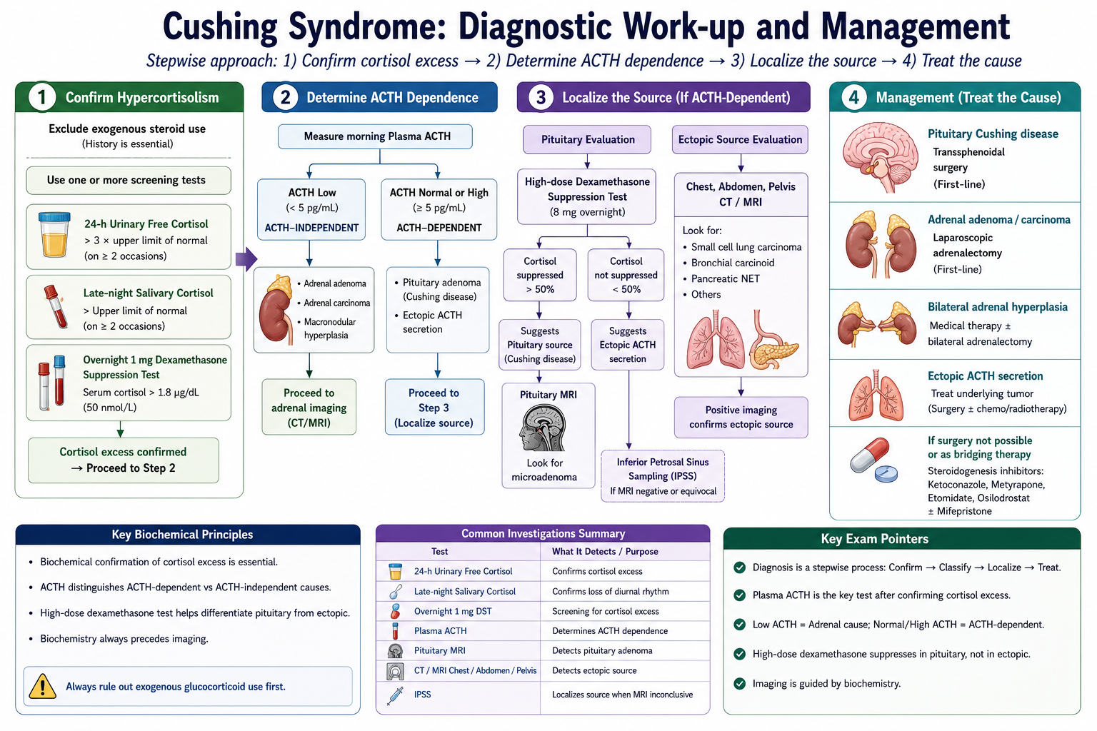
Investigation of suspected Cushing syndrome has two main purposes. First, the clinician must establish that pathological cortisol excess is actually present. Second, once hypercortisolism has been confirmed, its source must be identified. Imaging should therefore follow appropriate biochemical assessment rather than replacing it.
Step 1 — Exclude exogenous glucocorticoid exposure
A careful drug history is essential. Glucocorticoids may be taken orally, injected, inhaled or applied topically. If the clinical syndrome is caused by prescribed glucocorticoids, endogenous ACTH and cortisol production are often suppressed by negative feedback.
Step 2 — Demonstrate inappropriate cortisol excess
Normal cortisol secretion changes during the day and is suppressed by negative feedback. Tests for Cushing syndrome therefore assess either excessive total cortisol production, loss of the normal late-night fall in cortisol, or failure of cortisol to suppress appropriately after administration of dexamethasone.
- Urinary free cortisol: assesses cortisol production over an extended collection period.
- Late-night salivary cortisol: assesses whether the normal low nighttime cortisol level has been lost.
- Low-dose dexamethasone suppression testing: assesses whether cortisol is appropriately suppressed by glucocorticoid negative feedback.
Abnormal results should be interpreted in the clinical context because acute illness, major physiological stress and some medications can interfere with normal cortisol physiology or test performance.
Dexamethasone suppression test
Dexamethasone is a potent synthetic glucocorticoid. In a normal person, dexamethasone provides strong negative feedback to the hypothalamus and pituitary. ACTH secretion falls, so adrenal cortisol secretion also falls.
In endogenous Cushing syndrome, this normal suppressive response is impaired. Therefore, a low-dose dexamethasone suppression test is used as part of the assessment for autonomous cortisol excess.
A high-dose dexamethasone suppression test has historically been used after hypercortisolism is established. Pituitary corticotroph adenomas may retain some feedback responsiveness and therefore may show suppression at higher dexamethasone exposure, whereas ectopic ACTH production and autonomous adrenal cortisol production usually do not. However, this distinction is not completely reliable, so results must be interpreted together with ACTH measurement and other investigations.
Step 3 — Determine whether disease is ACTH dependent
Once endogenous cortisol excess has been established, plasma ACTH helps localize the abnormality.
| ACTH pattern | Interpretation | Next direction |
|---|---|---|
| Suppressed ACTH | Cortisol production is probably autonomous | Evaluate the adrenal glands |
| ACTH not suppressed | ACTH-dependent cortisol excess | Differentiate pituitary from ectopic ACTH production |
Pituitary imaging is appropriate when biochemical findings support ACTH-dependent disease. Adrenal imaging is appropriate when ACTH is suppressed. When pituitary and ectopic ACTH sources remain difficult to distinguish, specialist testing such as sampling of the inferior petrosal sinuses may be used.
Management principles
Treatment aims to remove or control the cause of cortisol excess while protecting the patient from complications such as hypertension, diabetes, infection, osteoporosis and thrombotic risk.
- Exogenous Cushing syndrome: reduce glucocorticoid exposure when clinically possible. Prolonged treatment should not be stopped abruptly because the hypothalamic–pituitary–adrenal axis may be suppressed.
- Pituitary Cushing disease: treatment is directed at the ACTH-secreting pituitary tumor, usually by specialist pituitary surgery.
- Adrenal cortisol-producing tumor: treatment is directed at the affected adrenal gland, usually by surgical removal when appropriate.
- Ectopic ACTH syndrome: identify and treat the underlying ACTH-producing tumor when possible.
- Persistent or severe hypercortisolism: drugs that inhibit cortisol synthesis or block glucocorticoid action may be used when definitive treatment is delayed, unsuccessful or unsuitable.
After successful treatment of longstanding endogenous hypercortisolism, endogenous ACTH–cortisol activity may remain suppressed temporarily. Patients therefore require appropriate endocrine monitoring and may need glucocorticoid replacement during recovery.
D. Primary Hyperaldosteronism
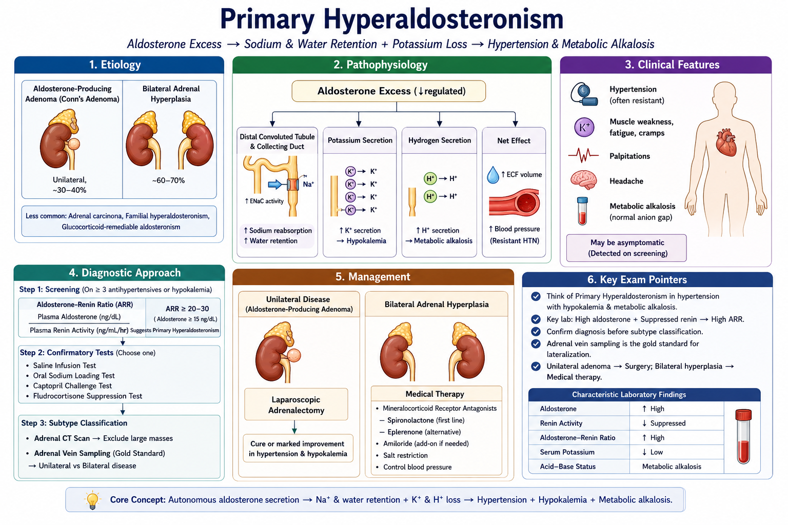
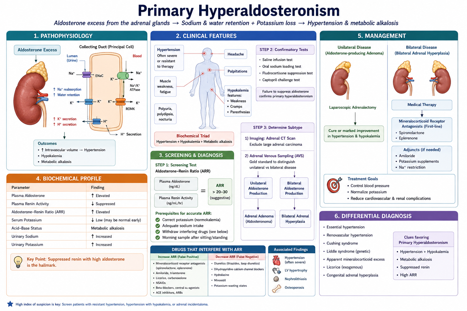
Primary hyperaldosteronism is a disorder in which the adrenal cortex secretes aldosterone autonomously or inappropriately despite suppression of the renin–angiotensin system. Aldosterone excess increases sodium reabsorption while increasing urinary potassium and hydrogen-ion loss. The resulting pattern is therefore hypertension with suppressed renin, with hypokalaemia and metabolic alkalosis developing in some patients.
Etiology
The major causes are autonomous aldosterone production from one adrenal gland or excessive secretion from both adrenal glands.
- Bilateral adrenal hyperplasia causing excessive aldosterone production.
- Aldosterone-producing adrenal adenoma.
- Familial forms of primary hyperaldosteronism.
- Rarely, an aldosterone-producing adrenal cortical carcinoma.
Pathophysiology
Aldosterone excess → increased potassium and hydrogen-ion secretion → hypokalaemia + metabolic alkalosis
Despite sodium retention, marked generalized edema is usually not a dominant feature. With continued volume expansion, compensatory mechanisms increase sodium excretion. This phenomenon is often described as aldosterone escape.
Clinical presentation
Hypertension is the central clinical feature. Hypokalaemia may occur spontaneously or become apparent during treatment with potassium-wasting diuretics, but a normal potassium concentration does not exclude primary hyperaldosteronism.
- Persistent or difficult-to-control hypertension.
- Hypokalaemia in some patients.
- Muscle weakness, cramps or fatigue when potassium depletion is significant.
- Polyuria and polydipsia may occur because marked potassium depletion impairs renal concentrating ability.
- Metabolic alkalosis due to increased hydrogen-ion secretion.
Diagnostic work-up
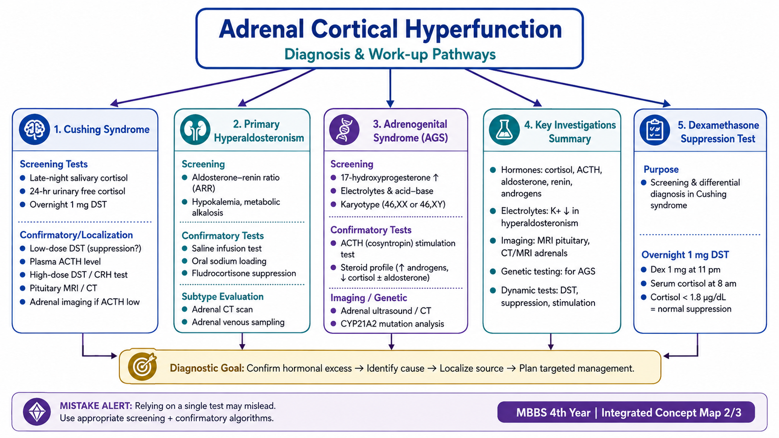
Investigation begins by demonstrating that aldosterone secretion is inappropriately high relative to renin. The aldosterone-to-renin relationship is therefore used for screening. A pattern of increased aldosterone activity together with suppressed renin raises suspicion of autonomous aldosterone secretion.
When screening is positive, a confirmatory suppression test may be used to demonstrate that aldosterone secretion fails to suppress normally. After biochemical confirmation, adrenal imaging assesses structural abnormalities.
Imaging alone cannot always determine whether one adrenal gland or both are responsible, particularly because incidental adrenal nodules become increasingly common with age. In selected patients being considered for surgery, adrenal venous sampling may therefore be used to determine whether aldosterone production is unilateral or bilateral.
Management
Treatment is determined mainly by whether the excessive aldosterone production is unilateral or bilateral.
- Unilateral aldosterone-producing disease: surgical removal of the affected adrenal gland is the definitive treatment when the patient is an appropriate surgical candidate.
- Bilateral adrenal hyperplasia: treatment is primarily medical with mineralocorticoid receptor blockade.
- Patients who are not suitable for surgery: mineralocorticoid receptor antagonists can control the effects of aldosterone excess.
Spironolactone is an important mineralocorticoid receptor antagonist used for this purpose and is discussed in detail later in the chapter.
E. Adrenogenital Syndrome
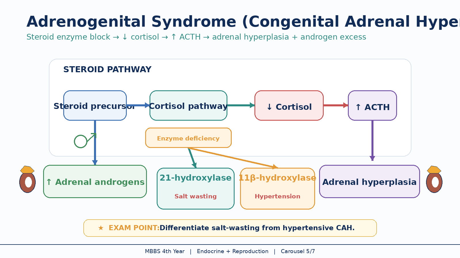
Adrenogenital syndrome refers to clinical manifestations produced by excessive adrenal androgen activity. An important cause is congenital adrenal hyperplasia (CAH), a group of inherited disorders in which deficiency of an enzyme required for cortisol synthesis alters adrenal steroid production. Reduced cortisol removes normal negative feedback on ACTH, so ACTH rises and chronically stimulates the adrenal cortex. Steroid precursors are then diverted into alternative pathways, commonly increasing androgen production.
Central mechanism
21-hydroxylase deficiency
21-hydroxylase deficiency is the major form of congenital adrenal hyperplasia associated with androgen excess. The enzyme is required in pathways leading to cortisol and aldosterone. Deficiency therefore reduces cortisol production and, in more severe disease, reduces aldosterone production. ACTH increases because cortisol feedback is lost, and steroid precursors are diverted toward adrenal androgen synthesis.
The clinical pattern depends on the severity of the enzyme deficiency and the patient’s sex.
- Genetic females: excessive fetal adrenal androgen exposure may produce varying degrees of virilization of the external genitalia.
- Genetic males: external genitalia may appear male at birth, so the disorder may initially be less obvious.
- Both sexes: excess androgen can cause rapid childhood growth and early sexual development, followed by premature epiphyseal maturation and reduced final adult height.
- Severe aldosterone deficiency: renal sodium loss can cause dehydration, hypotension, hyperkalaemia and potentially life-threatening salt-wasting adrenal crisis.
- Milder or non-classic disease: may present later with acne, hirsutism, menstrual irregularity or other manifestations of androgen excess.
11β-hydroxylase deficiency
Deficiency of 11β-hydroxylase also reduces cortisol production and increases ACTH-driven adrenal androgen production. However, steroid precursors with mineralocorticoid activity accumulate. Therefore, unlike the salt-wasting form of 21-hydroxylase deficiency, patients may develop hypertension and hypokalaemia together with androgen excess.
Diagnostic work-up
Diagnosis is based on the clinical pattern together with the steroid hormone profile. In suspected 21-hydroxylase deficiency, 17-hydroxyprogesterone is an important biochemical marker because it accumulates proximal to the enzymatic block. Electrolytes are particularly important when salt-wasting disease is suspected.
Assessment may include:
- 17-hydroxyprogesterone.
- Serum electrolytes and assessment of volume status.
- Adrenal androgen concentrations.
- Renin and aldosterone assessment where mineralocorticoid deficiency is suspected.
- Further endocrine and genetic assessment when required to define the enzyme defect.
Management
Treatment replaces deficient hormones and reduces excessive ACTH stimulation. Glucocorticoid replacement provides the missing glucocorticoid effect and restores negative feedback, which lowers ACTH and therefore reduces excessive adrenal androgen production.
- Glucocorticoid replacement is the main long-term therapy.
- Mineralocorticoid replacement is required when aldosterone deficiency is present.
- Patients with salt-wasting disease require appropriate sodium and fluid management.
- Acute adrenal crisis requires urgent glucocorticoid and fluid replacement.
- Long-term management requires monitoring of growth, development, androgen control and adequacy of hormone replacement.
- Patients with significant genital virilization require individualized multidisciplinary assessment and care.
F. Glucocorticoids: Classification, Pharmacology and Clinical Use
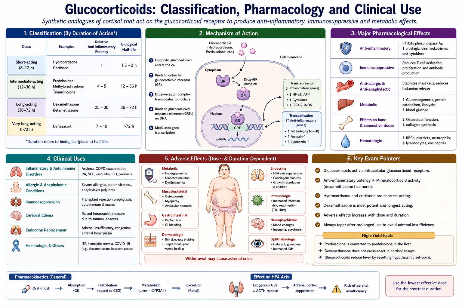
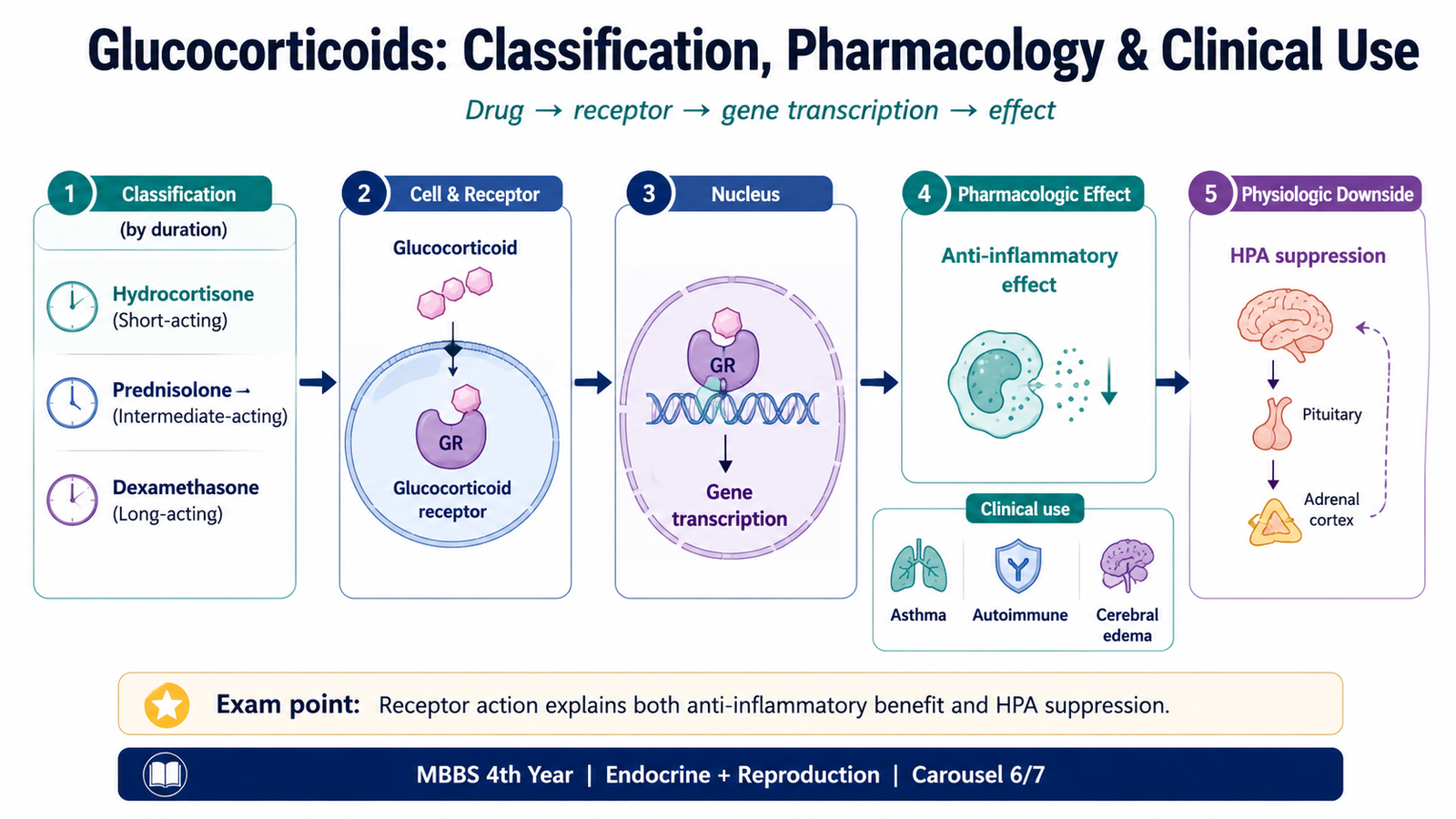
Glucocorticoid drugs reproduce some or most of the actions of endogenous cortisol. Their major clinical value comes from two properties: they can replace deficient glucocorticoid activity, and at higher pharmacological exposure they produce powerful anti-inflammatory and immunosuppressive effects. Different glucocorticoids vary in duration of action, glucocorticoid potency and mineralocorticoid activity.
Classification
| Duration group | Examples | Important principle |
|---|---|---|
| Short-acting | Hydrocortisone, cortisone | Hydrocortisone resembles endogenous cortisol and retains mineralocorticoid activity. |
| Intermediate-acting | Prednisone, prednisolone, methylprednisolone, triamcinolone | Commonly used for systemic anti-inflammatory and immunosuppressive therapy. |
| Long-acting | Dexamethasone, betamethasone | Potent glucocorticoid activity with very little mineralocorticoid activity. |
Mechanism of action
Glucocorticoids diffuse across the plasma membrane and bind to the intracellular glucocorticoid receptor. The activated receptor complex moves into the nucleus, where it changes transcription of many genes.
Glucocorticoids reduce production of inflammatory cytokines, decrease expression of several inflammatory enzymes and reduce migration and activation of inflammatory leukocytes. These actions explain why the drugs suppress inflammation across many different diseases rather than acting on only one inflammatory mediator.
Pharmacological effects
The same physiological actions that make glucocorticoids useful can cause toxicity when exposure is excessive or prolonged.
- Carbohydrate metabolism: increase hepatic glucose production and oppose insulin action, which can raise blood glucose.
- Protein metabolism: promote protein breakdown, contributing to muscle wasting and skin thinning during prolonged excess.
- Fat metabolism: alter fat distribution during chronic exposure.
- Inflammation: strongly suppress inflammatory mediator formation and leukocyte activity.
- Immune system: suppress cell-mediated and other immune responses.
- Bone: reduce bone formation and contribute to osteoporosis during long-term therapy.
- Vascular system: help maintain vascular responsiveness but excessive exposure contributes to hypertension.
- Hypothalamic–pituitary–adrenal axis: suppress CRH and ACTH through negative feedback.
Pharmacokinetic relevance
Many glucocorticoids can be given orally, while parenteral preparations are available when rapid or reliable systemic treatment is required. Local preparations may also be delivered by inhalation, topical application, intra-articular injection or other routes when treatment is directed toward a particular tissue.
Endogenous cortisol circulates largely bound to plasma proteins, particularly corticosteroid-binding globulin. Synthetic glucocorticoids differ in protein binding, metabolism and biological duration. They are mainly metabolized in the liver and their metabolites are eliminated predominantly through the kidneys.
Prednisone is converted in the body to the active drug prednisolone. Dexamethasone has a long biological duration and minimal mineralocorticoid effect, which makes it particularly useful when strong glucocorticoid action is required without significant sodium-retaining activity and when glucocorticoid negative feedback is being tested.
Clinical uses
Glucocorticoids are used when their physiological replacement effect or their anti-inflammatory and immunosuppressive action provides a clinical benefit.
- Hormone replacement: adrenal glucocorticoid deficiency and congenital adrenal hyperplasia.
- Inflammatory disorders: severe inflammatory diseases affecting different organ systems.
- Allergic and respiratory disease: suppression of significant airway or allergic inflammation.
- Autoimmune disease: reduction of damaging immune activity.
- Prevention or treatment of transplant-related immune reactions: as part of immunosuppressive therapy.
- Selected cerebral edema: dexamethasone may reduce vasogenic edema associated with intracranial tumors.
- Selected hematological and oncological conditions: glucocorticoids may be included in treatment regimens because of their lympholytic and anti-inflammatory actions.
Adverse effects
Toxicity is especially important with prolonged systemic therapy. Many adverse effects represent an exaggerated form of normal cortisol action.
- Cushingoid appearance and weight gain.
- Hyperglycaemia and worsening of diabetes.
- Hypertension and fluid retention, particularly with preparations having mineralocorticoid activity.
- Proximal muscle weakness and muscle wasting.
- Osteoporosis and fracture risk.
- Thin skin, bruising and poor wound healing.
- Increased susceptibility to infection and masking of inflammatory signs.
- Mood disturbance, insomnia and other neuropsychiatric effects.
- Peptic and gastrointestinal complications, particularly when additional risk factors are present.
- Cataract and raised intraocular pressure during prolonged exposure.
- Growth suppression in children.
- Suppression of the hypothalamic–pituitary–adrenal axis.
G. Drugs that Reduce Adrenocortical Hormone Action or Synthesis
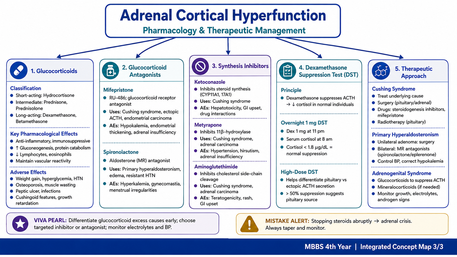
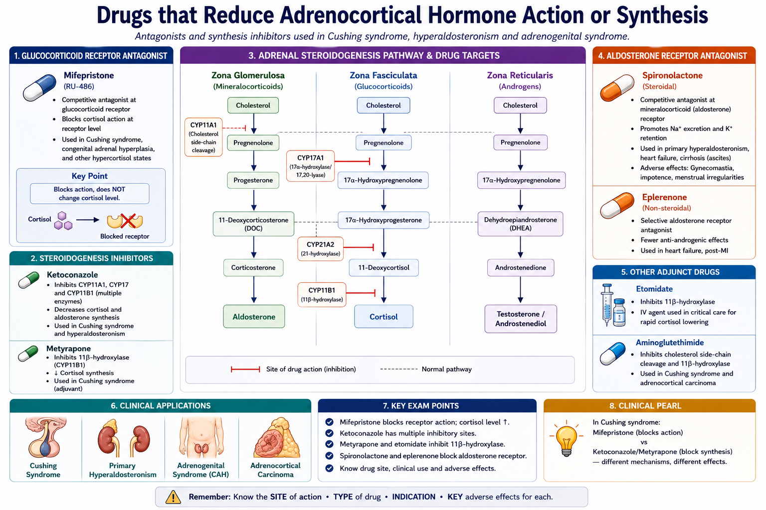
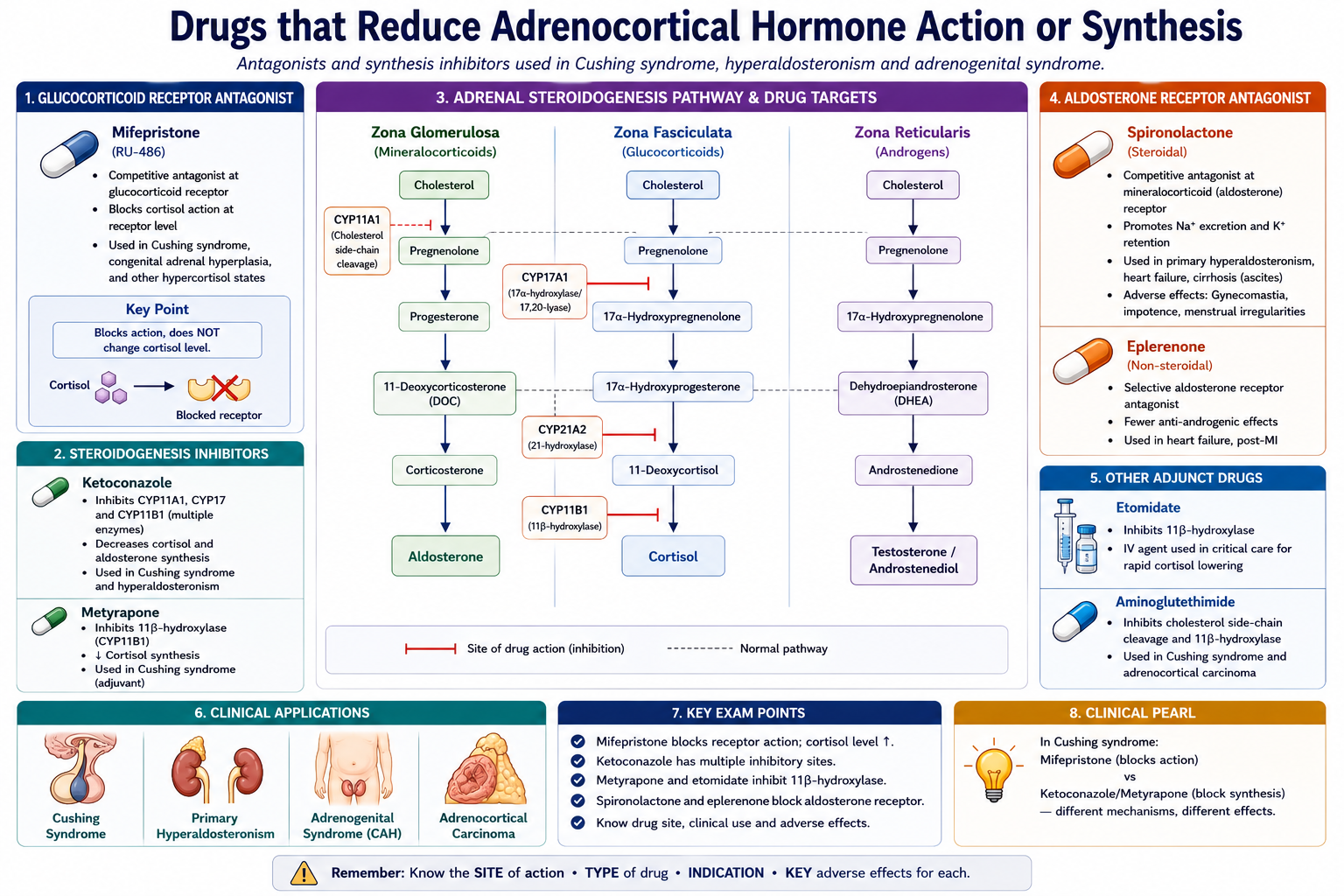
Hypercortisolism can be treated pharmacologically at two broad levels. A drug may reduce synthesis of adrenal steroids, or it may block the action of the excessive hormone at its receptor. These drugs are particularly useful when surgery cannot immediately correct the source of hormone excess, when disease persists after definitive treatment, or when surgery is unsuitable.
Classification
- Glucocorticoid receptor antagonist: mifepristone.
- Adrenal steroid-synthesis inhibitors: ketoconazole, metyrapone and aminoglutethimide.
- Mineralocorticoid receptor antagonist: spironolactone.
Mifepristone
Mifepristone acts as a glucocorticoid receptor antagonist. Instead of reducing cortisol production, it prevents cortisol from producing its usual effects at the receptor. This can improve manifestations of cortisol excess even though circulating ACTH and cortisol concentrations may remain elevated.
Mifepristone also antagonizes progesterone receptors. Its glucocorticoid-blocking action can be useful in selected patients with endogenous Cushing syndrome, particularly when clinically important metabolic manifestations persist and definitive treatment has failed or is unsuitable.
Important adverse effects include fatigue, nausea and manifestations related to excessive mineralocorticoid activity such as hypokalaemia and hypertension. Because cortisol remains elevated, cortisol concentration itself cannot be used as a simple marker of therapeutic effect. Its antiprogestational activity may also produce endometrial effects and uterine bleeding and has important implications in pregnancy.
Ketoconazole
Ketoconazole is an antifungal drug that, at clinically relevant systemic exposure, inhibits several cytochrome P450 enzymes involved in adrenal steroid synthesis. It therefore reduces cortisol production and may also reduce androgen synthesis.
It can be used to control endogenous cortisol excess when definitive treatment is delayed or unsuccessful. Important adverse effects include gastrointestinal symptoms and hepatotoxicity. Inhibition of androgen synthesis may contribute to gynecomastia and impaired gonadal function in men. Ketoconazole also has important drug-interaction potential through hepatic enzyme inhibition.
Metyrapone
Metyrapone inhibits adrenal 11β-hydroxylase, an enzyme required near the final step of cortisol synthesis. Cortisol production falls, while steroid precursors proximal to the blocked step increase.
It is used to reduce cortisol production in endogenous Cushing syndrome. Accumulation of steroid precursors can increase androgenic and mineralocorticoid effects. Consequently, adverse effects may include acne or hirsutism and worsening hypertension or hypokalaemia. Excessive suppression of cortisol can produce adrenal insufficiency.
Aminoglutethimide
Aminoglutethimide inhibits the conversion of cholesterol to pregnenolone, an early step required for synthesis of adrenal steroid hormones. It therefore has a relatively broad suppressive effect on adrenal steroidogenesis. It also inhibits aromatase.
Aminoglutethimide has historically been used to suppress adrenal steroid production, but its clinical role is now limited. Adverse effects include sedation, dizziness, skin rash and excessive adrenal suppression. Because the initial enzymatic step is inhibited, several adrenal steroid pathways may be affected rather than cortisol alone.
Spironolactone
Spironolactone is a competitive mineralocorticoid receptor antagonist. It acts mainly in aldosterone-sensitive parts of the distal nephron. By preventing aldosterone from activating its receptor, it reduces sodium reabsorption and decreases potassium and hydrogen-ion secretion.
Spironolactone is therefore particularly useful in primary hyperaldosteronism, especially bilateral adrenal hyperplasia or situations in which definitive surgery is not performed. It is also used in several other clinical conditions associated with harmful aldosterone effects.
The most important metabolic adverse effect is hyperkalaemia, because potassium excretion is reduced. Spironolactone can also interact with androgen and progesterone receptors, producing endocrine adverse effects such as gynecomastia, reduced libido or menstrual irregularities.
Integrated Mechanism Flow
Tumor, hyperplasia, exogenous steroid or enzyme defect
Cortisol, aldosterone or androgen abnormality
Metabolic, renal, vascular, skin, muscle or reproductive changes
Cushing syndrome, hyperaldosteronism or adrenogenital syndrome
Hormone pattern and regulatory feedback identify the pathway
Remove source, replace deficiency, inhibit synthesis or block receptor
Important Comparison
These three adrenal cortical disorders are easier to distinguish when the dominant hormone abnormality is identified first.
| Feature | Cushing syndrome | Primary hyperaldosteronism | Adrenogenital syndrome / CAH |
|---|---|---|---|
| Main abnormality | Excess glucocorticoid effect | Autonomous aldosterone excess | Steroid enzyme defect with excessive adrenal androgen production in major forms |
| Typical clinical clue | Central obesity, proximal weakness, purple striae | Hypertension with suppressed renin; hypokalaemia may occur | Virilization or premature androgen effects |
| Important mechanism | Cortisol excess causes catabolic, metabolic and immunosuppressive effects | Increased renal sodium retention with potassium and hydrogen-ion loss | Low cortisol → high ACTH → adrenal hyperplasia and precursor diversion |
| Key investigation principle | Confirm cortisol excess, then determine ACTH dependence | Demonstrate inappropriate aldosterone relative to suppressed renin | Steroid precursor profile; 17-hydroxyprogesterone is important in 21-hydroxylase deficiency |
| Management direction | Correct source of cortisol excess; pharmacological suppression/blockade when needed | Unilateral surgery or mineralocorticoid receptor blockade | Glucocorticoid replacement ± mineralocorticoid replacement |
⭐ AIM High-Yield Review
Adrenal Cortical Hyperfunction
Cushing Syndrome • Hyperaldosteronism • Adrenogenital Syndrome • Glucocorticoid Pharmacology
Watch the videos after reading the AIM Learning Material. Focus on the hormone abnormality, mechanism, characteristic clinical pattern, diagnostic logic and relevant pharmacology rather than trying to memorize every detail in the videos.
Cushing Syndrome — Diagnostic Work-up
Use this video to reinforce the causes of hypercortisolism and the diagnostic approach to suspected Cushing syndrome.
Educational source: Medicosis Perfectionalis
Hyperaldosteronism
Reinforce how excessive aldosterone alters renal sodium, potassium and hydrogen-ion handling and produces the characteristic biochemical and clinical pattern.
Educational source: Osmosis from Elsevier
Biochemistry of Congenital Adrenal Hyperplasia
This video is especially useful for understanding why enzyme defects cause low cortisol, increased ACTH and diversion of steroid precursors toward androgen production.
Educational source: Paul Bolin, M.D. — CRASH! Medical Review Series
Pharmacology of Glucocorticoids
Use this video to consolidate glucocorticoid receptor action, pharmacological effects, therapeutic uses and important adverse effects.
Educational source: Armando Hasudungan
Aldosterone Antagonism — Spironolactone
This video begins at the potassium-sparing diuretic section. Pay particular attention to spironolactone and how mineralocorticoid receptor blockade changes sodium and potassium handling.
Educational source: Armando Hasudungan
🎯 AIM Video Learning Targets
- Differentiate Cushing syndrome from Cushing disease.
- Understand how cortisol excess produces the characteristic Cushing phenotype.
- Follow the sequence: confirm hypercortisolism → determine ACTH dependence → localize the source.
- Connect aldosterone excess with hypertension, renin suppression and potassium loss.
- Explain why congenital adrenal enzyme defects produce increased ACTH and androgen excess.
- Connect glucocorticoid receptor action with therapeutic effects and long-term adverse effects.
- Explain why spironolactone is useful in primary hyperaldosteronism.
“`