
This chapter follows the KMU learning outcomes in a logical sequence and connects Pharmacology, Medicine and Pediatrics where the concepts naturally meet. First understand how insulin works and how metabolic emergencies develop; then use the final high-yield review for revision.
Topic 8 — Insulin Therapy and Acute Diabetic Emergencies: Hypoglycemia, DKA, HHS and Lactic Acidosis
Endocrine + Reproduction Module • Insulin pharmacology, recognition and treatment of acute metabolic emergencies, and early recognition of Type 1 diabetes in children.
1. Topic Introduction
Insulin therapy replaces or supplements the body’s own insulin and is essential in Type 1 diabetes and in several important situations in other forms of diabetes. Understanding insulin requires more than memorizing drug names: the student must know how different preparations behave, how insulin lowers blood glucose, and why excessive insulin can cause dangerous hypoglycemia. At the opposite extreme, severe lack of effective insulin can produce diabetic ketoacidosis or hyperosmolar hyperglycemic state. Lactic acidosis is another serious metabolic emergency that may occur in a patient with diabetes. This chapter also explains how Type 1 diabetes can be recognized early in children, including infants who may otherwise first present with severe dehydration, ketoacidosis, coma or death.
A. Insulin Preparations: Sources, Classification and Time-Action Profiles
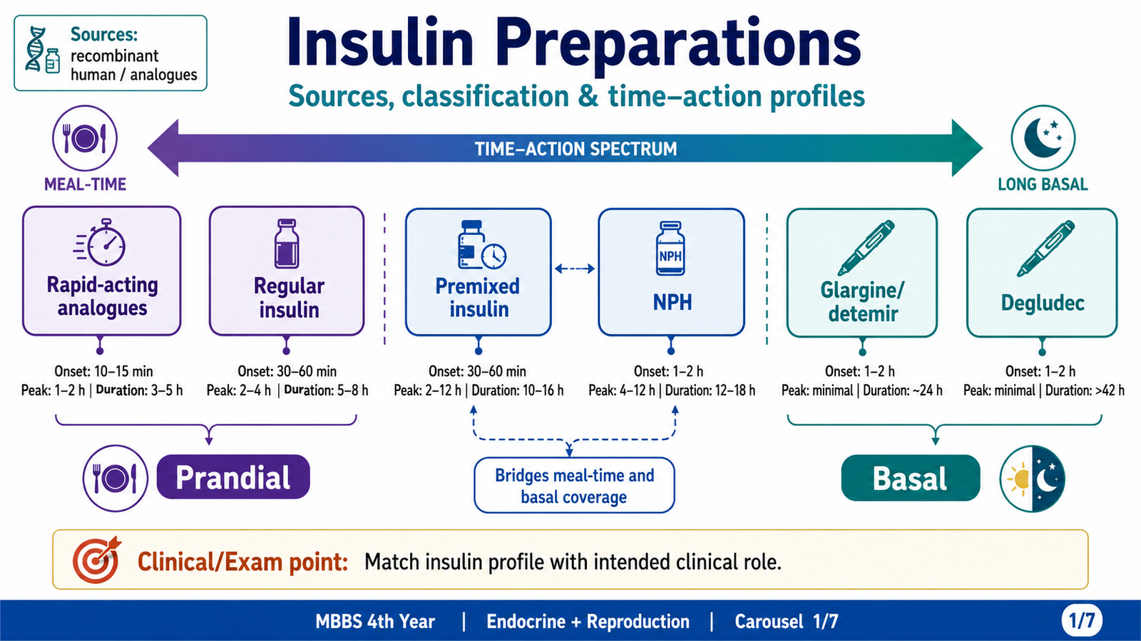
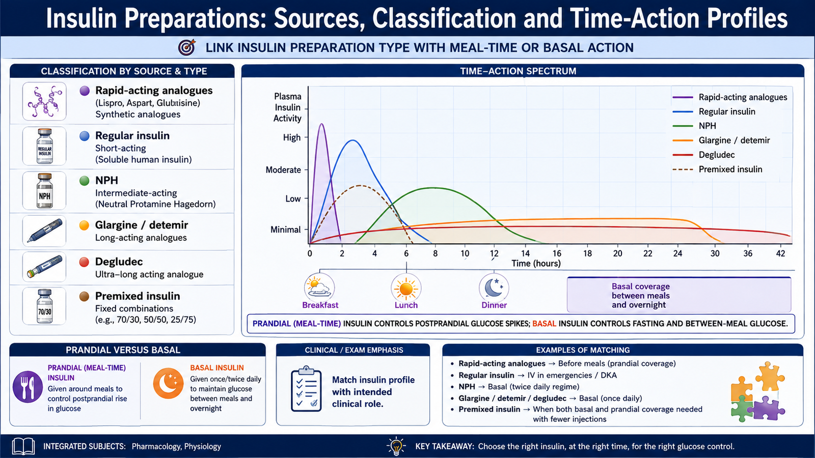
Insulin preparations are designed to reproduce two normal patterns of insulin secretion. Basal insulin controls glucose production between meals and during the night, while prandial or meal-time insulin controls the rise in glucose after food. Different preparations achieve these effects by changing how rapidly insulin is absorbed from the injection site and how long its action persists.
Sources of insulin
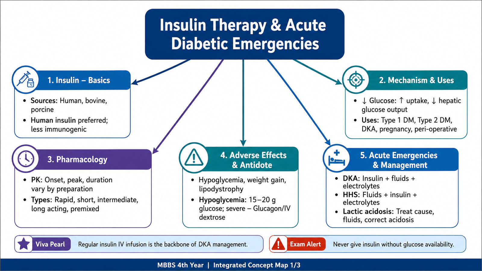
Insulin was historically extracted from bovine or porcine pancreas. Modern treatment mainly uses human insulin produced by recombinant DNA technology and recombinant insulin analogues. Recombinant human insulin has the same amino-acid sequence as naturally occurring human insulin, while analogues contain deliberate small sequence changes that alter absorption or duration of action.
| Source | Difference from human insulin | Clinical significance |
|---|---|---|
| Human insulin | Identical amino-acid sequence to endogenous human insulin | Low immunogenicity; modern standard preparation |
| Porcine insulin | Differs by one amino acid at B30: alanine replaces human threonine | Historically useful and less immunogenic than bovine insulin |
| Bovine insulin | Differs from human insulin at three amino-acid positions | More antigenic than porcine or human insulin; now largely historical |
The greater structural difference of bovine insulin explains its greater tendency to produce anti-insulin antibodies and allergic reactions. Animal-source insulin is therefore much less important in modern practice.
Classification by duration of action
| Class | Examples | Approximate profile | Main role |
|---|---|---|---|
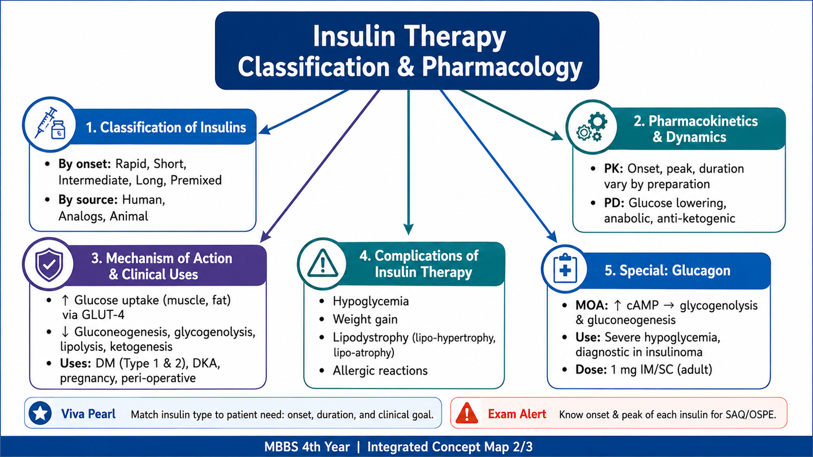
| Rapid-acting analogues | Lispro, aspart, glulisine | Begin within minutes, early peak, usually last about 3–5 hours | Meal-time glucose control |
| Short-acting | Regular insulin | Slower onset than rapid analogues; lasts several hours | Meal-time use; intravenous use in DKA |
| Intermediate-acting | NPH insulin | Delayed onset with a definite peak; commonly acts for much of the day or night | Basal coverage |
| Long-acting analogues | Glargine, detemir | Slow, relatively flat action with little or no pronounced peak | Basal insulin replacement |
| Ultra-long acting | Degludec | Very prolonged, relatively flat basal action extending beyond one day | Prolonged basal coverage |
| Premixed preparations | Fixed mixtures of prandial and intermediate components | Two components provide meal-related and background action | Simplified regimens but less flexibility |
Pharmacokinetic and pharmacodynamic principles
Insulin is a peptide hormone and would be digested in the gastrointestinal tract, so routine insulin is given parenterally, most commonly by subcutaneous injection. Absorption from subcutaneous tissue, rather than elimination from plasma, mainly determines the apparent duration of most injected preparations.
- Rapid analogues have structural changes that reduce self-association, allowing faster absorption.
- Regular insulin forms hexamers after injection and must dissociate before absorption, so its onset is slower.
- NPH is complexed with protamine, slowing absorption and producing an intermediate duration with a noticeable peak.
- Glargine forms a subcutaneous depot from which insulin is slowly released.
- Detemir is prolonged partly through albumin binding.
- Degludec forms a slowly absorbed subcutaneous depot and has a very prolonged action.
Subcutaneous absorption may vary with the injection site, local blood flow, exercise, temperature and injection technique. A pronounced insulin peak increases the possibility of hypoglycemia if food intake does not match insulin action.
B. Insulin Mechanism, Clinical Uses and Complications
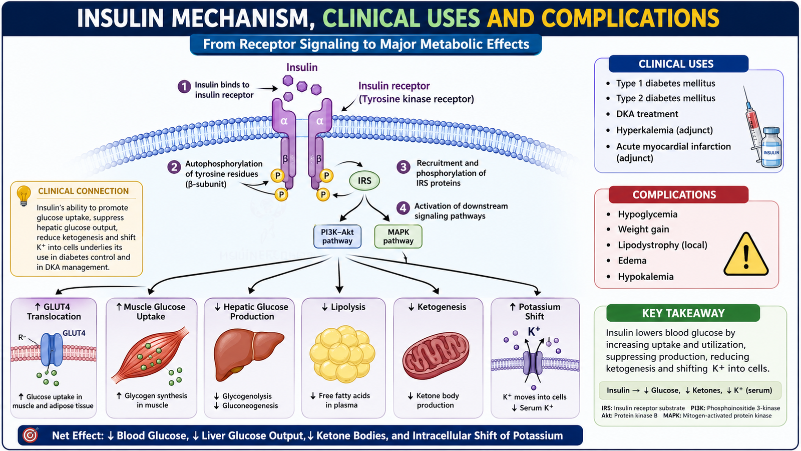
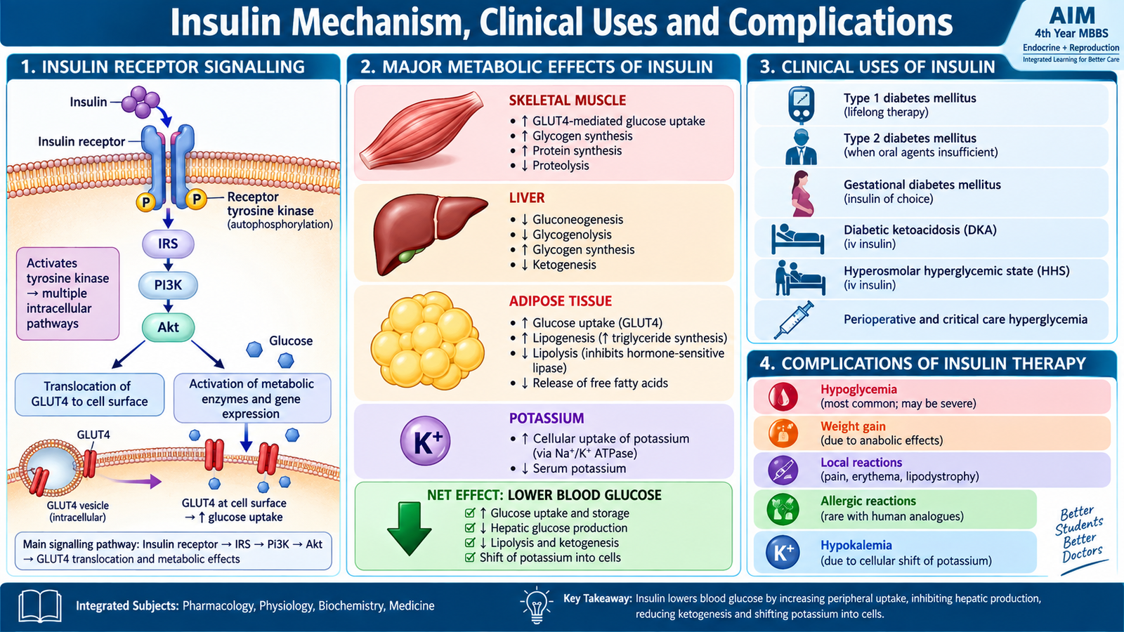
Insulin lowers blood glucose by coordinating the storage and use of nutrients. It does not simply “push glucose into cells.” It also reduces hepatic glucose production, suppresses fat breakdown and ketone formation, promotes protein synthesis and shifts potassium into cells.
Mechanism of action
Insulin has different but coordinated effects in major metabolic tissues:
- Skeletal muscle: increases glucose uptake, glycogen formation and protein synthesis.
- Adipose tissue: increases glucose uptake and triglyceride storage while suppressing lipolysis.
- Liver: promotes glycogen synthesis and suppresses gluconeogenesis, glycogen breakdown and ketogenesis.
- Potassium: promotes movement of potassium into cells, partly through stimulation of Na+/K+-ATPase activity.
The suppression of lipolysis and ketogenesis is particularly important in DKA. When insulin is severely deficient, free fatty acids reach the liver and are converted into ketone bodies, producing metabolic acidosis.
Clinical uses
Insulin is indispensable whenever the body cannot provide enough effective insulin to control metabolism.
- Type 1 diabetes mellitus: lifelong insulin replacement is essential because endogenous insulin production is severely deficient.
- Type 2 diabetes mellitus: insulin may be required when non-insulin therapy is insufficient, when hyperglycemia is severe or symptomatic, or during significant acute illness.
- Diabetic ketoacidosis: intravenous regular insulin suppresses ketogenesis and corrects the underlying insulin deficiency.
- Hyperosmolar hyperglycemic state: insulin is used after initial fluid resuscitation to control persistent severe hyperglycemia.
Complications of insulin therapy
The most important complication is hypoglycemia. It occurs when insulin action exceeds the amount of circulating glucose available from food or hepatic glucose production.
- Hypoglycemia: may progress from sweating and tremor to seizure, loss of consciousness and coma.
- Weight gain: results from improved glucose utilization, reduced urinary calorie loss and the anabolic action of insulin.
- Lipohypertrophy: repeated injections into the same site stimulate local fat accumulation and may make insulin absorption unpredictable.
- Lipoatrophy: loss of local subcutaneous fat is now uncommon with highly purified human insulin and analogues.
- Allergic reactions and insulin antibodies: much less common with modern human preparations.
- Hypokalemia: insulin moves potassium into cells and can lower extracellular potassium, especially during intensive treatment of DKA.
- Insulin edema: uncommon fluid retention may occur after initiation or intensification of insulin treatment.
C. Hypoglycemia, Hypoglycemic Coma and Glucagon
Hypoglycemia occurs when circulating glucose falls sufficiently to trigger autonomic symptoms or impair brain function. The brain depends heavily on circulating glucose, so severe or prolonged hypoglycemia can rapidly produce confusion, seizures, coma and neurological injury. In a person receiving insulin, treatment should not be delayed when severe hypoglycemia is strongly suspected.
Important causes
Insulin-induced hypoglycemia usually reflects a mismatch between the insulin dose and the patient’s immediate metabolic needs.
- Too much insulin or an incorrect insulin preparation.
- Delayed, missed or smaller-than-expected meal.
- Unplanned or prolonged exercise, because muscles consume more glucose.
- Vomiting or inability to eat after insulin has been given.
- Alcohol intake, particularly without food, because alcohol can suppress hepatic gluconeogenesis.
- Reduced insulin clearance, especially with renal impairment.
- Failure to adjust insulin during changing food intake or illness.
Clinical features
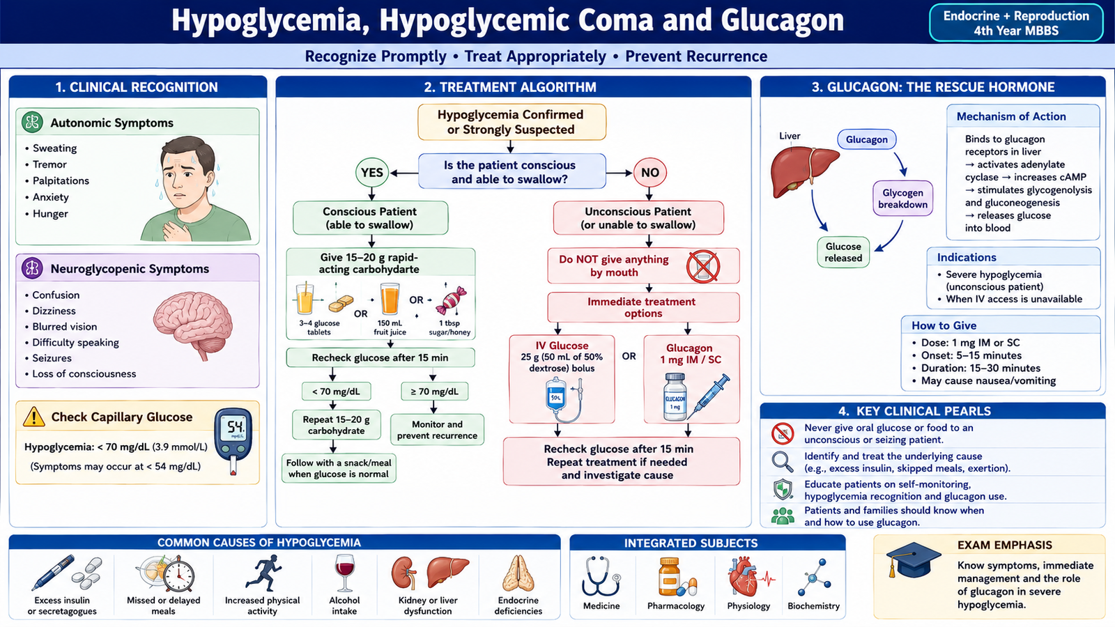
Symptoms occur through two main mechanisms. The counter-regulatory sympathetic response produces warning symptoms, while reduced glucose supply to the brain produces neuroglycopenia.
| Autonomic warning features | Neuroglycopenic features |
|---|---|
| Sweating, tremor, palpitations, hunger, anxiety | Difficulty concentrating, behavioral change, confusion, weakness, visual disturbance, seizure, coma |
A plasma glucose below about 70 mg/dL (3.9 mmol/L) is an important alert value in a person with diabetes, but severe hypoglycemia is defined clinically by the need for assistance rather than by one glucose number.
Management of insulin-induced hypoglycemia
Management depends mainly on whether the patient is able to swallow safely.
- Conscious and able to swallow: give rapidly absorbed oral glucose or another fast-acting carbohydrate, then reassess. Once recovered, further carbohydrate may be needed to prevent recurrence.
- Confused, unconscious, fitting or unable to swallow: do not give food or fluid by mouth because of aspiration risk. Give intravenous glucose when venous access is available.
- If intravenous access is not immediately available: glucagon can be used as rescue therapy.
- Recheck glucose and clinical response because hypoglycemia may recur, particularly after long-acting insulin.
- After recovery, identify and correct the cause: insulin error, missed food, exercise, renal dysfunction or another precipitating factor.
Glucagon: mechanism and clinical use
Glucagon is a physiological counter-regulatory hormone that raises blood glucose mainly by acting on the liver.
Its major clinical role in this topic is severe hypoglycemia when oral treatment is impossible and intravenous glucose is not immediately available. It may be given by an available parenteral or intranasal rescue preparation.
Glucagon depends substantially on hepatic glycogen stores. Its effect may therefore be reduced after prolonged fasting, severe liver disease or situations in which glycogen has been depleted. Nausea and vomiting may occur, so airway protection remains important in an unconscious patient.
D. Diabetic Ketoacidosis: Recognition, Diagnostic Work-Up and Treatment
Diabetic ketoacidosis (DKA) is an acute metabolic emergency caused by severe deficiency of effective insulin together with increased counter-regulatory hormones such as glucagon, catecholamines and cortisol. The combination causes hyperglycemia, dehydration, ketone production and metabolic acidosis. DKA is particularly associated with Type 1 diabetes, but it can occur in other forms of diabetes.
Precipitating factors
DKA usually develops because insulin has become insufficient for the patient’s metabolic needs.
- New presentation of Type 1 diabetes.
- Missed or interrupted insulin treatment.
- Infection, which increases counter-regulatory hormones and insulin requirements.
- Acute myocardial infarction, stroke, pancreatitis or other major physiological stress.
- Surgery or significant acute illness.
- Certain medications; SGLT2 inhibitors are an important example because DKA may occur with less striking hyperglycemia.
Pathophysiology
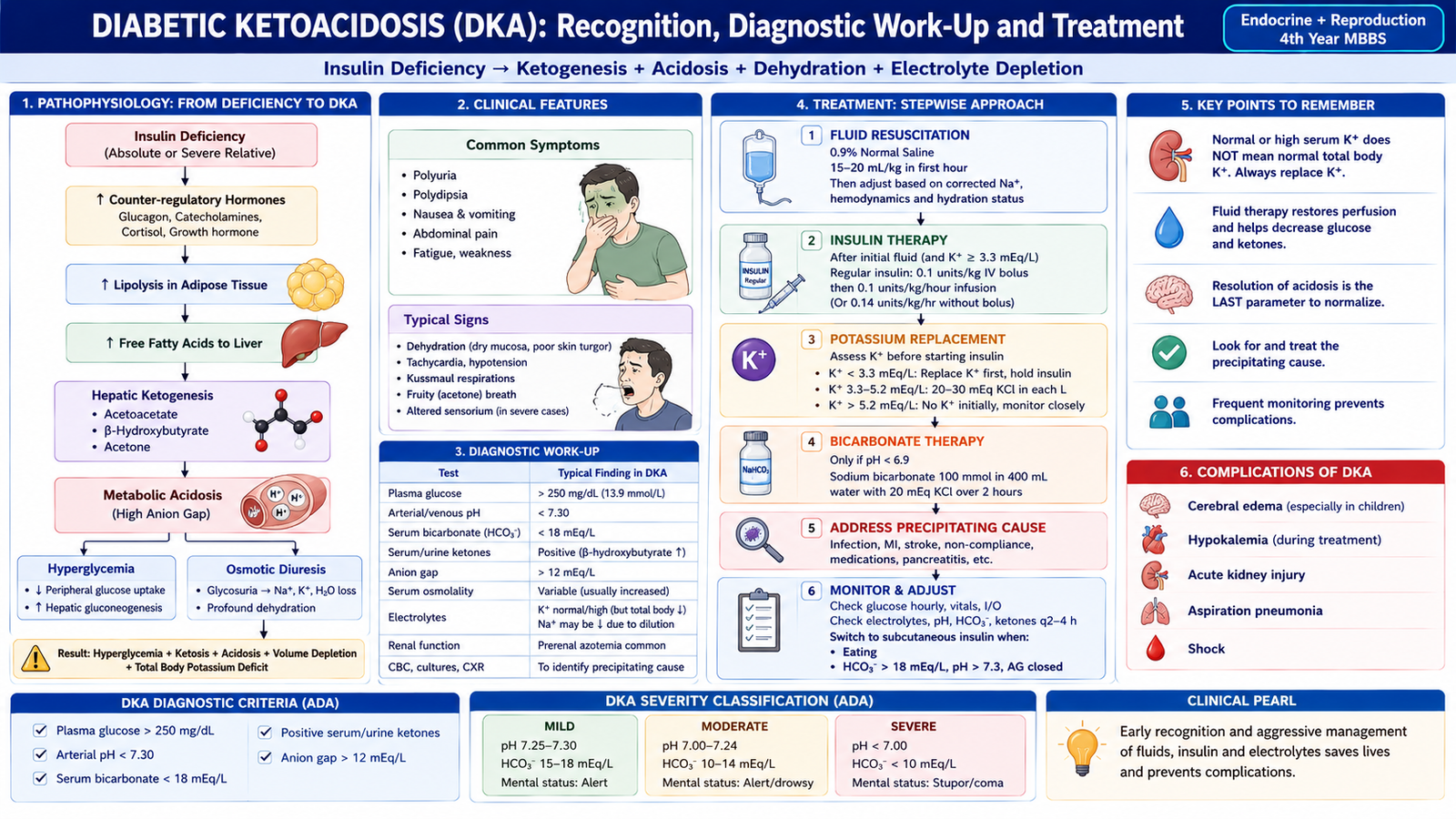
At the same time: insulin deficiency → lipolysis → free fatty acids → hepatic ketone production → metabolic acidosis.
Potassium deserves special attention. Osmotic diuresis causes substantial total-body potassium loss. However, acidosis and insulin deficiency shift potassium out of cells, so the initial serum potassium may be normal or even high. Once insulin is given, potassium moves back into cells and the serum level may fall rapidly.
Clinical features
- Polyuria and polydipsia from osmotic diuresis.
- Weight loss and weakness.
- Dehydration, dry mucosa, tachycardia and hypotension.
- Nausea, vomiting and abdominal pain.
- Kussmaul breathing: deep, rapid respiration produced as respiratory compensation for metabolic acidosis.
- Acetone or fruity odor of the breath may occur because volatile ketones are exhaled.
- Drowsiness, confusion and ultimately coma in severe disease.
Diagnostic work-up
The diagnostic work-up must demonstrate the three central abnormalities—hyperglycemia, ketosis and metabolic acidosis—while also determining dehydration, electrolyte disturbance and the precipitating cause.
- Blood glucose: demonstrates hyperglycemia, although severe hyperglycemia is not essential in every case.
- Blood ketones: measurement of β-hydroxybutyrate directly reflects the major circulating ketone and is useful for diagnosis and monitoring.
- Urinary ketones: may support diagnosis but do not always reflect current β-hydroxybutyrate accurately.
- Venous or arterial acid-base assessment: shows reduced pH and bicarbonate due to metabolic acidosis.
- Serum electrolytes: particularly sodium and potassium, guide safe fluid and potassium replacement.
- Urea and creatinine: help assess dehydration and renal function.
- Anion gap: is typically increased because unmeasured ketoacids accumulate.
- Investigations should also search for the trigger, such as infection or another acute illness.
Treatment and pharmacological management
DKA treatment corrects several problems simultaneously. The main principles are fluid replacement, insulin, potassium management, monitoring and treatment of the precipitating cause.
- Initial stabilization and fluids: assess airway, breathing and circulation and begin appropriate intravenous isotonic crystalloid replacement. Restoring intravascular volume improves renal perfusion and itself lowers glucose.
- Assess potassium before and during insulin therapy: severe potassium depletion must be corrected because insulin can cause a dangerous further fall in serum potassium.
- Intravenous regular insulin: suppresses hepatic ketogenesis, reduces glucose production and allows peripheral glucose utilization.
- Potassium replacement: is guided by repeated serum potassium measurements and renal function.
- Add glucose-containing fluid when necessary: as blood glucose falls, glucose may be added so that insulin can continue until ketosis and acidosis have resolved. Treatment must therefore not stop merely because the glucose concentration has normalized.
- Treat the precipitating cause: for example infection or interruption of insulin therapy.
- Monitor response: glucose, ketones, electrolytes, renal function and acid-base status must be followed repeatedly.
Bicarbonate is not a routine treatment for DKA. Insulin and restoration of circulation usually allow ketoacids to be metabolized and bicarbonate to regenerate. Bicarbonate is reserved for exceptional circumstances of extreme acidemia under specialist management.
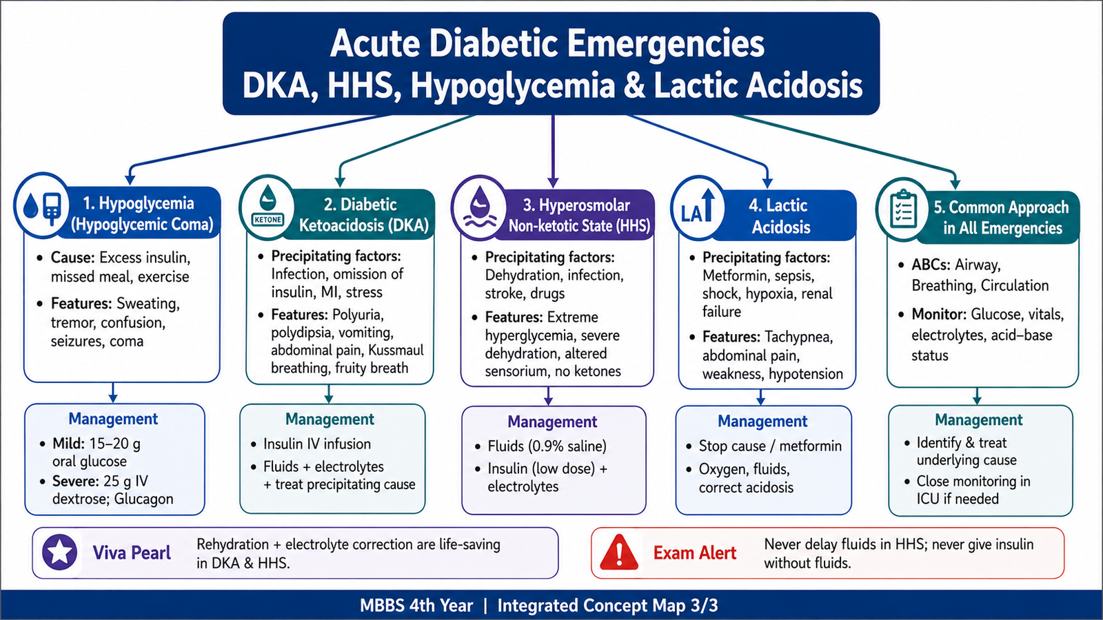
E. Hyperosmolar Hyperglycemic State: Recognition, Diagnostic Work-Up and Treatment
Hyperosmolar hyperglycemic state (HHS), historically called hyperosmolar non-ketotic diabetic coma, is characterized by severe hyperglycemia, profound dehydration and increased plasma osmolality. The modern term HHS is preferred because not every patient is comatose and small amounts of ketosis may occasionally be present.
Why HHS develops
In HHS there is usually enough insulin activity to suppress major lipolysis and ketone production, but not enough to control blood glucose. Hyperglycemia therefore becomes very severe. Large quantities of glucose are lost in the urine, pulling water and electrolytes with them.
Precipitating factors
- Infection.
- Previously undiagnosed or poorly controlled diabetes.
- Missed glucose-lowering treatment.
- Stroke, myocardial infarction or another major acute illness.
- Conditions that reduce access to water or impair the ability to respond to thirst.
- Drugs or illnesses that worsen hyperglycemia or dehydration.
Clinical features
The illness often evolves more gradually than DKA, allowing severe dehydration and hyperosmolality to develop before presentation.
- Polyuria and polydipsia early in the course.
- Marked dehydration, dry mucosa, tachycardia and hypotension.
- Weakness and profound volume depletion.
- Confusion, lethargy and reduced consciousness.
- Seizures or focal neurological abnormalities may occur because severe hyperosmolality affects brain function.
- Kussmaul respiration and prominent ketotic breath are less typical because major ketoacidosis is usually absent.
Diagnostic work-up
Investigations establish severe hyperglycemia and hyperosmolality while assessing electrolytes, renal function and the possibility of overlapping ketoacidosis.
- Blood glucose.
- Serum electrolytes, especially sodium and potassium.
- Serum osmolality or calculation of effective osmolality.
- Urea and creatinine to assess dehydration and renal function.
- Blood ketones and acid-base status to assess for DKA or mixed DKA/HHS.
- Investigations directed toward infection, myocardial infarction, stroke or another precipitating illness.
Treatment
The most important initial abnormality in HHS is usually profound water depletion. Fluid therapy therefore has an especially central role.
- Restore circulating volume with intravenous fluid. Correction should be controlled because very rapid changes in osmolality can be harmful.
- Monitor sodium, potassium, renal function and osmolality.
- Replace potassium when indicated.
- Use insulin after initial fluid therapy if substantial hyperglycemia persists. Fluids alone may initially produce a significant fall in glucose.
- Treat the underlying precipitant such as infection or acute vascular disease.
- Monitor neurological status and complications because severe hyperosmolality, thrombosis and circulatory compromise can be life-threatening.
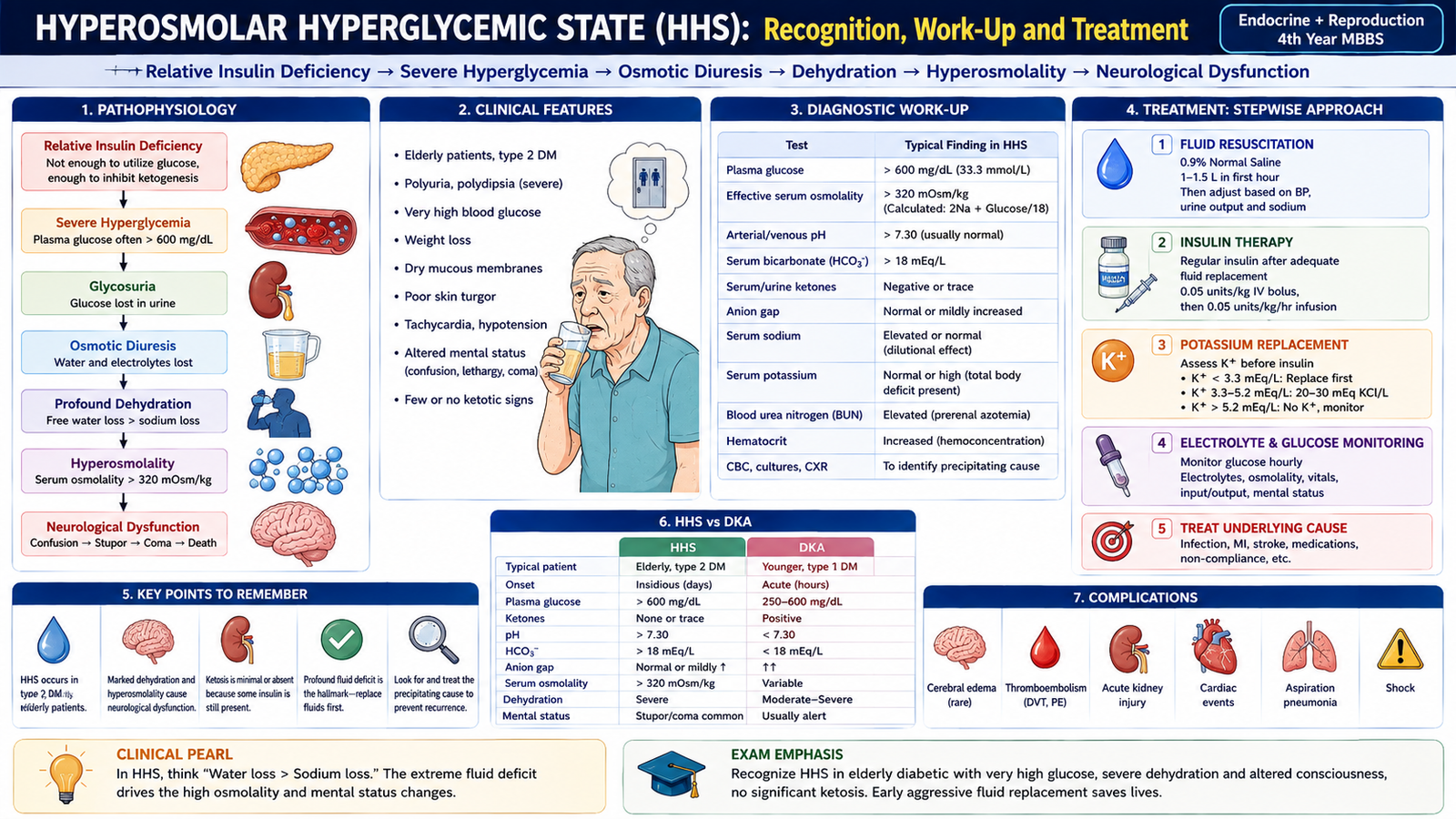
F. Lactic Acidosis in the Patient with Diabetes
Lactic acidosis is a high-anion-gap metabolic acidosis produced by accumulation of lactic acid. Lactate is normally formed during glucose metabolism and is cleared mainly by the liver and, to a lesser degree, the kidneys. It accumulates when production becomes excessive, clearance falls, or both occur together.
Precipitating factors
The most important distinction is whether lactate accumulation occurs because tissues are inadequately oxygenated or perfused, or despite the absence of obvious systemic hypoperfusion.
- Tissue hypoxia or hypoperfusion: shock, severe sepsis, major circulatory failure or severe hypoxemia increase anaerobic lactate production.
- Severe liver dysfunction: reduces lactate clearance.
- Renal failure: may contribute to impaired drug clearance and metabolic disturbance.
- Metformin-associated lactic acidosis: rare but important, particularly when metformin accumulates in renal dysfunction or when severe hypoxia, shock or another major illness is also present.
Clinical features
The manifestations often reflect both the acidosis and the illness causing it.
- Weakness and severe malaise.
- Nausea, vomiting or abdominal discomfort.
- Deep or rapid breathing as compensation for metabolic acidosis.
- Hypotension or poor peripheral perfusion when shock is present.
- Confusion, drowsiness or coma in severe cases.
Diagnostic work-up
Diagnosis requires evidence of metabolic acidosis together with an elevated blood lactate concentration and assessment of the underlying cause.
- Blood lactate.
- Venous or arterial blood gas for pH and bicarbonate.
- Serum electrolytes and calculation of the anion gap.
- Blood glucose and ketones to distinguish or identify accompanying DKA.
- Renal and liver function.
- Assessment for sepsis, shock, hypoxemia or another cause of impaired tissue perfusion.
- Medication history, particularly metformin use and circumstances that could reduce its clearance.
Treatment
The central treatment is correction of the process causing lactate accumulation rather than simply attempting to neutralize the acid.
- Stabilize airway, breathing and circulation.
- Correct hypoxemia and restore adequate tissue perfusion when present.
- Treat sepsis, shock or another underlying illness.
- Stop metformin or another relevant precipitating medication when drug-associated lactic acidosis is suspected.
- Correct fluid, electrolyte and glucose abnormalities appropriately.
- Severe metformin-associated lactic acidosis with significant drug accumulation or organ failure may require renal replacement therapy under specialist care.
Routine bicarbonate administration does not correct the underlying cause of lactate production and is therefore not a substitute for restoring circulation and treating the precipitating illness.
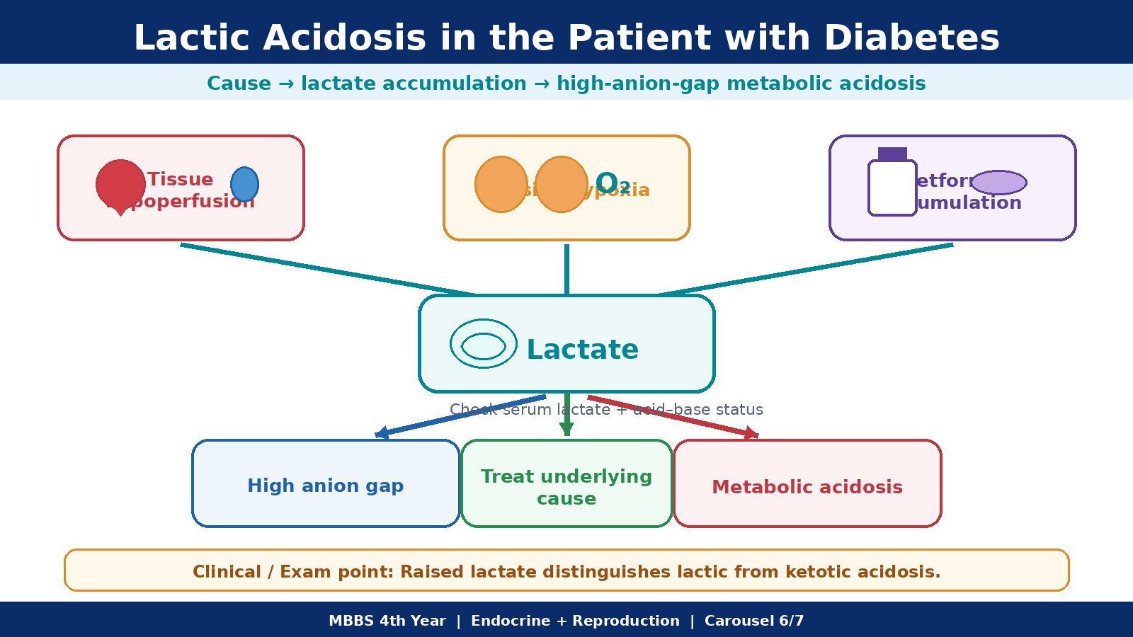
G. Type 1 Diabetes in Children: Early Recognition, Investigation and Initial/Continuing Care
Type 1 diabetes in children can progress rapidly from apparently simple symptoms to severe dehydration and DKA. Early recognition is therefore particularly important. Older children often describe classic symptoms clearly, but infants and young children may show only nonspecific illness, excessive wet nappies, weight loss or altered behavior.
Blood-glucose parameters useful for recognition
A bedside capillary glucose measurement is an effective immediate screening test when diabetes is suspected. Formal diagnosis is based on plasma glucose or an appropriately standardized HbA1c measurement.
| Parameter | Value indicating diabetes | Interpretation |
|---|---|---|
| Random plasma glucose | ≥200 mg/dL (11.1 mmol/L) | Diagnostic when accompanied by classic symptoms or hyperglycemic crisis |
| Fasting plasma glucose | ≥126 mg/dL (7.0 mmol/L) | Supports diagnosis when appropriately confirmed |
| 2-hour plasma glucose during oral glucose tolerance testing | ≥200 mg/dL (11.1 mmol/L) | Another accepted diagnostic criterion |
| HbA1c | ≥6.5% | Reflects chronic hyperglycemia when an appropriate standardized assay is used |
When hyperglycemia is unequivocal and a child has classic symptoms or a hyperglycemic emergency, urgent treatment should not be delayed for repeated diagnostic testing. In an asymptomatic child without unequivocal hyperglycemia, abnormal diagnostic testing generally requires confirmation.
Early clinical recognition
- Polyuria: glucose in urine produces osmotic diuresis.
- Polydipsia: increased thirst compensates for urinary water loss.
- New nocturia or secondary enuresis: a previously dry child may begin wetting the bed.
- Weight loss: calories are lost in urine and insulin deficiency promotes fat and protein breakdown.
- Fatigue and reduced school performance: reflect dehydration and impaired cellular energy utilization.
- Increased appetite may occur despite weight loss because glucose cannot be used normally by insulin-dependent tissues.
Presentation in infants and young children
Very young children cannot describe thirst, polyuria or weakness. Their presentation can therefore be mistaken for infection, gastroenteritis or another common childhood illness.
- Unusually heavy or frequently wet nappies.
- Persistent thirst or unusually frequent feeding for fluids.
- Poor weight gain or weight loss.
- Irritability, lethargy or reduced activity.
- Recurrent candidal diaper-area infection may occur in the presence of glycosuria.
- Vomiting, abdominal pain and dehydration as DKA develops.
- Deep rapid breathing, fruity breath, drowsiness, seizures or coma indicate advanced metabolic decompensation.
Investigations in a newly diagnosed child
The immediate question is not only whether the child has diabetes but also whether DKA is already present.
- Immediate bedside glucose followed by appropriate plasma glucose confirmation.
- Urine or blood ketones, especially if the child is unwell or glucose is markedly elevated.
- Venous blood gas and bicarbonate if DKA is suspected.
- Serum sodium, potassium, urea and creatinine to assess electrolyte disturbance and dehydration.
- HbA1c to estimate the degree of preceding hyperglycemia.
- Diabetes-associated autoantibodies may support Type 1 diabetes when confirmation of diabetes type is clinically necessary.
Management of newly diagnosed Type 1 diabetes
A child with DKA requires emergency management. A stable child without DKA still requires prompt initiation of insulin because Type 1 diabetes represents major insulin deficiency.
- Begin an appropriate insulin replacement regimen, commonly using basal and meal-time insulin components.
- Teach the family blood-glucose monitoring and interpretation.
- Teach recognition and immediate treatment of hypoglycemia.
- Explain the relationship between insulin, meals and physical activity.
- Teach injection technique and rotation of injection sites.
- Provide a sick-day plan, including more frequent glucose monitoring and ketone testing when the child is unwell.
- Ensure the family knows when vomiting, rising ketones, deep breathing or altered consciousness requires urgent medical assessment.
Assessment of a previously known diabetic child
When a child already receiving insulin becomes unwell or has poor glucose control, assessment should identify whether the problem is insufficient insulin, excess insulin, illness, food intake, activity or treatment technique.
- Review glucose records and recurrent patterns of hyperglycemia or hypoglycemia.
- Ask about missed insulin, timing of injections, meals and physical activity.
- Inspect injection sites for lipohypertrophy.
- Assess growth, weight and general wellbeing.
- During illness, check glucose frequently and test ketones when hyperglycemia or symptoms suggest insulin deficiency.
- Do not simply stop basal insulin during illness. Insulin deficiency can rapidly lead to ketogenesis and DKA.
- Persistent vomiting, inability to maintain hydration, significant ketones, deep breathing or altered consciousness requires urgent hospital assessment.
Children with DKA require particularly careful fluid, electrolyte and neurological monitoring because cerebral edema is a feared pediatric complication. New headache, slowing heart rate, altered behavior, worsening consciousness or other neurological deterioration during treatment requires immediate senior assessment and emergency management.
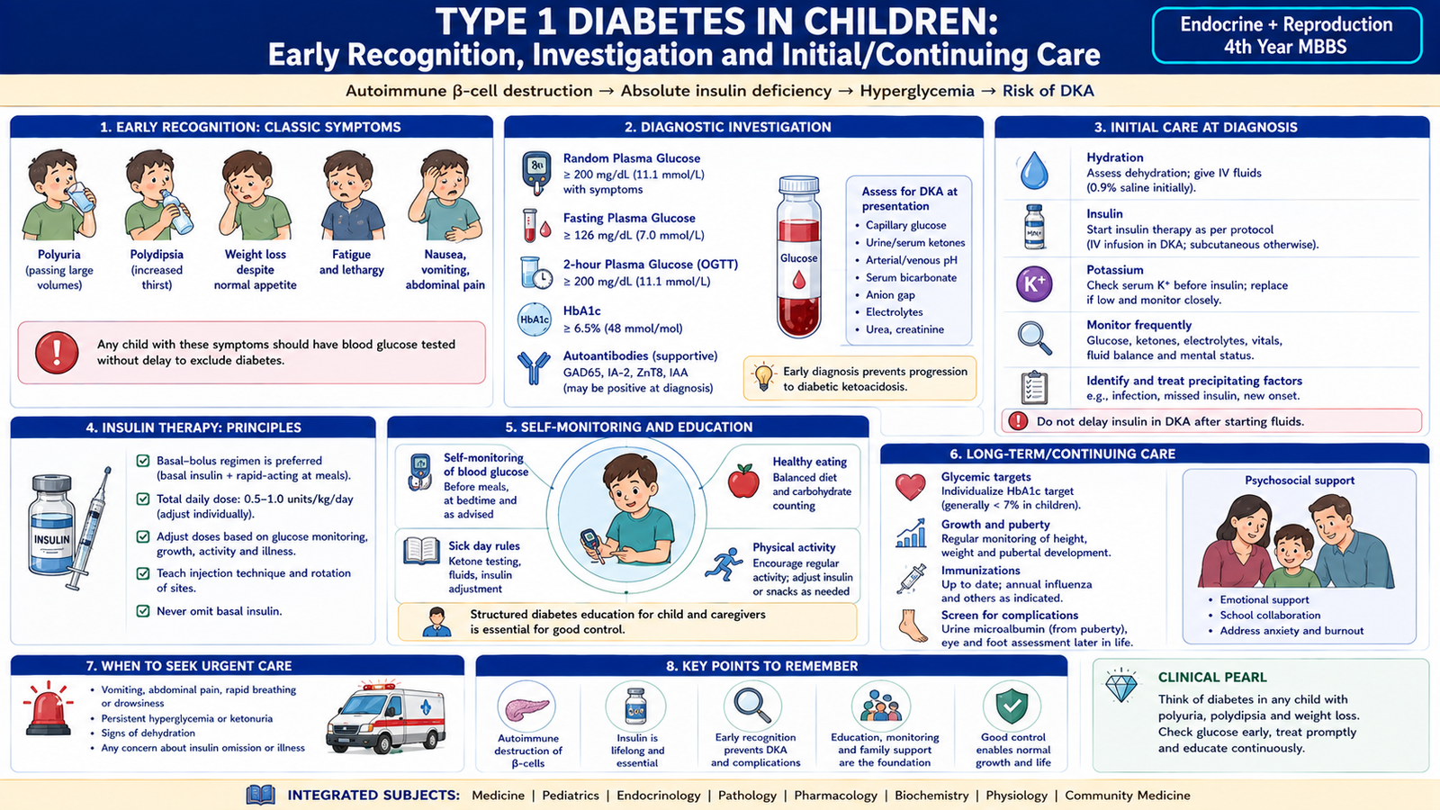
3. Integrated Mechanism Flow
Too much insulin or severe/relative insulin deficiency
Glucose falls in hypoglycemia or rises in insulin deficiency
Absolute deficiency causes ketogenesis; relative deficiency favors severe hyperosmolarity
Dehydration, potassium loss, acidosis or hyperosmolality
Confusion, seizure, neurological deterioration or coma
Glucose rescue, fluids, insulin, potassium and treatment of the precipitating cause
4. Important Comparison — DKA versus HHS
DKA and HHS are both hyperglycemic emergencies caused by inadequate insulin effect, but the degree of insulin deficiency determines whether severe ketogenesis or extreme hyperosmolality becomes dominant.
| Feature | DKA | HHS |
|---|---|---|
| Typical diabetes context | Common in Type 1 diabetes | More often associated with Type 2 diabetes |
| Effective insulin | Severely deficient | Insufficient for glucose control but often enough to limit major ketogenesis |
| Development | Usually more rapid | Often develops more gradually |
| Hyperglycemia | Present but not necessarily extreme | Usually very marked |
| Ketones | Prominent | Absent or relatively mild |
| Metabolic acidosis | Major feature | Usually absent or mild unless another process coexists |
| Dehydration | Significant | Usually more profound |
| Neurological disturbance | Occurs with severe illness | Often prominent because of marked hyperosmolality |
| Treatment emphasis | Fluids + insulin to stop ketogenesis + potassium management | Careful fluid replacement is especially important; insulin follows initial fluids |
⭐ 5. AIM High-Yield Review
Insulin Therapy & Acute Diabetic Emergencies
Use these focused videos after completing the learning material to reinforce insulin pharmacology, hypoglycemia, DKA and HHS.
Insulins — Pharmacology
Covers insulin physiology, mechanism of action, major preparations, time-action profiles and important adverse effects.
Diabetes Mellitus & Diabetic Ketoacidosis
Explains the metabolic basis of diabetes and connects insulin deficiency with lipolysis, ketogenesis, metabolic acidosis and DKA.
Hyperosmolar Hyperglycemic State
Covers precipitating factors, severe hyperglycemia, hyperosmolality, dehydration, neurological manifestations, investigations and management.
Approach to Hypoglycemia
Reinforces clinical recognition of hypoglycemia, severe neuroglycopenia and the emergency approach including glucose treatment.
“`