
Hyperthyroidism and Graves Disease: Pathology, Clinical Evaluation and Antithyroid Pharmacotherapy
1. Topic Introduction
Hyperthyroidism is a state in which the thyroid gland produces excessive thyroid hormones. The resulting increase in thyroid-hormone action affects almost every organ system, especially the cardiovascular, nervous and metabolic systems. Graves disease is the most important autoimmune cause of hyperthyroidism and has additional characteristic features such as diffuse thyroid enlargement and ophthalmopathy. To understand the topic clearly, we will first review normal thyroid structure, hormone synthesis and pituitary regulation. We will then connect the causes and pathogenesis of hyperthyroidism with morphology, clinical features and investigations. Finally, we will study the major antithyroid therapies and the special mechanism and management principles of Graves ophthalmopathy.
A. Normal Thyroid Structure, Hormone Synthesis and Regulation
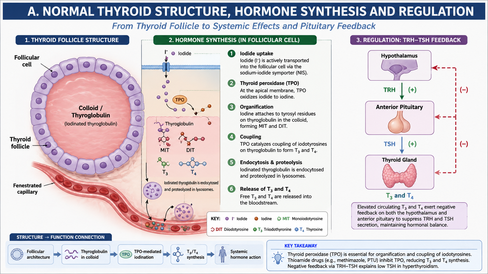
Understanding hyperthyroidism becomes much easier when the normal thyroid gland is understood first. The thyroid is an endocrine gland in the anterior neck. Its functional units are thyroid follicles, which manufacture, store and release the thyroid hormones thyroxine (T4) and triiodothyronine (T3). T4 is produced in greater quantity, while T3 is biologically more active.
Gross and Microscopic Structure
The thyroid consists of right and left lobes connected by an isthmus. Microscopically, the gland is composed of many spherical follicles. Each follicle is lined by follicular epithelial cells and contains central colloid, which is rich in thyroglobulin. Thyroglobulin acts as the protein framework on which thyroid hormones are synthesized and stored.
Parafollicular or C cells lie between or beside follicles and produce calcitonin. They are different from follicular cells and are not responsible for T3 and T4 synthesis.
How Thyroid Hormones Are Synthesized
Thyroid-hormone synthesis is a multistep process. Each step is important because several antithyroid drugs work by interrupting it.
Thyroid peroxidase (TPO) is particularly important. It participates in oxidation of iodide, organification of iodine and coupling reactions. This explains why inhibition of TPO by thioamide drugs reduces new thyroid-hormone synthesis.
Most circulating thyroid hormone is T4. In peripheral tissues, particularly the liver and other organs, some T4 is converted to the more active T3 by deiodinase enzymes.
Hypothalamic-Pituitary-Thyroid Regulation
The thyroid is controlled through a negative-feedback system. The hypothalamus releases thyrotropin-releasing hormone (TRH), which stimulates the anterior pituitary to release thyroid-stimulating hormone (TSH). TSH then stimulates thyroid follicular cells, increasing hormone synthesis, release and glandular growth.
When circulating free T3 and T4 become excessive, normal negative feedback suppresses TSH. Therefore, a low TSH is an important biochemical clue in primary hyperthyroidism.
Major Actions of Thyroid Hormones
Thyroid hormones increase metabolic activity and influence cardiovascular function, growth and nervous-system activity. Excess hormone therefore exaggerates these normal effects.
- Increase basal metabolic activity and oxygen consumption in many tissues.
- Increase heat production, explaining heat intolerance and sweating in hyperthyroidism.
- Increase cardiac responsiveness to catecholamines, contributing to tachycardia and palpitations.
- Increase metabolic turnover of carbohydrates, fats and proteins.
- Support normal growth and central nervous system development.
B. Hyperthyroidism and Graves Disease: Etiology and Pathogenesis
Hyperthyroidism means excessive synthesis and secretion of thyroid hormones by the thyroid gland. Thyrotoxicosis is the clinical state produced by excessive thyroid-hormone action, regardless of where the hormone comes from. The terms are often used together, but they are not completely identical. For example, thyroiditis may cause thyrotoxicosis by leakage of preformed hormone without increased hormone synthesis.
Important Causes
The cause determines both the investigation pattern and the treatment approach. Graves disease causes generalized stimulation of the thyroid, whereas autonomous nodules produce hormone independently of normal pituitary control.
- Graves disease: autoimmune stimulation of the TSH receptor, usually producing diffuse hyperthyroidism.
- Toxic multinodular goitre: multiple autonomously functioning thyroid nodules produce excessive hormone.
- Toxic adenoma: a single autonomously functioning thyroid nodule produces excess hormone.
- Thyroiditis: damaged follicles release stored hormone, producing thyrotoxicosis without sustained increased synthesis.
- Exogenous thyroid hormone: excessive intake may produce thyrotoxicosis without an overactive thyroid gland.
Graves Disease: Core Mechanism
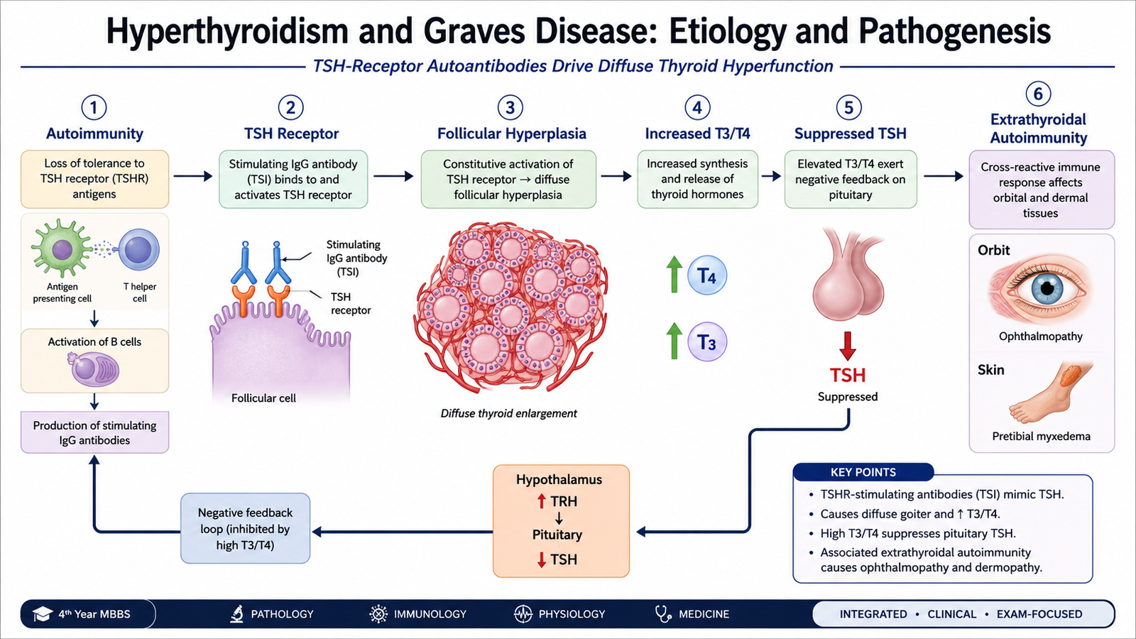
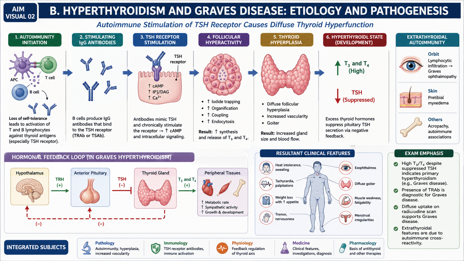
Graves disease is an autoimmune disorder. The immune system produces IgG autoantibodies directed against the TSH receptor on thyroid follicular cells. Instead of destroying the receptor, these antibodies stimulate it. The receptor therefore behaves as if it is continuously receiving a TSH signal.
The thyroid therefore becomes diffusely enlarged even though circulating pituitary TSH is low. This apparent paradox is important: the gland is being stimulated by autoantibodies rather than TSH itself.
The autoimmune process in Graves disease also affects tissues outside the thyroid. Orbital fibroblasts and other orbital cells participate in immune-mediated inflammation, producing Graves ophthalmopathy. Similar connective-tissue changes may produce localized dermopathy, classically over the shins.
C. Morphology and Clinical Features of Graves Hyperthyroidism
The pathological changes in Graves disease reflect continuous stimulation of thyroid follicular cells. Increased cell activity produces diffuse enlargement and characteristic microscopic changes. The increased circulating thyroid hormones then produce widespread metabolic and adrenergic manifestations.
Gross Morphology
The thyroid is usually diffusely and symmetrically enlarged. The enlargement results from hypertrophy and hyperplasia of follicular epithelial cells rather than the formation of a single dominant mass.
- The gland is enlarged throughout both lobes.
- The capsule generally remains intact.
- The gland may appear soft and fleshy because of increased cellularity and vascularity.
- Increased blood flow may be clinically appreciated as a thyroid bruit in an active gland.
Microscopic Morphology
Persistent receptor stimulation makes the follicular cells highly active. Instead of being flat or cuboidal, they become tall and crowded.
- Tall, crowded follicular epithelial cells indicate active hormone synthesis.
- The epithelium may form papillary infoldings into the follicular lumen. These lack the true fibrovascular cores of neoplastic papillae.
- Colloid is reduced and may show a scalloped margin because active follicular cells are rapidly taking it up.
- The interstitium may contain lymphocytes and lymphoid follicles with germinal centres, reflecting the autoimmune nature of the disease.
Clinical Features of Excess Thyroid Hormone
Most manifestations can be understood by remembering two effects: increased metabolic activity and increased sensitivity to catecholamines.
| System | Important Features | Why They Occur |
|---|---|---|
| General/metabolic | Weight loss despite preserved or increased appetite, heat intolerance, sweating | Increased metabolic rate and thermogenesis |
| Cardiovascular | Palpitations, tachycardia, increased pulse pressure; atrial fibrillation may occur | Increased β-adrenergic responsiveness and cardiac workload |
| Nervous system | Fine tremor, anxiety, irritability, restlessness, brisk reflexes | Enhanced adrenergic and neuronal activity |
| Muscle | Proximal muscle weakness | Increased protein turnover and catabolism |
| Gastrointestinal | Frequent bowel movements | Increased gastrointestinal motility |
| Reproductive | Menstrual disturbance and reduced fertility may occur | Excess thyroid hormone alters normal reproductive endocrine function |
Features Suggesting Graves Disease
Clinical hyperthyroidism can occur from several causes, but some findings particularly support Graves disease.
- Diffuse goitre: the whole gland is enlarged rather than a single nodule.
- Thyroid bruit: may be present because of increased vascularity.
- Graves ophthalmopathy: immune-mediated orbital disease producing proptosis and other ocular abnormalities.
- Pretibial myxedema: localized infiltrative dermopathy, usually over the anterior lower legs.
Lid retraction and lid lag may occur because of increased sympathetic activity and can accompany thyrotoxicosis from different causes. True Graves ophthalmopathy, however, results from an autoimmune orbital process and is therefore more specific to Graves disease.
D. Clinical Evaluation and Investigations
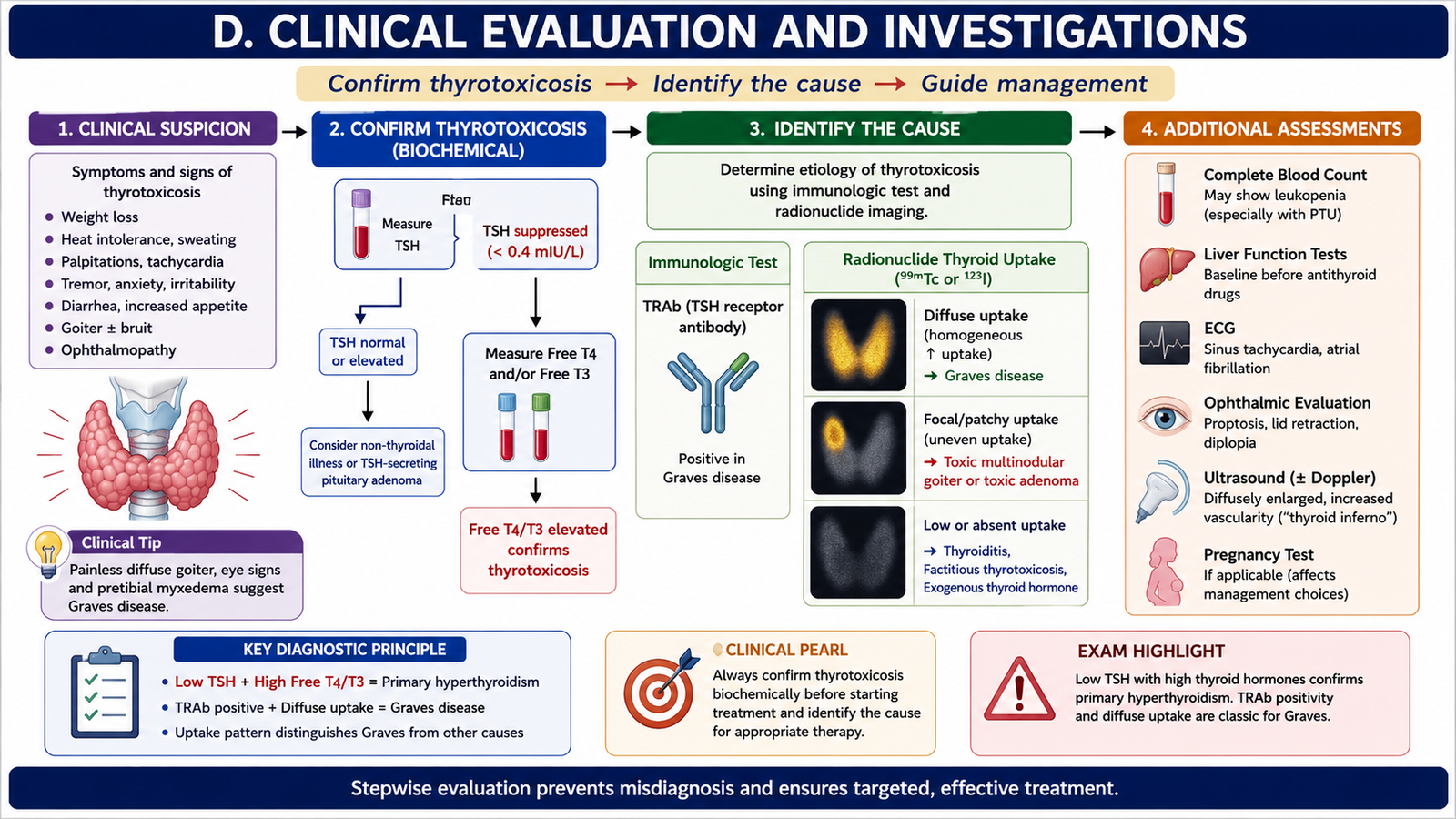
The clinical evaluation has two main goals: first, establish whether the patient truly has biochemical thyrotoxicosis; second, determine its cause. Symptoms alone are not enough because anxiety, weight loss, palpitations and tremor may occur in many other conditions.
History and Examination
History should identify the pattern and duration of symptoms and look for features that suggest the cause. Examination should confirm the physiological effects of excess thyroid hormone and assess the thyroid gland and eyes.
- Ask about weight change, appetite, heat intolerance and excessive sweating.
- Ask about palpitations, exercise intolerance and symptoms suggesting an arrhythmia.
- Assess tremor, nervousness, irritability and muscle weakness.
- Ask about bowel and menstrual changes where relevant.
- Look for tachycardia, fine tremor, warm skin and brisk reflexes.
- Examine the thyroid for diffuse or nodular enlargement and, when appropriate, increased vascularity.
- Examine the eyes for lid retraction, proptosis, ocular-movement limitation and other features of Graves ophthalmopathy.
Thyroid Function Tests
In primary hyperthyroidism, excessive thyroid hormone suppresses pituitary TSH by negative feedback. Therefore, the usual biochemical pattern is a suppressed TSH with increased free T4 and/or T3.
High circulating thyroid hormone → stronger negative feedback on anterior pituitary → low TSH.
Some patients have predominant elevation of T3 while T4 is less clearly increased. Therefore, measurement of T3 may help when clinical suspicion remains high despite a less striking T4 result.
TSH-Receptor Antibodies
Detection of TSH-receptor antibodies (TRAb) supports the diagnosis of Graves disease because these antibodies directly reflect the autoimmune process responsible for thyroid stimulation.
Radioactive Iodine Uptake and Thyroid Scintigraphy
Radioactive iodine uptake helps determine whether the gland is actively synthesizing excess hormone. A gland producing large quantities of new hormone takes up more iodine, whereas thyrotoxicosis caused by leakage of stored hormone shows reduced uptake.
- Graves disease: diffuse increased uptake throughout the gland.
- Toxic adenoma: focal increased uptake in an autonomous nodule with suppression of surrounding tissue.
- Toxic multinodular goitre: irregular or patchy areas of increased uptake.
- Thyroiditis or exogenous hormone: low uptake because new hormone synthesis is not driving the thyrotoxicosis.
Ultrasound
Ultrasound is particularly useful when the thyroid is nodular, when structural abnormalities need clarification, or when a palpable lesion requires characterization. Doppler assessment may demonstrate increased vascularity in Graves disease, but biochemical tests remain central to diagnosing hyperthyroidism.
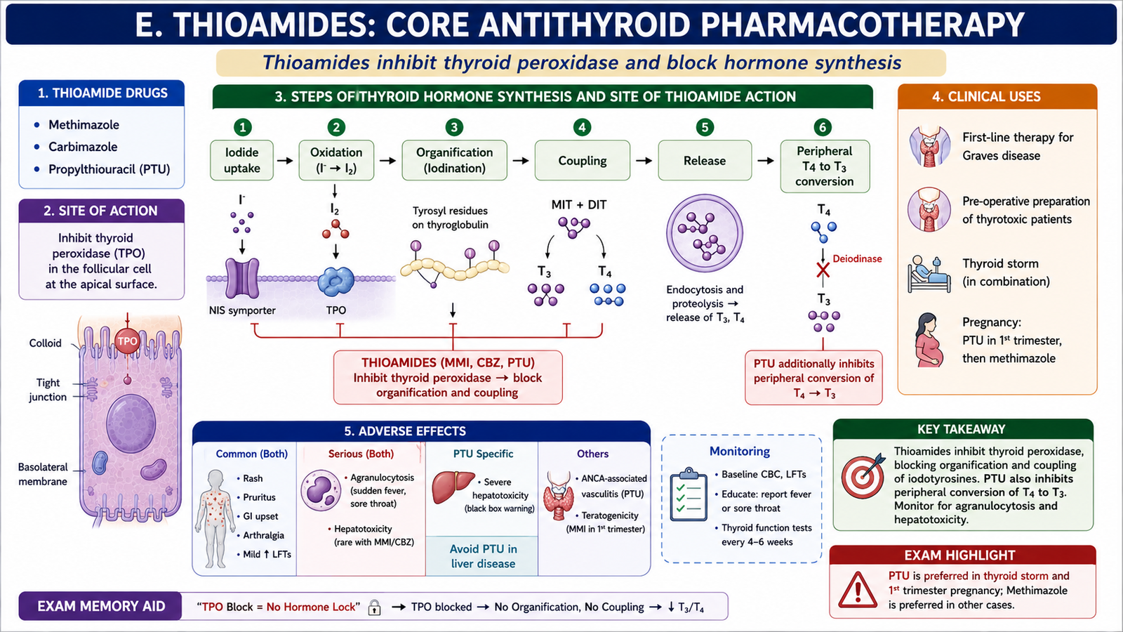
E. Thioamides: Core Antithyroid Pharmacotherapy
Thioamides are major drugs for reducing thyroid-hormone synthesis. The important drugs are methimazole, its prodrug carbimazole, and propylthiouracil (PTU). Their therapeutic effect develops gradually because they prevent synthesis of new hormone but do not immediately remove hormone already stored in thyroid colloid.
Classification
- Methimazole
- Carbimazole — converted to methimazole in the body
- Propylthiouracil (PTU)
Mechanism of Action
Thioamides inhibit thyroid peroxidase. This blocks important reactions needed to create new T3 and T4.
PTU has an additional action: it reduces peripheral conversion of T4 to T3 by inhibiting a deiodination step. This additional action is useful when rapid reduction of active T3 formation is desirable.
Clinical Uses and Drug-Selection Logic
Thioamides are particularly useful when ongoing synthesis of thyroid hormone needs to be suppressed, especially in Graves disease. They may be used as primary medical treatment or as part of preparation for definitive treatment.
- Graves hyperthyroidism: suppresses continued hormone synthesis while the disease is medically controlled.
- Before thyroid surgery: may be used to bring the patient toward a euthyroid state before operation.
- Severe thyrotoxicosis or thyroid storm: PTU may be useful because it inhibits both thyroid-hormone synthesis and peripheral T4-to-T3 conversion.
Methimazole or carbimazole is generally convenient for routine medical treatment because its action is longer lasting and severe hepatic toxicity is less prominent than with PTU. PTU remains important in selected situations, including circumstances in which its additional inhibition of peripheral T4-to-T3 conversion is useful.
Adverse Effects
Most patients tolerate thioamides, but several adverse effects are examination-important because some can be serious.
- Skin rash and pruritus: relatively common hypersensitivity-type effects.
- Gastrointestinal upset: may occur during treatment.
- Agranulocytosis: a rare but serious fall in granulocytes, producing marked susceptibility to infection. Fever or sore throat during therapy therefore requires urgent clinical assessment.
- Hepatotoxicity: particularly important with PTU, which may rarely produce severe liver injury.
- Cholestatic hepatic injury: is more associated with methimazole than PTU.
- Teratogenic effects: methimazole exposure during early fetal development has characteristic potential fetal risks, which influences drug selection during pregnancy.
F. Iodide, Radioactive Iodine, β-Blockers and Overall Treatment
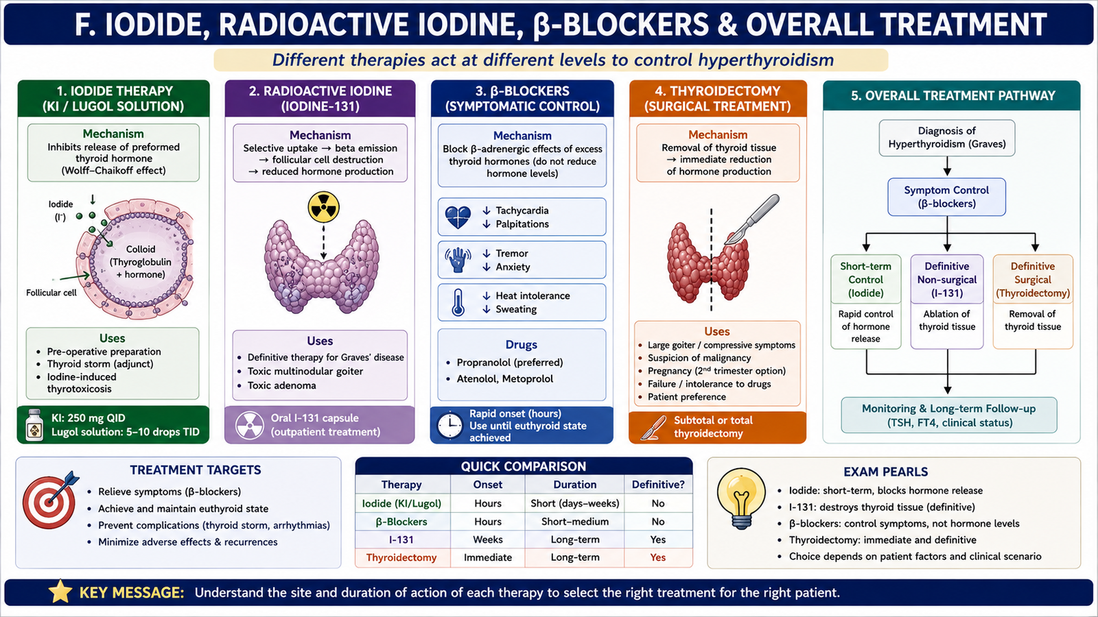
Treatment of hyperthyroidism works at different levels. Some therapies suppress new hormone synthesis, some rapidly reduce hormone release, some destroy overactive thyroid tissue, and others mainly control symptoms. Understanding where each treatment acts makes drug selection much easier.
Potassium Iodide
Although iodine is normally required for thyroid-hormone synthesis, a large pharmacological iodide load temporarily suppresses thyroid activity. This is an important example of a substance having different effects at physiological and pharmacological concentrations.
This acute inhibition is often called the Wolff-Chaikoff effect. The effect is temporary because the thyroid eventually adapts or “escapes” from sustained iodide inhibition. Therefore, iodide is useful for short-term control rather than long-term treatment of hyperthyroidism.
Clinical Uses of Iodide
- Preoperative preparation: reduces thyroid vascularity and hormone release before thyroid surgery.
- Severe thyrotoxicosis or thyroid storm: provides rapid inhibition of hormone release as part of combined treatment.
- When used during severe thyrotoxicosis, iodide is given after a drug that blocks new hormone synthesis so that the supplied iodine is not used as substrate for additional hormone production.
Lugol Iodine Solution
Lugol solution is an aqueous preparation containing elemental iodine together with potassium iodide. Potassium iodide helps iodine dissolve in water. In hyperthyroidism, it provides a concentrated iodide source for short-term inhibition of thyroid-hormone release and reduction of thyroid vascularity.
Adverse Effects of Iodide
Excess iodide can produce a group of symptoms known as iodism.
- Metallic or unpleasant taste
- Soreness of the mouth or gums
- Salivary-gland swelling
- Gastrointestinal irritation
- Skin eruptions or hypersensitivity reactions
Radioactive Iodine
Radioactive iodine, particularly iodine-131, is taken up by thyroid follicular cells in the same way as ordinary iodine. Once concentrated within the thyroid, it emits radiation that damages and destroys functioning thyroid tissue. Its effect therefore develops progressively rather than immediately.
Clinical Uses of Radioactive Iodine
Radioactive iodine is a definitive treatment for many patients with persistent hyperthyroidism, including Graves disease and autonomous toxic thyroid tissue, when it is clinically appropriate.
Because it destroys thyroid tissue, a major long-term consequence is hypothyroidism. This is not simply an adverse laboratory finding; it means the patient may subsequently require thyroid-hormone replacement. Radioactive iodine may also cause temporary thyroid inflammation and can worsen Graves ophthalmopathy in susceptible patients.
Role of β-Blockers
Many distressing manifestations of hyperthyroidism result from increased adrenergic responsiveness. β-blockers provide rapid symptomatic relief while the underlying thyroid disorder is being controlled.
- Reduce tachycardia and palpitations.
- Reduce tremor.
- Reduce adrenergic anxiety and restlessness.
- Help control cardiovascular symptoms during the period before antithyroid treatment becomes fully effective.
Propranolol is particularly useful because, in addition to β-adrenergic blockade, it can reduce peripheral conversion of T4 to T3 at sufficiently high therapeutic exposure. However, β-blockers do not correct the underlying thyroid overproduction and should be understood mainly as symptomatic therapy.
Overall Treatment Strategy and Prognosis
Treatment is selected according to the cause of hyperthyroidism, severity, gland characteristics, associated eye disease and individual clinical circumstances. The main therapeutic approaches are medical suppression of hormone synthesis, radioactive destruction of thyroid tissue or surgical removal of thyroid tissue.
- β-blocker: controls adrenergic symptoms rapidly.
- Thioamide: reduces new thyroid-hormone synthesis.
- Iodide: provides short-term inhibition of hormone release and reduces thyroid vascularity.
- Radioactive iodine: provides definitive reduction of functioning thyroid tissue.
- Thyroidectomy: offers definitive treatment when surgery is clinically preferable, such as significant goitre, compressive disease, concerning structural abnormalities or situations in which other definitive therapies are unsuitable.
Most patients with appropriately treated hyperthyroidism have a good prognosis. Recurrence can occur after a course of medical antithyroid therapy, whereas definitive treatment with radioactive iodine or surgery can result in permanent hypothyroidism. Long-term follow-up therefore includes assessment of thyroid function even after hyperthyroid symptoms have resolved.
G. Graves Ophthalmopathy: Pathogenesis, Features and Management
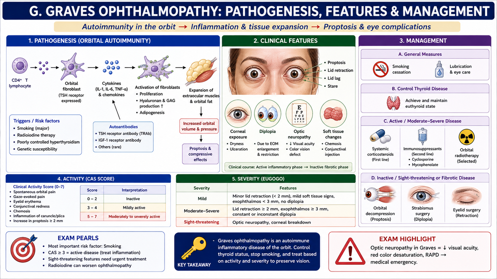
Graves ophthalmopathy is an autoimmune inflammatory disorder of the orbit associated with Graves disease. It is not simply the result of excess circulating thyroid hormone. This distinction explains why eye disease may not exactly parallel the biochemical severity of hyperthyroidism.
Pathogenesis
The autoimmune reaction involves orbital fibroblasts and immune cells. Orbital fibroblasts can participate in immune responses related to TSH-receptor autoimmunity. Activated T lymphocytes release cytokines that stimulate fibroblasts and promote production of hydrophilic glycosaminoglycans.
Because the bony orbit cannot expand, increased soft-tissue volume pushes the eye forward. Extraocular-muscle enlargement may restrict movement and produce diplopia. In severe disease, orbital pressure can threaten the optic nerve.
Clinical Features
- Proptosis or exophthalmos: forward displacement of the globe because orbital tissue volume is increased.
- Periorbital swelling: results from inflammation and edema.
- Gritty, dry or irritated eyes: occur because exposure and altered eyelid closure disturb the ocular surface.
- Diplopia: results from impaired movement of enlarged or fibrotic extraocular muscles.
- Restricted ocular movements: reflects mechanical limitation of affected muscles.
- Conjunctival redness or chemosis: may reflect orbital and ocular-surface inflammation.
Lid retraction may accompany Graves disease, but it is important to separate this adrenergic feature from the deeper autoimmune orbital disease responsible for true proptosis and extraocular-muscle dysfunction.
Assessment
Evaluation includes the severity and activity of eye symptoms, visual function, ocular movements, corneal exposure and signs of optic-nerve compromise. Imaging of the orbit may be required when significant orbital disease, muscle enlargement or optic-nerve compression is suspected.
Management Principles
Management depends on the severity and activity of ophthalmopathy. Control of thyroid dysfunction is important, but simply normalizing thyroid-hormone levels does not directly remove established autoimmune orbital inflammation.
- Mild disease: supportive ocular care may be sufficient, including protection of the ocular surface and lubrication when needed.
- Active inflammatory disease: specialist assessment is required; anti-inflammatory and immunomodulatory treatment may be needed according to severity.
- Significant exposure or diplopia: requires ophthalmic assessment and treatment directed at the affected ocular structures.
- Optic-nerve compromise: is an ophthalmic emergency requiring urgent treatment to relieve the threat to vision.
- Radioactive iodine consideration: radioactive iodine may aggravate ophthalmopathy in susceptible patients, so existing eye disease influences treatment planning.
Integrated Mechanism Flow
The central sequence of Graves hyperthyroidism can be summarized in six linked steps:
TSH-receptor stimulating IgG autoantibodies develop.
Persistent receptor activation stimulates follicular cells despite low pituitary TSH.
Diffuse hypertrophy and hyperplasia produce an enlarged, hypervascular thyroid.
Increased T3/T4 raises metabolism and adrenergic responsiveness.
Weight loss, heat intolerance, tremor, tachycardia and other hyperthyroid features appear.
β-blockade controls symptoms; thioamides reduce synthesis; iodide reduces release; radioactive iodine or surgery reduces thyroid tissue.
Important Comparison — Major Antithyroid Treatment Approaches
The easiest way to differentiate the major therapies is to identify where each one acts and whether it controls symptoms, hormone synthesis, hormone release or functioning thyroid tissue.
| Therapy | Main Action | Speed/Role | Important Point |
|---|---|---|---|
| Thioamides | Inhibit TPO and new hormone synthesis | Gradual control | PTU additionally reduces peripheral T4 → T3 conversion |
| Potassium iodide / Lugol solution | Acutely inhibits hormone release and thyroid activity | Rapid, short-term | Effect is temporary because escape occurs |
| Radioactive iodine | Destroys functioning follicular tissue | Definitive, delayed effect | Hypothyroidism is a common long-term outcome; avoid in pregnancy/breastfeeding |
| β-Blockers | Block adrenergic effects | Rapid symptomatic control | Do not stop thyroid-hormone synthesis |