
This chapter follows the supplied KMU learning outcomes and moves from understanding breast lesions to investigation, treatment and prevention. First understand the explanations and clinical links; then use the final high-yield review for rapid revision.
Topic 23 — Breast Lump and Breast Cancer: Benign Disease, Investigation, Management and Prevention
ENDOCRINE + REPRODUCTION
An integrated approach to benign breast lesions, carcinoma breast, imaging, triple assessment, treatment, hormone receptors, SERMs, screening, prevention and communication of a cancer diagnosis.
Topic Introduction
A breast lump may arise from normal developmental or hormonal changes, a benign lesion or a malignant tumor. The important skill is therefore not simply to memorize individual diseases, but to understand how their pathology produces characteristic clinical and imaging findings and how a breast lesion is assessed safely. In this chapter, you will learn the important benign breast conditions, the development and presentation of breast carcinoma, the principles of staging, triple assessment, ultrasound and mammography, treatment based on disease extent and receptor status, tamoxifen and other selective estrogen-receptor modulators, breast-cancer screening and prevention, and a structured approach to communicating a cancer diagnosis.
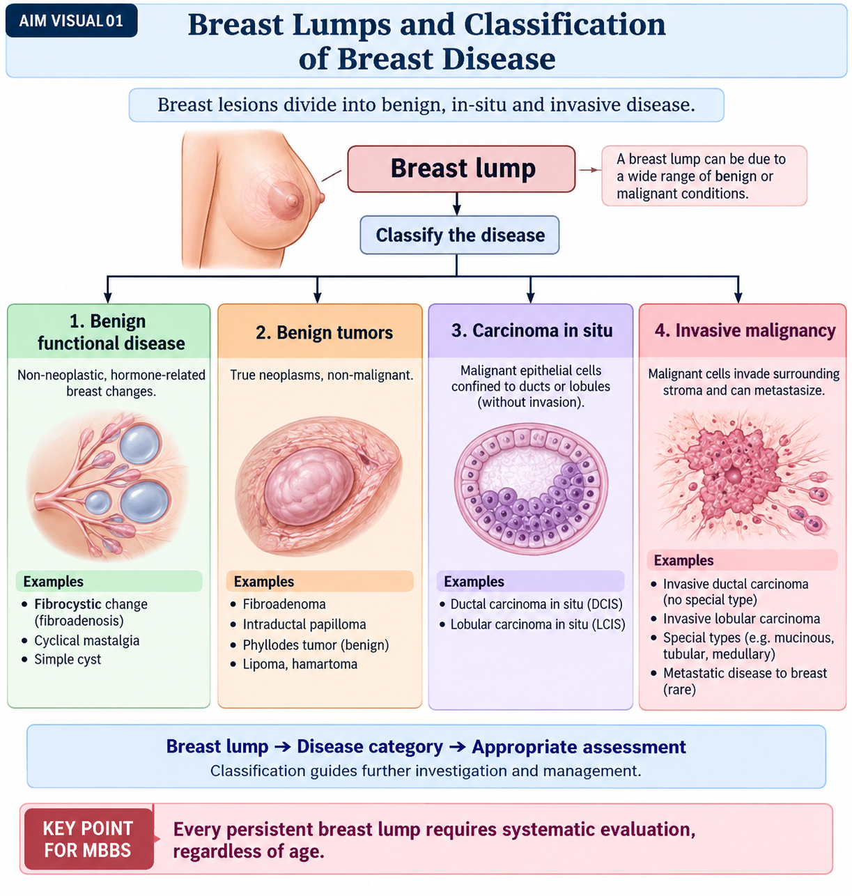
A. Breast Lumps and Classification of Breast Disease
A breast lump is a localized area that feels or appears different from the surrounding breast tissue. Some lumps result from benign hormonal or structural changes, while others represent neoplasia. Because clinical appearance alone cannot always establish the diagnosis, classification provides a framework for deciding what needs investigation.
Classification of benign breast disease
Benign breast disorders include functional changes, benign tumors, inflammatory conditions and other non-malignant lesions. Important conditions within the present topic include:
- Fibrocystic changes: a spectrum that includes cyst formation, fibrosis, epithelial hyperplasia and adenosis.
- Fibroadenoma: a benign biphasic tumor composed of stromal and epithelial elements.
- Intraductal papilloma: a benign papillary proliferation arising within a breast duct.
- Inflammatory and related lesions: mastitis, breast abscess and duct ectasia.
- Traumatic/reactive lesions: particularly fat necrosis, which may clinically mimic malignancy.
- Gynecomastia: benign enlargement of the male breast due to an imbalance between estrogenic and androgenic influence.
Classification of malignant breast disease
Most malignant breast tumors arise from mammary epithelium. They can first be divided according to whether the abnormal cells remain confined by the basement membrane or have invaded the surrounding stroma.
- Carcinoma in situ: ductal carcinoma in situ (DCIS) and lobular carcinoma in situ (LCIS).
- Invasive carcinoma: invasive carcinoma of no special type and invasive lobular carcinoma are major forms; several less common special histological types also occur.
- Paget disease of the nipple: usually reflects extension of an underlying ductal carcinoma into the epidermis of the nipple.
- Other rare malignancies: sarcomas, lymphomas and metastatic tumors involving the breast.
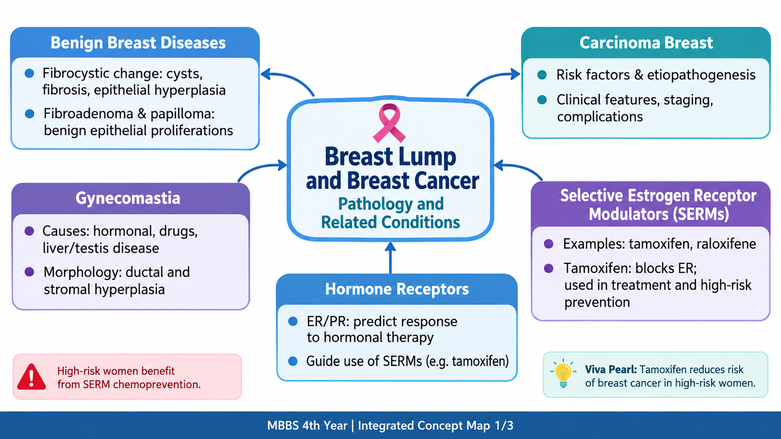
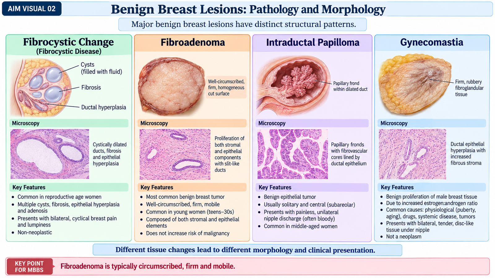
B. Benign Breast Lesions: Pathology and Morphology
Benign breast lesions are common and may present as a lump, pain or nipple discharge. Their importance lies both in distinguishing them from carcinoma and in understanding that different benign proliferative patterns carry different relationships with future breast-cancer risk.
Fibrocystic changes
Fibrocystic change is a collective term for several non-neoplastic alterations that usually develop in hormonally responsive breast tissue. The major components required in this topic are cysts, fibrosis, epithelial hyperplasia and adenosis.
1. Cysts
Cysts form when terminal ducts and acini become dilated. Small cysts may enlarge and become visible grossly. Their lining may show apocrine metaplasia, in which epithelial cells acquire abundant eosinophilic cytoplasm.
2. Fibrosis
Cysts may rupture and release their contents into surrounding tissue. The resulting chronic inflammatory reaction promotes stromal fibrosis. This fibrosis contributes to the firm and sometimes irregular texture of the breast.
3. Epithelial hyperplasia
Hyperplasia means an increase in the number of epithelial cells lining ducts or lobules. In usual epithelial hyperplasia, several layers of cells may partially fill and bridge the ductal lumen. Hyperplasia without atypia is distinct from atypical hyperplasia, which has greater importance as a marker of increased carcinoma risk.
4. Adenosis
Adenosis is an increase in the number of acini within a lobule. In sclerosing adenosis, proliferating acini become compressed and distorted by stromal fibrosis. This can produce a firm lesion and may resemble carcinoma clinically, radiologically or microscopically, but the basic lobular architecture and myoepithelial layer are preserved.
Fibroadenoma
Fibroadenoma is a benign tumor containing both glandular epithelium and proliferating fibrous stroma. It commonly occurs in younger women and is hormonally responsive. Clinically, it often forms a firm, smooth, well-defined and freely mobile lump.
Gross morphology
- Well-circumscribed, rounded or oval mass.
- Firm or rubbery consistency.
- Usually sharply demarcated from surrounding breast tissue.
- Cut surface is solid, grey-white and may show slit-like spaces.
Microscopic morphology
The proliferating stroma surrounds and compresses benign ducts. The ducts remain lined by epithelial and myoepithelial cells. Depending on how the stroma grows around the ducts, the pattern may appear pericanalicular, with rounded ducts, or intracanalicular, where ducts become compressed into elongated slit-like spaces.
Intraductal papilloma
An intraductal papilloma is a benign papillary growth arising within a breast duct. A central papilloma often develops in a large duct beneath the nipple and may present with spontaneous serous or bloody nipple discharge because the delicate papillary fronds can bleed.
- Grossly: a small friable papillary mass lies within a dilated duct.
- Microscopically: branching fibrovascular cores are covered by epithelial and myoepithelial cells.
Gynecomastia
Gynecomastia is enlargement of the male breast caused by increased estrogenic stimulation relative to androgenic action. It may be unilateral or bilateral and usually forms a subareolar swelling.
Important causes
- Physiological: puberty and aging.
- Reduced androgen effect: testicular failure or antiandrogen therapy.
- Increased estrogen effect: some testicular or other hormone-producing tumors and increased peripheral estrogen formation.
- Systemic disease: particularly chronic liver disease and some endocrine disorders.
- Drugs: examples include spironolactone, cimetidine, ketoconazole and some antiandrogenic drugs.
Morphology
The ducts proliferate within increased stroma. Early lesions show periductal stromal edema, while established lesions become more fibrotic. Unlike the normal female breast, fully developed lobules are usually absent.
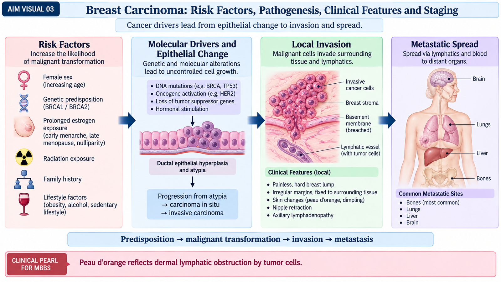
C. Breast Carcinoma: Risk Factors, Pathogenesis, Clinical Features and Staging
Breast carcinoma develops when mammary epithelial cells acquire genetic and regulatory abnormalities that allow uncontrolled growth, survival, invasion and eventually metastasis. Its behavior is influenced not only by the anatomical extent of disease but also by biological features such as estrogen receptor, progesterone receptor and HER2 status.
Risk factors
Breast-cancer risk is produced by interaction between age, inherited susceptibility, hormonal exposure and acquired breast changes. Important factors include:
- Increasing age.
- Strong family history of breast or ovarian cancer.
- Inherited pathogenic variants, especially BRCA1 and BRCA2.
- Previous carcinoma of one breast.
- Proliferative breast disease with atypia.
- Longer lifetime exposure to estrogenic stimulation, such as early menarche or late menopause.
- Nulliparity or later first full-term pregnancy.
- Postmenopausal obesity, which can increase peripheral estrogen production.
- Previous significant therapeutic irradiation involving the breast region.
Etiopathogenesis
Breast carcinoma may be hereditary or sporadic. Genetic changes alter pathways controlling proliferation, DNA repair and growth signaling. In many cancers, hormone-receptor signaling drives tumor growth; in others, amplification or overactivity of growth-factor pathways such as HER2 becomes important.
Inherited or acquired genetic alteration → abnormal epithelial proliferation → atypia/in-situ disease → breach of basement membrane → stromal invasion → lymphatic or hematogenous spread.
Once malignant cells cross the basement membrane, the lesion becomes invasive. Invasive cells can enter lymphatics and blood vessels. Regional lymph nodes, particularly axillary nodes, are therefore important for staging and prognosis.
Clinically useful biological groups
- Hormone-receptor positive: ER and/or PR expressed; endocrine therapy can suppress hormone-dependent growth.
- HER2-positive: HER2 overexpression or amplification provides a target for anti-HER2 therapy.
- Triple-negative: ER-negative, PR-negative and HER2-negative; endocrine and HER2-directed therapies are not effective.
Clinical features
Early breast carcinoma is often painless. As the tumor infiltrates surrounding tissue, it becomes less mobile and may distort the skin or nipple.
- Hard, irregular or poorly defined breast lump.
- Fixation to surrounding breast tissue, skin or deeper structures in advanced disease.
- Skin dimpling or tethering due to involvement and shortening of supporting fibrous structures.
- Nipple retraction due to fibrosis and traction on involved ducts.
- Peau d’orange caused by cutaneous edema from obstruction of dermal lymphatics while hair follicles remain tethered.
- Ulceration in locally advanced disease.
- Abnormal nipple discharge in some tumors.
- Enlarged regional lymph nodes.
- Eczematous change of the nipple in Paget disease.
Spread and complications
Breast carcinoma spreads locally, through lymphatics and through blood vessels. Axillary nodes are common regional sites. Distant metastases commonly involve organs such as bone, lung, liver and brain.
- Local skin and chest-wall invasion.
- Ulceration, bleeding and secondary infection.
- Lymphatic obstruction and edema.
- Regional lymph-node involvement.
- Bone metastasis with pain or pathological fracture.
- Organ dysfunction from distant metastases.
Principles of staging
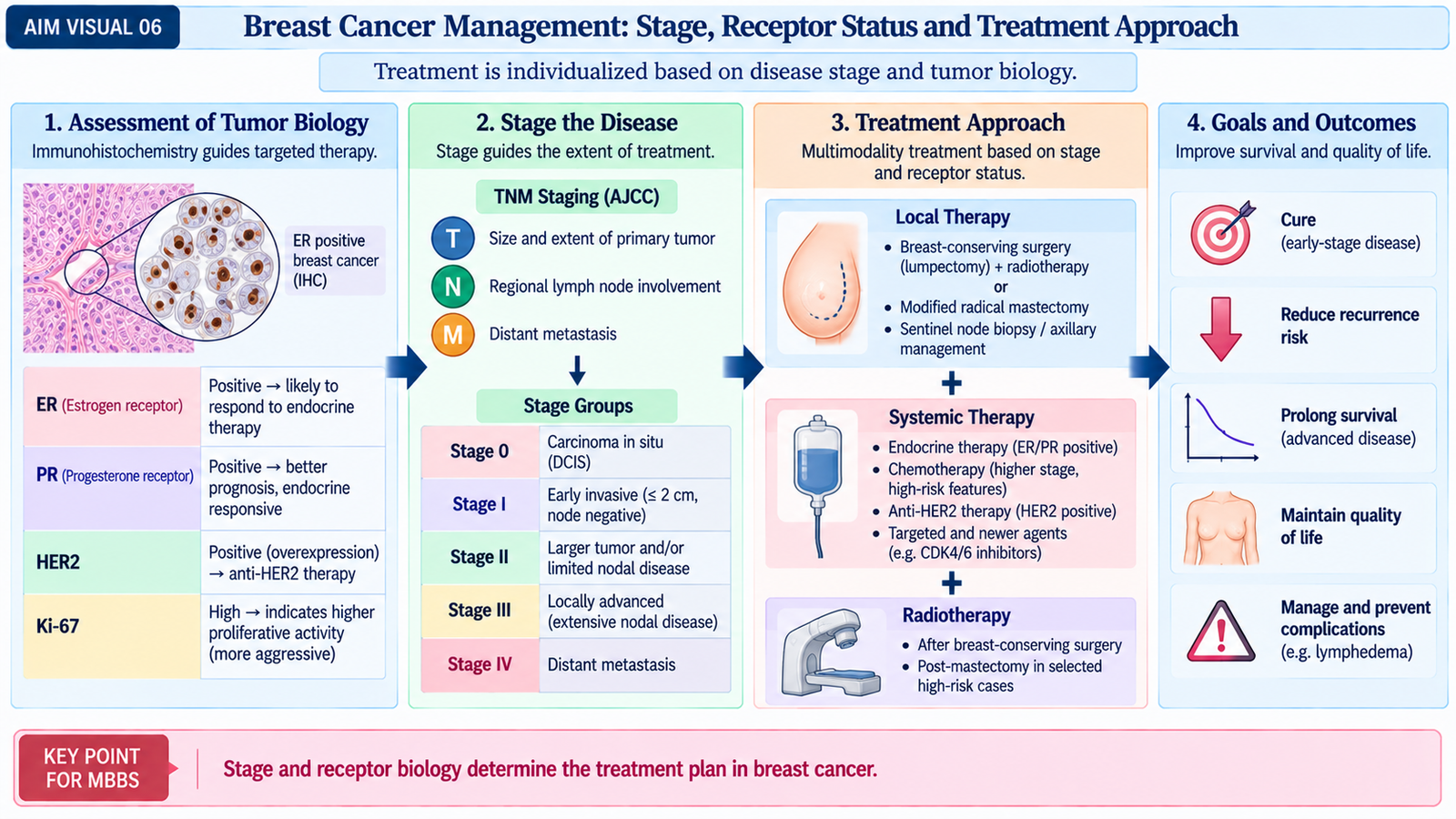
Breast cancer is staged principally through the TNM system. T describes the primary tumor and local extension, N describes regional lymph-node involvement, and M identifies distant metastasis. Modern clinical decisions also consider tumor biology.
- Stage 0: in-situ disease.
- Stages I–II: broadly represent earlier invasive disease with limited local or regional spread.
- Stage III: locally advanced disease, such as extensive regional nodal disease or major local extension.
- Stage IV: distant metastatic disease.
For undergraduate understanding, remember that increasing tumor extent, nodal involvement and distant metastasis progressively worsen stage and prognosis.
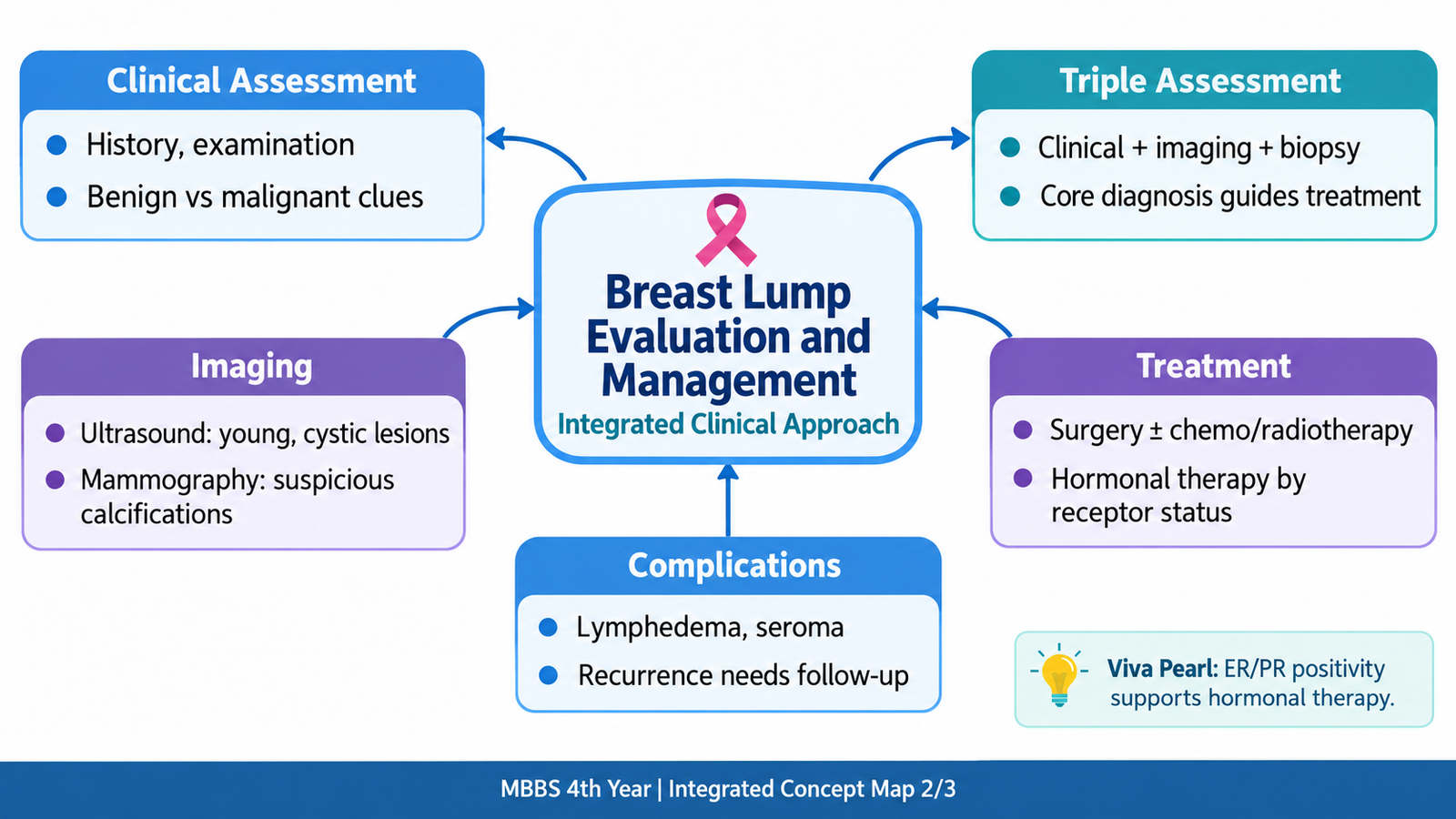
D. Clinical Evaluation and Triple Assessment of a Breast Lesion
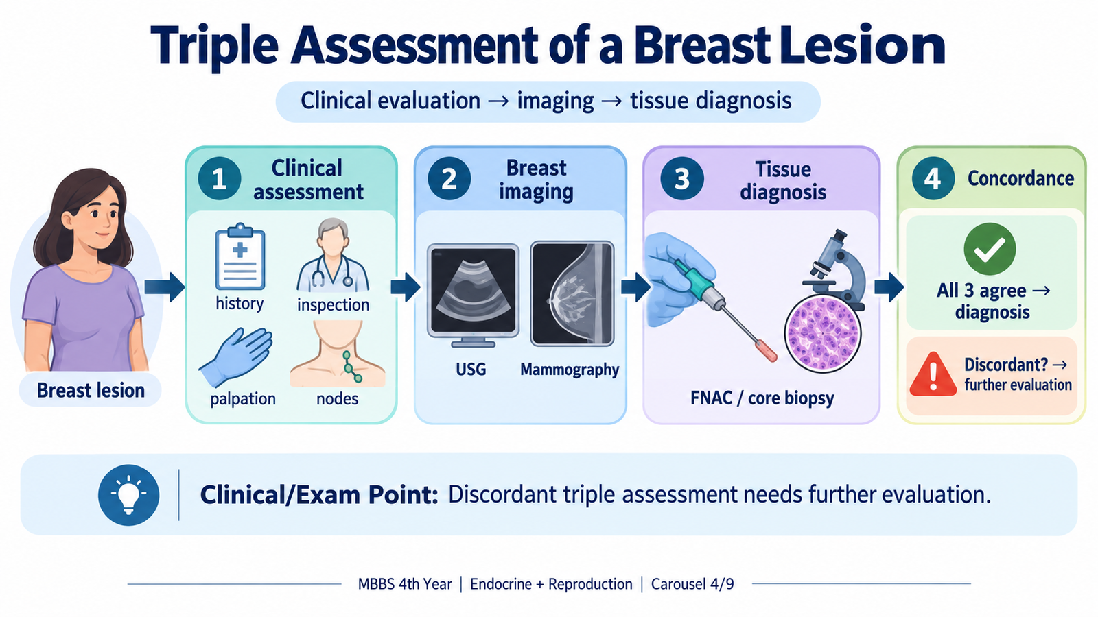
No single examination or test should be used in isolation when a suspicious breast lesion is being evaluated. The safest diagnostic approach is triple assessment: clinical assessment, appropriate imaging and pathological sampling. Each component answers a different question, and agreement among the three provides much greater diagnostic confidence.
1. Clinical assessment
History
History helps estimate the probability of benign or malignant disease and guides the choice of investigation.
- Age and duration of the lump.
- Change in size and whether symptoms vary with the menstrual cycle.
- Pain or tenderness.
- Nipple discharge, especially spontaneous or bloody discharge.
- Recent pregnancy or lactation where relevant.
- Previous breast disease or biopsy.
- Family history of breast or ovarian malignancy.
- Relevant hormonal and drug history.
Examination
Both breasts and regional lymph-node areas should be examined. The lump is assessed for site, size, shape, surface, margin, consistency, mobility and relationship to the skin, nipple and deeper structures.
- A smooth, mobile, well-circumscribed mass favors a benign process such as fibroadenoma.
- A hard, irregular or fixed lesion increases concern for malignancy.
- Skin tethering, nipple retraction and peau d’orange are important malignant signs.
- Axillary and other relevant regional lymph nodes should be assessed.
2. Imaging
Imaging characterizes the lesion and looks for features that cannot be assessed reliably by palpation. Ultrasound and mammography are complementary rather than interchangeable. The choice depends on the clinical situation, breast composition and purpose of imaging.
3. Pathological diagnosis
A suspicious solid lesion requires tissue diagnosis because imaging suggests probability but does not by itself establish histological type. Core-needle biopsy provides tissue architecture and usually allows assessment of invasive disease and important biomarkers. Fine-needle aspiration cytology may be useful in selected situations but provides cells rather than tissue architecture.
Clinical examination asks what the lesion feels and looks like.
Imaging asks what structural pattern the lesion has.
Biopsy asks what the lesion actually is histologically.
If the clinical, imaging and pathological findings do not agree, the discordance must be resolved rather than simply accepting the most reassuring result.
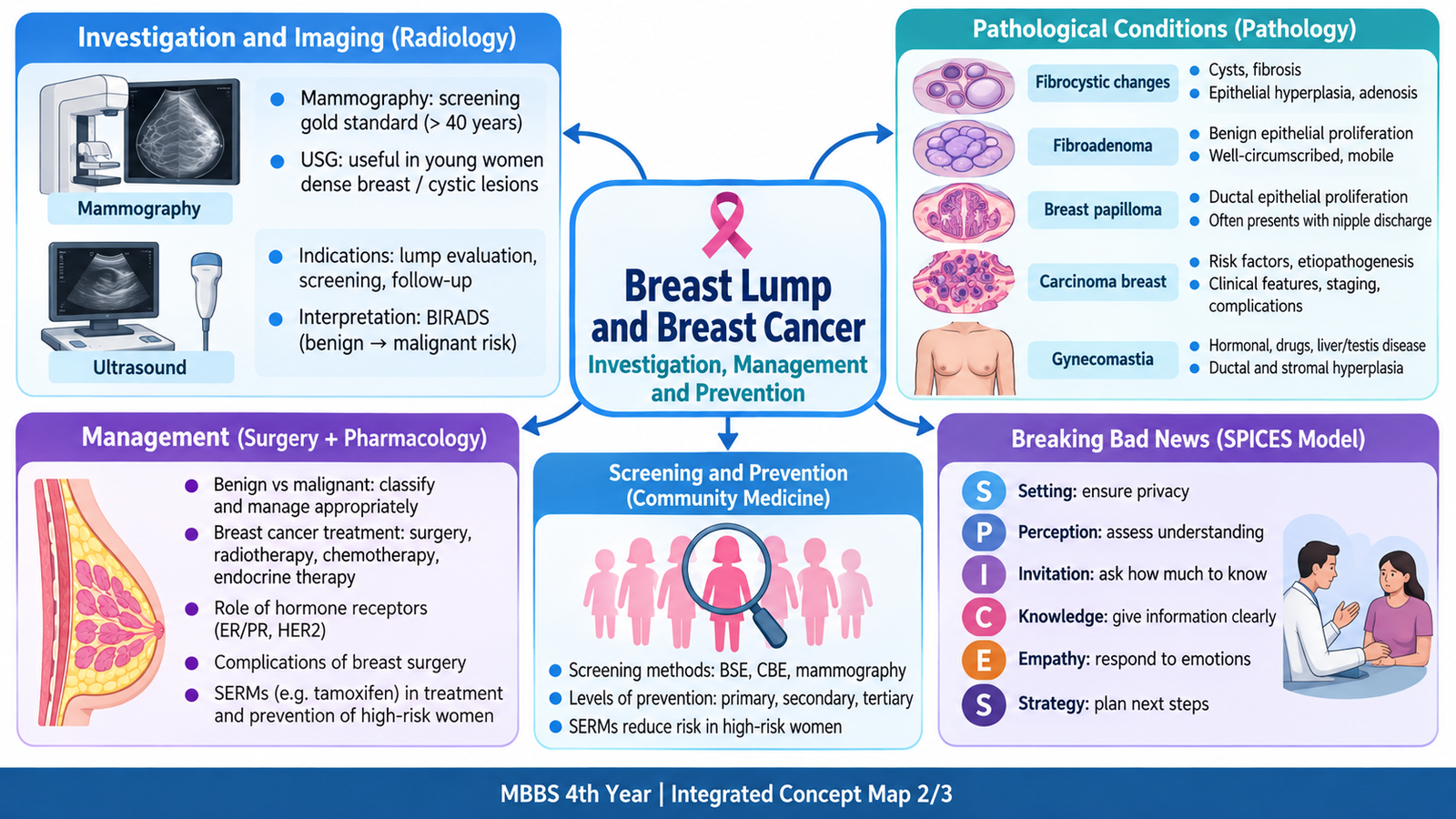
E. Ultrasound and Mammography in the Evaluation of Breast Lumps
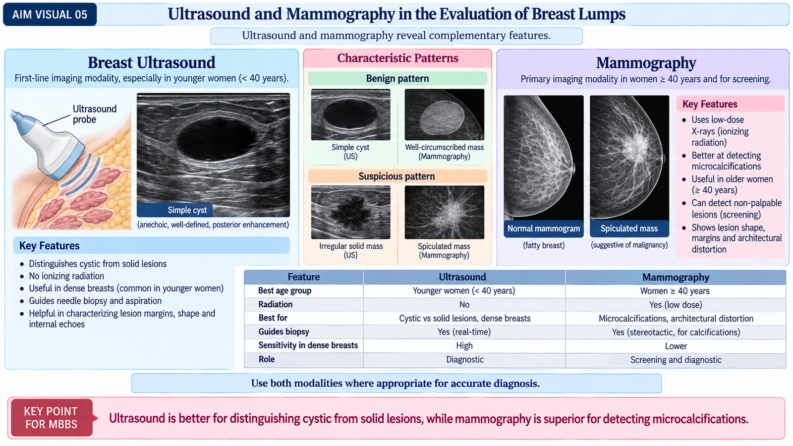
Ultrasound and mammography provide different information about a breast lesion. Ultrasound uses sound waves and is especially effective for distinguishing fluid-filled from solid lesions. Mammography uses low-dose X-rays and is particularly valuable for detecting architectural distortion and calcifications that may be clinically impalpable.
Breast ultrasound
Principle and procedure
A high-frequency ultrasound transducer is moved over the breast using coupling gel. Reflected sound waves create real-time images of the breast tissue. Doppler assessment may sometimes be added to examine vascularity.
Important indications
- Characterization of a palpable breast lump.
- Distinguishing a cystic lesion from a solid lesion.
- Assessment of dense breast tissue where mammographic characterization may be limited.
- Evaluation of lesions in younger patients.
- Guidance for needle aspiration or biopsy.
- Assessment of regional lymph nodes.
Interpretation
- Simple cyst: usually well-defined and anechoic, with posterior acoustic enhancement.
- Fibroadenoma: often oval, circumscribed, relatively homogeneous and oriented parallel to the skin.
- Suspicious malignant mass: may be irregular, poorly defined, non-parallel, heterogeneous or associated with posterior acoustic shadowing.
Mammography
Principle and procedure
Mammography uses low-dose X-rays to produce detailed images of compressed breast tissue. Compression reduces tissue overlap and improves image quality. Standard assessment commonly includes craniocaudal and mediolateral-oblique projections.
Important indications
- Population breast-cancer screening where mammographic screening is appropriate.
- Diagnostic evaluation of a clinically detected abnormality.
- Detection of suspicious calcification or architectural distortion.
- Assessment of the opposite breast after a cancer diagnosis.
Important mammographic patterns
Features associated with malignancy include an irregular or spiculated mass, architectural distortion and suspicious clustered microcalcifications. Skin thickening or nipple retraction may also be seen in advanced disease.
Some benign lesions are sharply circumscribed. An involuting fibroadenoma may develop coarse calcification. Imaging findings must always be interpreted together with the clinical presentation.
F. Breast Cancer Management, Hormone Receptors, Prognosis and Surgical Complications
Breast-cancer treatment is individualized according to the anatomical stage of disease and the biological characteristics of the tumor. Management therefore combines local treatment of the breast and regional nodes with systemic therapy directed at possible microscopic disease elsewhere.
Role of hormone receptors and HER2
Biopsy tissue is assessed for estrogen receptor (ER), progesterone receptor (PR) and HER2. These markers are important because they influence both prognosis and treatment selection.
- ER/PR-positive tumors: depend partly on estrogen signaling and can respond to endocrine therapy.
- HER2-positive tumors: may respond to drugs that specifically target HER2 signaling.
- Triple-negative tumors: lack ER, PR and HER2, so endocrine therapy and HER2-directed therapy are not useful.
Local treatment
Local treatment aims to control the tumor within the breast and regional lymphatic area.
- Breast-conserving surgery: removes the tumor with an adequate margin while preserving most of the breast. It is generally combined with radiotherapy when appropriate.
- Mastectomy: removes the breast and is selected when breast conservation is unsuitable or when other clinical factors favor mastectomy.
- Axillary staging: sentinel lymph-node assessment can determine whether regional lymphatic spread is present while limiting unnecessary extensive axillary surgery in suitable patients.
- Radiotherapy: improves local control after breast-conserving treatment and is used after mastectomy in selected patients according to recurrence risk.
Systemic pharmacological treatment
Systemic treatment reaches tumor cells beyond the breast. Selection is based on stage, recurrence risk and tumor biology.
- Endocrine therapy: used for hormone-receptor-positive cancer. Tamoxifen and aromatase inhibitors are major examples.
- Cytotoxic chemotherapy: used when the expected benefit from systemic cell-killing treatment is significant. It may be given before surgery or after surgery.
- Anti-HER2 therapy: agents such as trastuzumab are used in appropriately selected HER2-positive disease.
- Other targeted or immune-based treatments: may be used in selected breast-cancer subgroups under specialist management.
Neoadjuvant versus adjuvant therapy
Neoadjuvant treatment is given before definitive surgery. It can reduce tumor size, improve operability and provide information about treatment response. Adjuvant treatment is given after surgery to eradicate microscopic residual disease and reduce recurrence risk.
Advanced or metastatic disease
When distant metastasis is present, systemic therapy becomes the main treatment. The goals are disease control, relief of symptoms, maintenance of function and prolongation of useful survival. Local treatment may still be used for specific symptoms or complications.
Prognostic factors
The extent of disease remains a major determinant of prognosis. Important factors include:
- Tumor stage and size.
- Regional lymph-node involvement.
- Presence or absence of distant metastasis.
- Histological grade.
- ER, PR and HER2 status.
- Response to systemic treatment where applicable.
In general, earlier-stage, node-negative and hormone-receptor-positive tumors have a more favorable outlook than advanced or biologically aggressive disease, although targeted therapy has improved outcomes for several biologically defined groups.
Complications of breast-cancer surgery
Surgical complications result from wound healing, removal of breast tissue and disruption of axillary nerves or lymphatics.
- Bleeding or hematoma.
- Wound infection.
- Seroma: accumulation of lymphatic or serous fluid in the surgical space.
- Skin-flap or wound-healing problems.
- Altered sensation or numbness around the upper arm or surgical field.
- Shoulder stiffness and reduced range of movement.
- Arm lymphedema: caused by impaired lymphatic drainage following axillary treatment.
- Chronic postoperative pain.
- Rare nerve injuries affecting surrounding muscles.
- Psychological and body-image consequences requiring supportive care.
G. Selective Estrogen-Receptor Modulators and Tamoxifen
Selective estrogen-receptor modulators, or SERMs, bind estrogen receptors but act differently in different tissues. A SERM may block estrogen action in one organ while partially reproducing estrogen effects in another. This tissue selectivity explains both the benefits and adverse effects of tamoxifen.
Examples of SERMs
- Tamoxifen
- Raloxifene
- Toremifene
- Clomiphene
- Bazedoxifene
Mechanism of action of tamoxifen
Tamoxifen binds to intracellular estrogen receptors and alters estrogen-responsive gene transcription. Its effect depends on the tissue.
In breast tissue, tamoxifen acts mainly as an estrogen antagonist. In some other tissues, including bone and endometrium, it has partial estrogen-agonist activity. This explains why it can protect bone in some patients while also stimulating the endometrium.
Clinical uses
- Treatment of ER-positive breast cancer.
- Adjuvant endocrine therapy after local treatment of hormone-sensitive cancer.
- Treatment of selected recurrent or metastatic hormone-receptor-positive disease.
- Reduction of breast-cancer risk in appropriately selected high-risk women.
Important adverse effects and cautions
- Hot flushes and other anti-estrogenic symptoms.
- Increased risk of venous thromboembolic events.
- Endometrial stimulation, which can produce hyperplasia and increase the risk of endometrial carcinoma.
- Other less common adverse effects may occur and require clinical monitoring.
SERMs in breast-cancer prevention
Breast-cancer chemoprevention is considered for selected women whose predicted risk is sufficiently high to justify preventive medication. Tamoxifen reduces the development of estrogen-receptor-positive breast cancers because it blocks estrogen-mediated proliferation in breast tissue. Raloxifene is another preventive option in selected postmenopausal women.
SERMs are not routine preventive drugs for every woman. The expected reduction in breast-cancer risk must be balanced against adverse effects such as thromboembolism and, with tamoxifen, endometrial stimulation.
H. Breast-Cancer Screening and Levels of Prevention
Screening aims to detect breast cancer before symptoms develop, when treatment is more likely to be effective. Prevention is broader: it includes reducing preventable risk, detecting disease early and limiting disability after diagnosis and treatment.
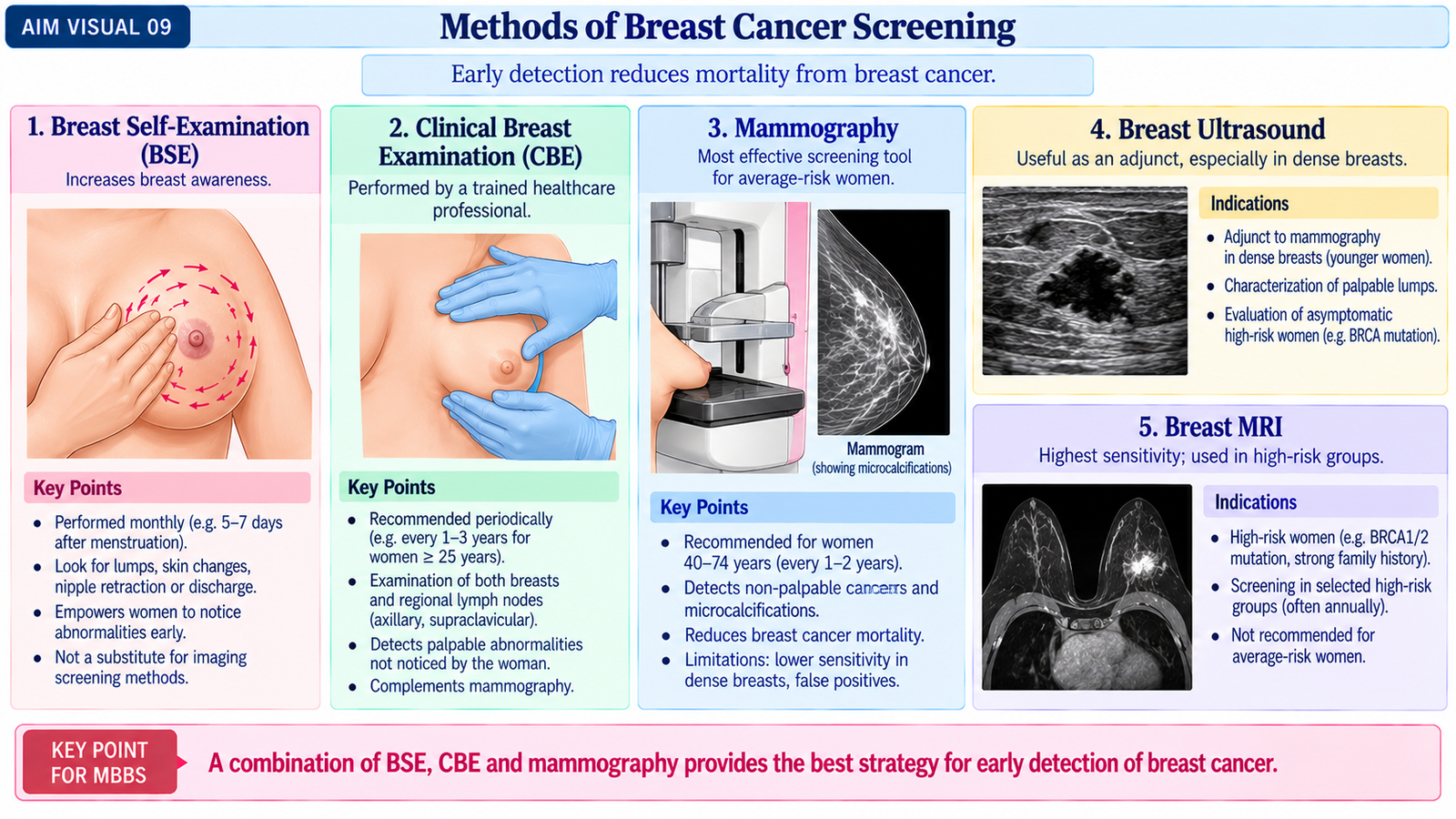
Methods of breast-cancer screening
Mammographic screening
Mammography is the principal imaging method used for organized breast-cancer screening. It can detect small masses, architectural distortion and suspicious microcalcifications before a lesion becomes palpable.
Clinical breast examination
Clinical examination may identify palpable masses, nipple changes, skin abnormalities and regional lymphadenopathy. Its role varies according to the screening system and available resources, and it does not replace diagnostic investigation of a detected abnormality.
Breast self-awareness
Women should be able to recognize significant changes in their own breasts and seek assessment when a new lump, persistent nipple change, abnormal discharge or skin change develops. Breast self-awareness is not a substitute for appropriate screening or diagnostic imaging.
Screening of high-risk women
Women with particularly high inherited or familial risk may require an intensified surveillance strategy. In selected high-risk individuals, breast MRI can be added to mammography because of its high sensitivity. Ultrasound is usually an adjunct rather than a replacement for standard mammographic screening.
Levels of prevention
Primordial prevention
Primordial prevention aims to prevent the development of modifiable risk factors in the population by encouraging healthy patterns of physical activity, weight control and other protective health behaviors from an early stage.
Primary prevention
Primary prevention acts before cancer develops. It includes reducing modifiable risk factors where possible and, in carefully selected high-risk women, considering preventive approaches such as SERMs or risk-reducing surgery after specialist assessment.
Secondary prevention
Secondary prevention means detecting disease early. Breast-cancer screening and prompt investigation of a newly discovered breast abnormality belong to this level.
Tertiary prevention
Tertiary prevention aims to reduce disability, recurrence-related morbidity and treatment complications after cancer has developed. Examples include definitive treatment, rehabilitation of shoulder movement, management of lymphedema, psychological support and appropriate care of advanced disease.
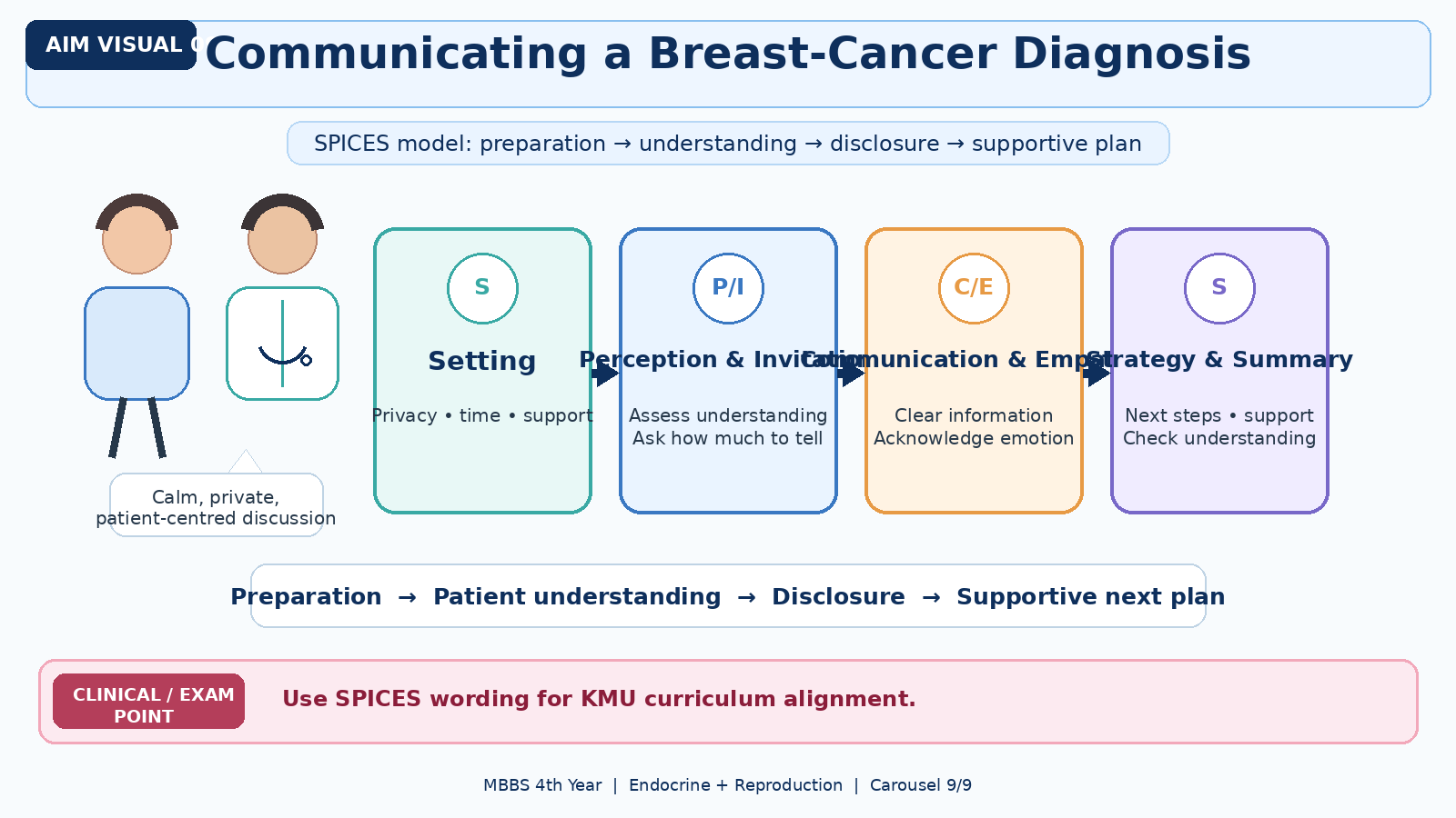
I. Communicating a Breast-Cancer Diagnosis: SPICES Model of Breaking Bad News
Bad news is information that seriously and negatively changes a patient’s understanding of the future. A cancer diagnosis must therefore be communicated truthfully but sensitively, allowing the patient to understand the information, express emotion and participate in the next plan of care.
For KMU alignment, retain the term SPICES when referring to the curriculum outcome while understanding the following structured communication principles.
1. Setting
Prepare the conversation properly. Ensure privacy, sit at an appropriate level, minimize interruptions and allow enough time. Ask whether the patient wishes a trusted relative or support person to be present.
2. Perception
Before giving new information, determine what the patient already understands. Open questions reveal misconceptions, expectations and how much of the situation the patient has already recognized.
3. Invitation
Ask how much information the patient wishes to receive. Most patients want clear information, but the physician should respect the individual’s preference regarding detail and involvement of family members.
4. Giving clear information
Give the diagnosis in simple language and in manageable pieces. Avoid unnecessary jargon. A short warning statement can prepare the patient for difficult information. Pause after important information and check understanding instead of delivering a long uninterrupted explanation.
5. Emotions and empathy
Allow silence and emotional reactions. Identify the emotion and respond with empathy rather than immediately returning to technical information. Acknowledging fear, sadness or shock helps preserve trust and shows that the patient has been heard.
6. Strategy and summary
Summarize what has been discussed and explain the immediate next step. The patient should leave the encounter knowing what happens next, who will remain involved in care and when further discussion will occur. Honest information should be combined with realistic support rather than false reassurance.
Integrated Mechanism Flow
Inherited, hormonal or acquired risk
Abnormal epithelial proliferation
Malignant cells remain confined
Basement membrane is breached
Nodes or distant organs involved
Stage + ER/PR/HER2 guide therapy
Important Comparison — Benign versus Suspicious/Malignant Breast Lump
| Feature | More suggestive of benign lesion | More suspicious for malignancy |
|---|---|---|
| Margin | Smooth, circumscribed | Irregular or poorly defined |
| Mobility | Often freely mobile | May be fixed or tethered |
| Consistency | Soft, cystic or rubbery | Often hard |
| Skin/nipple | Usually unchanged | Tethering, retraction, peau d’orange or ulceration |
| Ultrasound | Oval/circumscribed or simple cyst pattern | Irregular, non-parallel or shadowing mass |
| Mammography | Circumscribed benign-appearing lesion | Spiculation, distortion or suspicious microcalcification |
| Final diagnosis | Clinical appearance alone is insufficient when concern remains; triple assessment determines the diagnosis. | |
⭐ AIM High-Yield Review
Breast Cancer — Clinical Medicine
Review breast-cancer risk factors, pathophysiology, clinical features, diagnosis, imaging, staging, receptor status and treatment.