
Topic 12 — Adrenal and Neuroendocrine Tumors: Pheochromocytoma, MEN and GEP-NETs
1. Topic Introduction
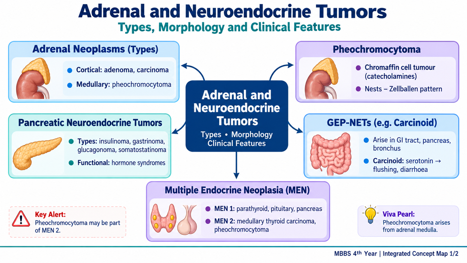
Adrenal and neuroendocrine tumors arise from hormone-producing cells and may present either because they secrete excessive hormones or because the growing tumor produces a mass effect. This chapter focuses on adrenal cortical tumors, pheochromocytoma, multiple endocrine neoplasia syndromes and gastro-entero-pancreatic neuroendocrine tumors, including carcinoid tumors. The central idea is that the cell from which a tumor develops largely determines the hormone it may produce, its characteristic microscopic appearance and the clinical syndrome it causes. You will learn how these tumors are classified, how their important morphological features are recognized, how hormonal syndromes guide investigation and how biochemical testing, imaging, pathology and appropriate treatment are combined in clinical practice.
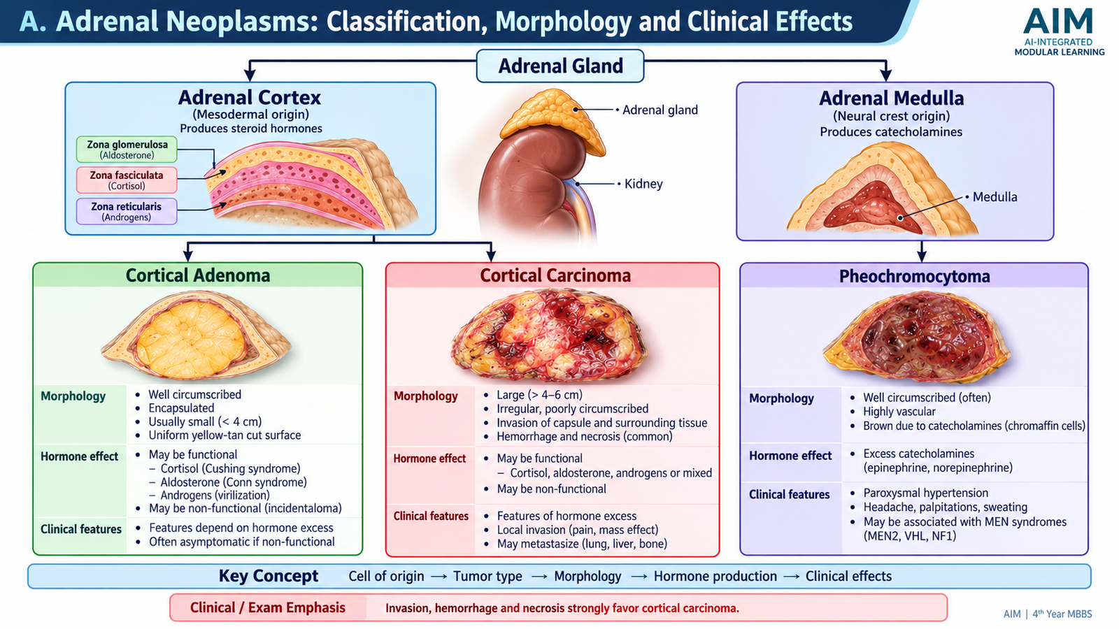
A. Adrenal Neoplasms: Classification, Morphology and Clinical Effects
The adrenal gland contains two functionally different regions. The adrenal cortex produces steroid hormones, while the adrenal medulla contains chromaffin cells that produce catecholamines. Adrenal tumors therefore behave differently depending on their cell of origin. Some tumors are discovered because they cause a recognizable hormone-excess syndrome, whereas others are found incidentally or after they become large enough to produce local symptoms.
Classification
For undergraduate understanding, the important primary adrenal neoplasms in this topic can be grouped according to their origin:
- Adrenal cortical adenoma — a benign neoplasm of adrenal cortical cells.
- Adrenal cortical carcinoma — a malignant cortical neoplasm.
- Pheochromocytoma — a neuroendocrine tumor of adrenal medullary chromaffin cells.
Cortical adenomas and carcinomas may be functional, meaning that they secrete excessive steroid hormones, or nonfunctional. Functional tumors may therefore present with manifestations of cortisol excess, aldosterone excess or androgen excess. A nonfunctional tumor is more likely to be detected incidentally or because of its size.
Adrenal Cortical Adenoma
An adrenal cortical adenoma is a benign, usually well-circumscribed tumor composed of cells resembling normal adrenal cortical cells. Many adenomas are clinically silent, but some produce excessive hormones.
- Usually well circumscribed and often surrounded by a thin capsule.
- The cut surface is commonly yellow because cortical cells contain abundant lipid.
- Large areas of hemorrhage and necrosis are generally not characteristic of an uncomplicated adenoma.
- Uniform cortical-type cells arranged in nests or cords.
- Cells may contain clear lipid-rich cytoplasm.
- Marked pleomorphism, destructive invasion and frequent atypical mitoses are not expected.
Adrenal Cortical Carcinoma
Adrenal cortical carcinoma is an uncommon malignant tumor of cortical cells. It is clinically important because it may both invade surrounding tissues and produce excessive adrenal steroids. Functional carcinomas may produce a mixed hormonal picture, particularly cortisol and androgen excess.
Grossly, cortical carcinomas tend to be larger and less uniform than adenomas. Their cut surface may be variegated because rapid tumor growth can outstrip its blood supply, producing hemorrhage and necrosis. Invasion into surrounding tissues or veins strongly supports malignant behavior.
Microscopically, malignant features may include disordered architecture, nuclear atypia, increased mitotic activity, atypical mitoses, necrosis and vascular or capsular invasion. No single isolated microscopic feature should be interpreted without the overall pathological picture.
Clinical Effects of Adrenal Cortical Tumors
The symptoms of a functional cortical tumor reflect the hormone produced. Cortisol secretion produces features of Cushing syndrome, aldosterone secretion produces hypertension with mineralocorticoid effects, and excess androgen secretion may produce virilization. A nonfunctional carcinoma may instead present with abdominal or flank discomfort, a palpable mass, constitutional symptoms or manifestations of metastatic disease.
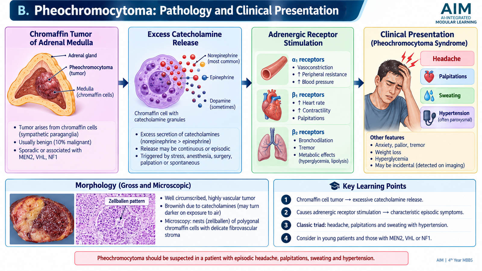
B. Pheochromocytoma: Pathology and Clinical Presentation
Pheochromocytoma is a catecholamine-producing neuroendocrine tumor that usually arises from chromaffin cells of the adrenal medulla. Its clinical importance is disproportionate to its frequency because excessive catecholamine release can produce severe hypertension, cardiac complications and potentially life-threatening crises.
How the Clinical Syndrome Develops
Catecholamine release may be continuous or episodic. Therefore, hypertension may be sustained in one patient and paroxysmal in another. Sudden episodes can sometimes be precipitated by stress, anesthesia, certain drugs or manipulation of the tumor.
Morphology
Grossly, pheochromocytomas are usually well-defined masses. Their appearance may range from yellow-tan to brown, and larger tumors may contain areas of hemorrhage, cystic change or necrosis.
Microscopically, the tumor is composed of polygonal chromaffin cells with granular cytoplasm. The cells characteristically form nests known as a Zellballen pattern. These nests are surrounded by a delicate vascular network and supporting sustentacular cells. The nuclei may show considerable variation in size and appearance without necessarily indicating malignant behavior.
Clinical Features
The symptoms result from excessive adrenergic stimulation. The classical symptom combination is episodic headache, palpitations and profuse sweating, usually associated with hypertension. Not every patient has the complete triad.
- Hypertension — may be sustained or occur in sudden attacks because catecholamines produce intense vasoconstriction.
- Headache — commonly accompanies sudden rises in blood pressure.
- Palpitations and tachycardia — result from cardiac adrenergic stimulation.
- Profuse sweating — reflects sympathetic activation.
- Tremor, anxiety and pallor — may occur during catecholamine surges.
- Hyperglycemia may develop because catecholamines promote hepatic glucose production and interfere with normal insulin-mediated glucose control.
Hereditary Associations
Pheochromocytoma may occur sporadically or as part of an inherited tumor syndrome. Important associations include MEN type 2, von Hippel-Lindau syndrome, neurofibromatosis type 1 and hereditary syndromes involving succinate dehydrogenase genes. A hereditary background becomes particularly important when disease is bilateral, multifocal, occurs at a young age or is accompanied by a suggestive family history or another syndrome-associated tumor.
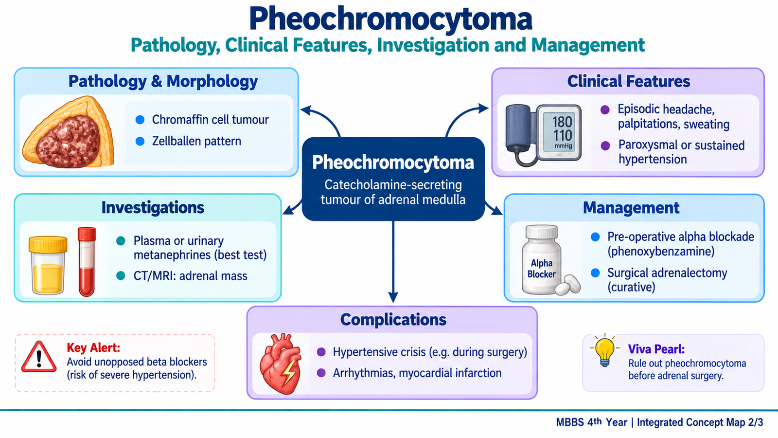
C. Pheochromocytoma: Diagnostic Work-up, Management and Complications
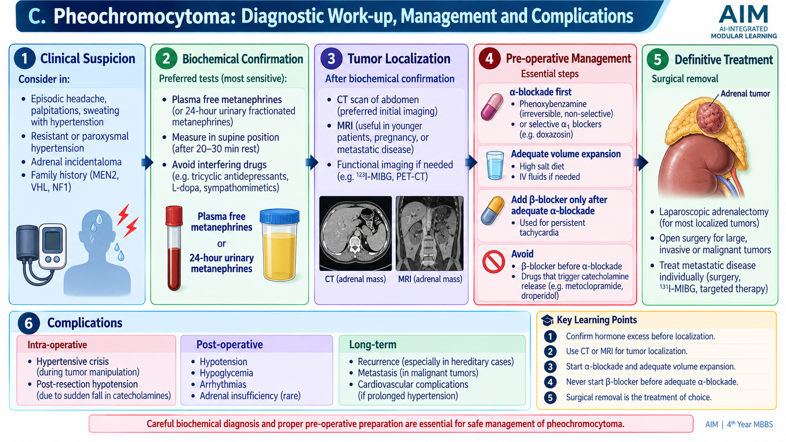
The diagnostic approach follows an important sequence. First establish that excessive catecholamine production is present. Only then should the tumor be localized anatomically. This order prevents an incidental adrenal mass from being incorrectly assumed to be a functioning pheochromocytoma.
1. Biochemical Confirmation
The most useful initial biochemical assessment is measurement of fractionated metanephrines, either in plasma or in a timed urine collection. Metanephrines are metabolites of catecholamines and are useful because the tumor continuously metabolizes catecholamines even when the patient’s symptoms are intermittent.
An elevated result must be interpreted in the clinical context because physiological stress, illness and some medications can interfere with catecholamine-related testing. Borderline or unexpected results therefore require careful reassessment rather than immediate labeling of the patient as having a tumor.
2. Tumor Localization
After biochemical evidence has established catecholamine excess, CT or MRI is used to identify the adrenal lesion and assess its anatomical relationships. Functional imaging based on neuroendocrine or catecholamine-related uptake may be used when disease is multifocal, metastatic, hereditary or not adequately localized by conventional imaging.
3. Assessment for Hereditary Disease
A patient with a young age at presentation, bilateral or multifocal tumors, a positive family history or features of an inherited endocrine syndrome should be evaluated for a hereditary cause. This matters because the result may influence surveillance for additional tumors and may have implications for relatives.
Management
Definitive treatment of a localized pheochromocytoma is usually surgical removal. However, operating on an untreated catecholamine-secreting tumor can provoke massive catecholamine release and severe cardiovascular instability. Preoperative medical preparation is therefore essential.
α-adrenergic blockade → restoration of adequate circulating volume → β-blockade only if still required for tachycardia → definitive surgery
Alpha blockade comes first because it reduces catecholamine-induced vasoconstriction. If clinically important tachycardia persists, a beta blocker may subsequently be added. Giving beta blockade before adequate alpha blockade is dangerous because beta-mediated vasodilatation is removed while alpha-mediated vasoconstriction remains unopposed, potentially causing a severe hypertensive reaction.
Patients with advanced, recurrent or metastatic disease require specialist management. Treatment may combine control of catecholamine effects with surgery, radionuclide-based treatment or other systemic approaches depending on tumor biology and extent of disease.
Complications
Complications are produced mainly by intense or prolonged catecholamine effects on the cardiovascular and cerebrovascular systems.
- Hypertensive crisis from sudden marked vasoconstriction.
- Cardiac arrhythmias from excessive adrenergic stimulation.
- Myocardial ischemia or infarction because severe hypertension and tachycardia increase myocardial oxygen demand.
- Catecholamine-related cardiomyopathy and acute heart failure.
- Stroke related to severe blood-pressure elevation.
- Perioperative cardiovascular collapse if an unsuspected tumor is manipulated without appropriate preparation.
D. Multiple Endocrine Neoplasia (MEN) Syndromes
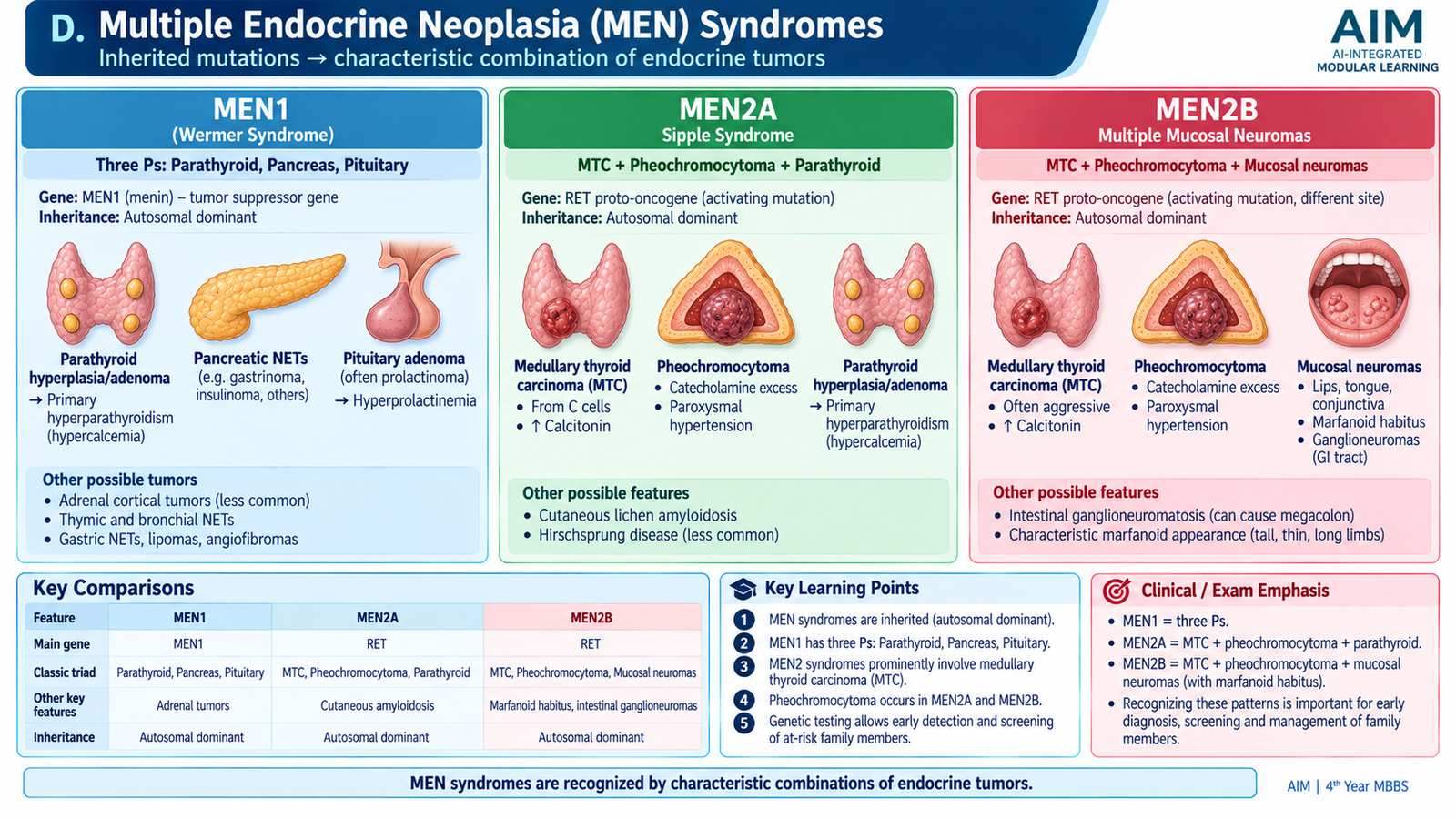
Multiple endocrine neoplasia syndromes are inherited disorders in which a person has a strong predisposition to develop tumors or hyperplasia in more than one endocrine organ. The major classical syndromes are MEN1 and MEN2; MEN2 is further divided into MEN2A and MEN2B. Recognizing the pattern is important because discovering one endocrine tumor may indicate that another tumor should be actively sought.
MEN1
MEN1 results from abnormalities involving the MEN1 tumor-suppressor gene, which encodes the protein menin. Loss of normal tumor-suppressor function allows endocrine cells to proliferate abnormally. The major organs classically involved are remembered as the three Ps:
- Parathyroid — multigland hyperplasia or multiple adenomas can produce primary hyperparathyroidism.
- Pancreatic and duodenal neuroendocrine tissue — tumors may be multiple and may produce hormones such as gastrin or insulin.
- Pituitary — adenomas may produce prolactin or other pituitary hormones, or may be nonfunctioning.
The endocrine lesions in MEN1 are often multiple rather than single isolated tumors. Clinical presentation therefore depends on which hormone excess develops first. A patient may present with hypercalcemia from parathyroid disease, recurrent peptic ulceration from gastrinoma, hypoglycemia from insulinoma or symptoms of a pituitary adenoma.
MEN2A
MEN2 syndromes result from activating abnormalities of the RET proto-oncogene. MEN2A is characterized principally by:
- Medullary thyroid carcinoma, commonly arising in a background of C-cell hyperplasia and tending to be multifocal or bilateral in hereditary disease.
- Pheochromocytoma, which may also be bilateral or multifocal.
- Parathyroid hyperplasia or adenomatous disease causing primary hyperparathyroidism.
MEN2B
MEN2B is also caused by activating RET abnormalities but has a distinct clinical pattern. It includes medullary thyroid carcinoma and pheochromocytoma, together with characteristic mucosal neuromas and a marfanoid body habitus. Intestinal ganglioneuromatous lesions may also occur. Primary hyperparathyroidism, an important component of MEN2A, is not a typical feature of MEN2B.
Diagnostic Approach
The diagnosis begins by recognizing the characteristic combination of endocrine tumors. Biochemical testing is then directed toward the potentially involved glands. For example, suspected MEN2 requires attention to medullary thyroid carcinoma and pheochromocytoma, while MEN1 requires evaluation for parathyroid, pituitary and pancreatic/duodenal neuroendocrine disease.
Genetic testing and genetic counseling are central to inherited MEN syndromes because identifying a pathogenic germline abnormality can confirm the syndrome and allows appropriate assessment of at-risk relatives.
Management Principles
Management is not directed toward a single organ only. Each endocrine tumor is treated according to its biological behavior and hormonal effects, while the patient remains under long-term surveillance for additional syndrome-associated tumors. In MEN2, pheochromocytoma must be identified and appropriately managed before procedures likely to provoke catecholamine release. Medullary thyroid carcinoma requires specialist thyroid management, and MEN1-associated pancreatic, parathyroid and pituitary disease is treated according to the organ and hormone involved.
E. Pancreatic Neuroendocrine Tumors: Types, Morphology and Clinical Syndromes
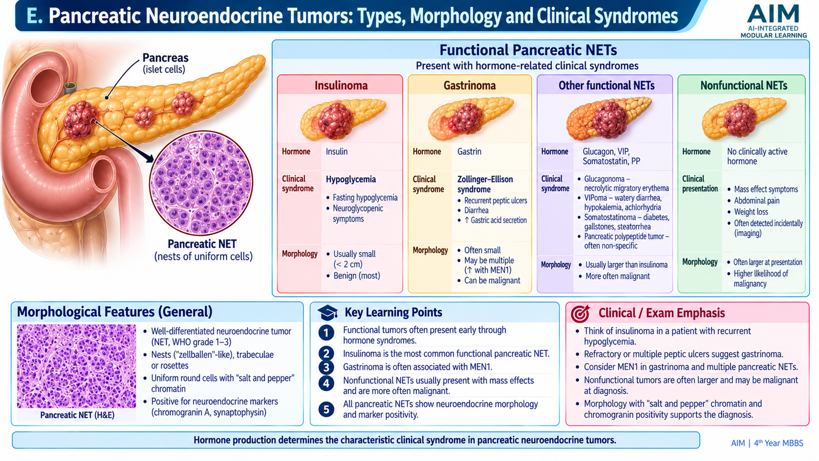
Pancreatic neuroendocrine tumors arise from neuroendocrine cells of the pancreas. They differ from the much more common pancreatic ductal adenocarcinoma in both morphology and biological behavior. The most useful clinical classification divides them into functional tumors, which produce a recognizable hormone syndrome, and nonfunctional tumors, which do not produce a clinically obvious hormone-excess syndrome.
General Morphology
Well-differentiated pancreatic neuroendocrine tumors are generally composed of relatively uniform cells arranged in nests, cords, trabeculae or gland-like structures. Their nuclei characteristically show finely granular, evenly dispersed chromatin often described as “salt-and-pepper” chromatin. The tumor cells express neuroendocrine markers such as synaptophysin and chromogranin.
The biological behavior of a pancreatic NET cannot be judged from hormone production alone. Pathological assessment therefore considers differentiation, mitotic activity and cellular proliferative activity, together with evidence of invasion or metastasis.
Insulinoma
An insulinoma is an insulin-secreting pancreatic neuroendocrine tumor. Excess insulin lowers blood glucose, especially during fasting. The clinical manifestations therefore result from both sympathetic activation caused by falling glucose and inadequate glucose supply to the brain.
- Adrenergic symptoms may include sweating, tremor, palpitations and hunger.
- Neuroglycopenic symptoms may include confusion, abnormal behavior, visual disturbance, seizures or loss of consciousness.
- Symptoms classically improve after glucose is given.
Most insulinomas are small and well differentiated, and many behave in a relatively indolent manner. Some occur as part of MEN1.
Gastrinoma
A gastrinoma produces excessive gastrin, which stimulates marked gastric acid secretion. The resulting syndrome is called Zollinger-Ellison syndrome. Excess acid produces recurrent or difficult-to-control peptic ulceration and may cause diarrhea by interfering with normal intestinal digestion and absorption.
Gastrinomas may arise in the duodenum or pancreas and are an important pancreatic/duodenal neuroendocrine manifestation of MEN1.
Glucagonoma
A glucagonoma secretes excessive glucagon. Increased hepatic glucose production contributes to hyperglycemia or diabetes. A particularly important clinical clue is necrolytic migratory erythema, a characteristic inflammatory skin eruption. Weight loss may also occur.
VIPoma
A VIPoma produces vasoactive intestinal peptide. Excess VIP causes marked intestinal secretion of water and electrolytes and reduces gastric acid secretion. The characteristic syndrome therefore includes profuse watery diarrhea, hypokalemia and reduced gastric acidity. Severe fluid and electrolyte depletion may become the immediate clinical problem.
Somatostatinoma
A somatostatinoma produces excess somatostatin, which inhibits several gastrointestinal and pancreatic hormones. Important manifestations include diabetes or glucose intolerance, steatorrhea and gallstones. Because these findings are individually nonspecific, the diagnosis may be less clinically obvious than insulinoma or gastrinoma.
Nonfunctional Pancreatic NETs
Nonfunctional tumors do not produce a clear endocrine syndrome. They may therefore remain unnoticed until they are large or metastatic. Presentation may include abdominal discomfort, weight loss, a pancreatic mass or manifestations of liver metastases.
F. Gastro-Entero-Pancreatic NETs and Carcinoid Tumors
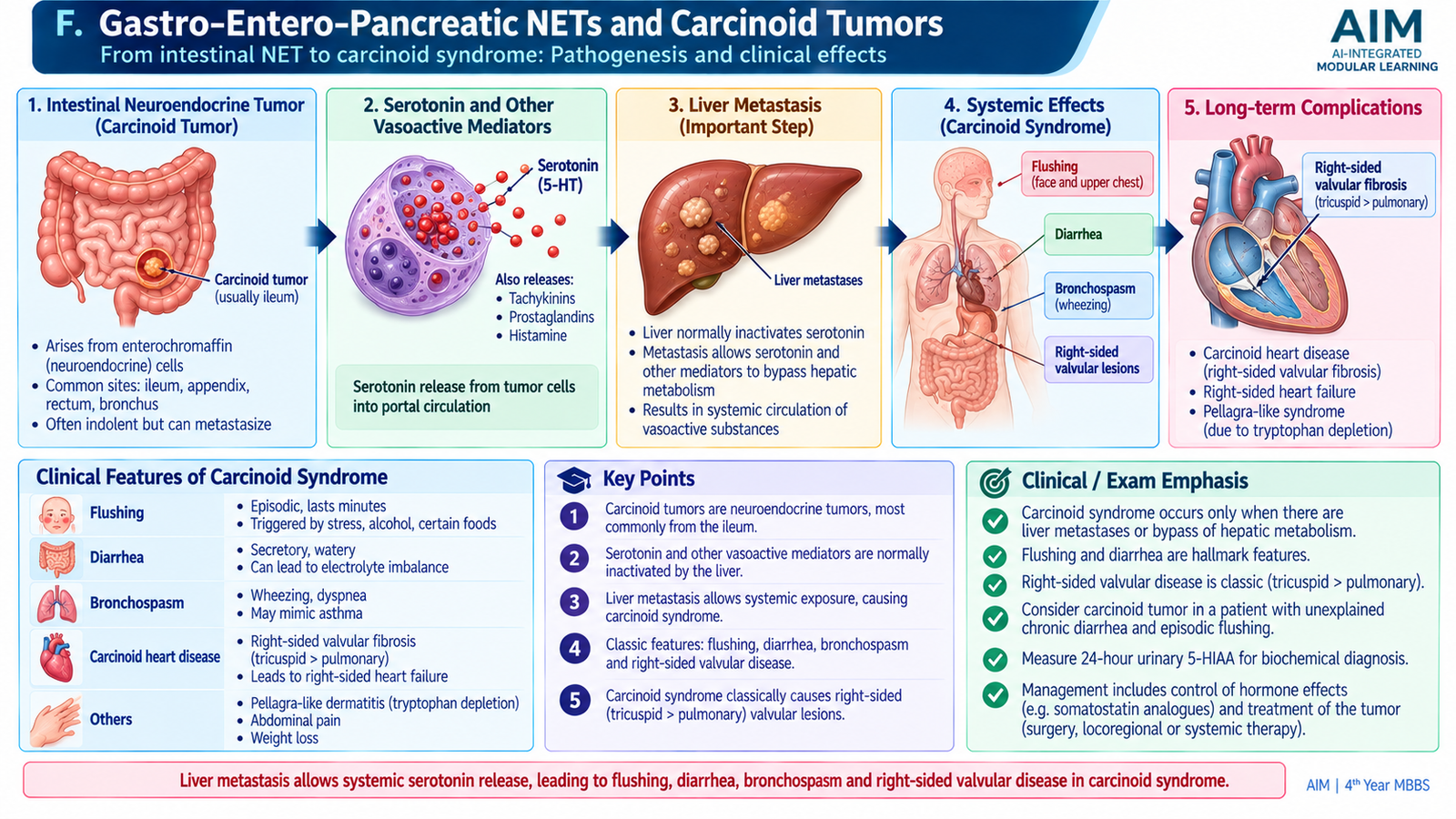
Gastro-entero-pancreatic neuroendocrine tumors (GEP-NETs) are neuroendocrine tumors arising within the gastrointestinal tract or pancreas. Many older texts use the term carcinoid tumor particularly for well-differentiated gastrointestinal neuroendocrine tumors. These tumors may remain localized for a long period, but their clinical behavior varies with site, size, differentiation, proliferative activity, hormone production and metastatic spread.
Morphology
Well-differentiated gastrointestinal NETs are often submucosal lesions. Microscopically, they contain relatively uniform neuroendocrine cells arranged in nests, cords or trabeculae with the characteristic finely stippled salt-and-pepper nuclear chromatin. Neuroendocrine differentiation is supported by markers such as synaptophysin and chromogranin.
Some serotonin-producing small-intestinal NETs stimulate a prominent desmoplastic fibrotic reaction in the mesentery. This fibrosis can tether or narrow the bowel and its blood supply, contributing to intestinal obstruction or ischemic symptoms.
Clinical Presentation
A GEP-NET may present because of the local tumor, metastatic disease or hormone secretion. Depending on location, patients may develop abdominal pain, altered bowel function, gastrointestinal obstruction or bleeding. Some lesions are discovered incidentally.
Carcinoid Syndrome
Some gastrointestinal NETs produce serotonin and other vasoactive substances. When these substances enter the portal circulation, the liver normally metabolizes much of them before they reach the systemic circulation. Consequently, a gastrointestinal NET commonly produces the full carcinoid syndrome only after tumor spread to the liver allows large amounts of vasoactive products to reach the systemic circulation.
The characteristic manifestations include:
- Episodic flushing caused by circulating vasoactive mediators.
- Secretory diarrhea due to effects on intestinal secretion and motility.
- Bronchospasm in some patients.
- Right-sided cardiac valvular fibrosis, particularly affecting tricuspid and pulmonary valves, after prolonged exposure to circulating serotonin-related mediators.
The right side of the heart is preferentially affected because circulating mediators reach the right heart before passing through the lungs, where many vasoactive substances are metabolized. This produces plaque-like fibrous thickening of right-sided endocardial and valvular surfaces and may ultimately cause right-sided heart failure.
Important Complications
- Local bowel obstruction or ischemic complications from tumor and mesenteric fibrosis.
- Hepatic metastases.
- Persistent secretory diarrhea with fluid and electrolyte loss.
- Carcinoid heart disease with progressive right-sided valvular dysfunction.
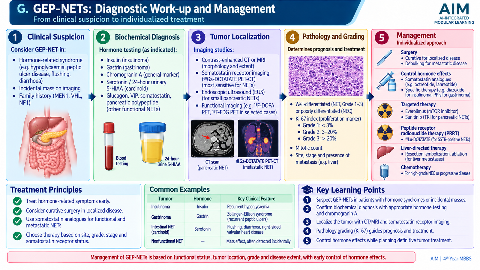
G. GEP-NETs: Diagnostic Work-up and Management
Investigation of a suspected GEP-NET is guided by the patient’s clinical syndrome. A functional tumor is first investigated by demonstrating inappropriate hormone production, whereas a nonfunctional tumor may initially be detected on imaging. Once a neuroendocrine tumor is suspected, the clinician must establish its location, determine its extent and obtain pathological information about differentiation and proliferative activity.
Biochemical Investigation
Hormonal testing should be selected according to the suspected syndrome rather than ordering every neuroendocrine marker indiscriminately.
- Suspected insulinoma: demonstrate inappropriate endogenous insulin secretion during documented hypoglycemia. Measurement of insulin together with C-peptide and related markers helps distinguish endogenous insulin production from administration of exogenous insulin.
- Suspected gastrinoma: assess fasting gastrin in an appropriate clinical and gastric-acid context. Further physiological testing may be required when the diagnosis remains uncertain.
- Suspected VIPoma, glucagonoma or somatostatinoma: measure the corresponding hormone when the clinical syndrome strongly suggests it.
- Suspected serotonin-producing carcinoid syndrome: urinary measurement of the serotonin metabolite 5-hydroxyindoleacetic acid (5-HIAA) is a classic biochemical investigation.
General neuroendocrine markers may support the diagnosis in selected situations but are not a substitute for syndrome-directed hormone testing, imaging and pathological confirmation.
Localization and Staging
Cross-sectional imaging with CT or MRI is used to locate the primary tumor and look for regional or distant spread, particularly liver metastases. Small pancreatic lesions may require more focused localization methods such as endoscopic ultrasonography. Many well-differentiated NETs express somatostatin receptors, allowing somatostatin-receptor imaging to help identify primary, multifocal or metastatic disease.
Pathological Confirmation
Histopathology establishes neuroendocrine differentiation and provides information about tumor differentiation and proliferative activity. Typical well-differentiated tumors have an organoid architecture and salt-and-pepper chromatin. Immunohistochemical expression of neuroendocrine markers supports the diagnosis. Mitotic activity and the proliferative marker Ki-67 contribute to grading and therefore help estimate biological behavior.
Management of Localized Disease
When disease is localized and technically resectable, surgical removal provides the main opportunity for cure. The exact operation depends on the site and extent of the tumor and is planned by the relevant specialist team rather than by a single standard procedure for every NET.
Control of Functional Syndromes
Hormonal symptoms often require control even before definitive tumor treatment. Therapy is directed toward the specific physiological disturbance:
- Insulinoma: prevent and treat hypoglycemia while the tumor is localized and definitive therapy is arranged; agents that reduce insulin release may be used in selected patients.
- Gastrinoma: strong gastric-acid suppression is important because acid hypersecretion causes the major morbidity.
- VIPoma: fluid and electrolyte replacement is essential because severe secretory diarrhea can cause marked dehydration and hypokalemia.
- Carcinoid syndrome and several other functioning NETs: somatostatin analogues can reduce hormone release and improve secretory symptoms.
Advanced or Metastatic Disease
Patients with unresectable or metastatic well-differentiated GEP-NETs require multidisciplinary care. Depending on the tumor site, grade, hormone activity and receptor expression, treatment may include somatostatin analogues, surgical or liver-directed approaches, molecularly targeted treatment, peptide-receptor radionuclide therapy or systemic antitumor therapy. At undergraduate level, the key principle is that treatment is individualized according to tumor biology, extent of disease and functional status.
3. Integrated Mechanism Flow
The different tumors in this chapter can be understood through one common sequence: a neuroendocrine or endocrine tumor produces either excessive hormone activity or progressive tumor growth, which then determines the clinical presentation and the diagnostic approach.
Endocrine or neuroendocrine cell proliferation
Hormone secretion or nonfunctional growth
Hormonal syndrome or mass effect
Syndrome-directed biochemical testing
Imaging and pathological assessment
Hormone control plus tumor-directed treatment
4. Important Comparison
MEN Syndromes
| Feature | MEN1 | MEN2A | MEN2B |
|---|---|---|---|
| Major gene | MEN1 / menin | RET | RET |
| Thyroid | Not defining | Medullary thyroid carcinoma | Medullary thyroid carcinoma |
| Pheochromocytoma | Not typical | Present | Present |
| Parathyroid disease | Very important component | May occur | Not typical |
| Other hallmark | Pituitary + pancreatic/duodenal NETs | Three endocrine-gland pattern | Mucosal neuromas + marfanoid habitus |
Important Functional Pancreatic NET Syndromes
| Tumor | Main hormone | Major clinical clue |
|---|---|---|
| Insulinoma | Insulin | Fasting hypoglycemic symptoms relieved by glucose |
| Gastrinoma | Gastrin | Recurrent/severe peptic ulceration with diarrhea |
| Glucagonoma | Glucagon | Hyperglycemia + necrolytic migratory erythema |
| VIPoma | VIP | Watery diarrhea + hypokalemia |
| Somatostatinoma | Somatostatin | Diabetes, steatorrhea and gallstones |
⭐ AIM High-Yield Review
Pheochromocytoma — Clinical Medicine
Review the pathophysiology, classical presentation, complications, diagnostic approach and management of pheochromocytoma.