
Topic 18 — Early Pregnancy Disorders: Miscarriage, Ectopic Pregnancy and Gestational Trophoblastic Disease
A structured introduction to pregnancy loss, ectopic implantation and trophoblastic disease, with emphasis on causes, clinical recognition, diagnosis, complications and undergraduate management principles.
1. Topic Introduction
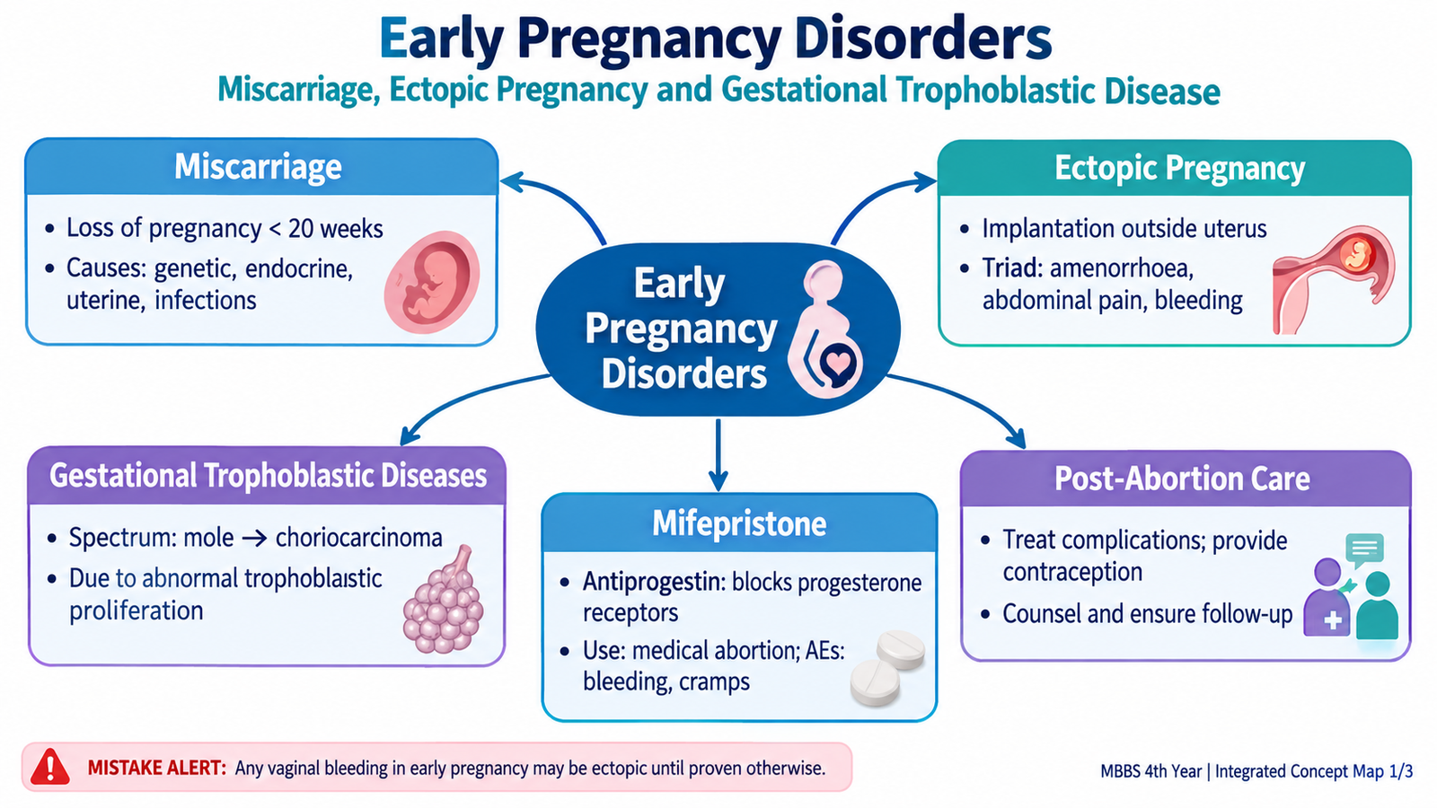
Early pregnancy disorders include conditions in which a pregnancy is lost, implants outside the uterine cavity, or develops abnormal placental trophoblastic tissue. The three major groups in this chapter are miscarriage, ectopic pregnancy and gestational trophoblastic disease. Although vaginal bleeding may occur in all three, their underlying mechanisms and clinical risks are different. Miscarriage mainly concerns loss of an intrauterine pregnancy, ectopic pregnancy may cause life-threatening internal hemorrhage, and trophoblastic disease may persist or become malignant. You will learn how these conditions develop, how they are recognized and investigated, their major complications, and the principles of management. Mifepristone and post-abortion care are included where they directly relate to early pregnancy care.
A. Miscarriage: Definition, Types, Etiology and Risk Factors
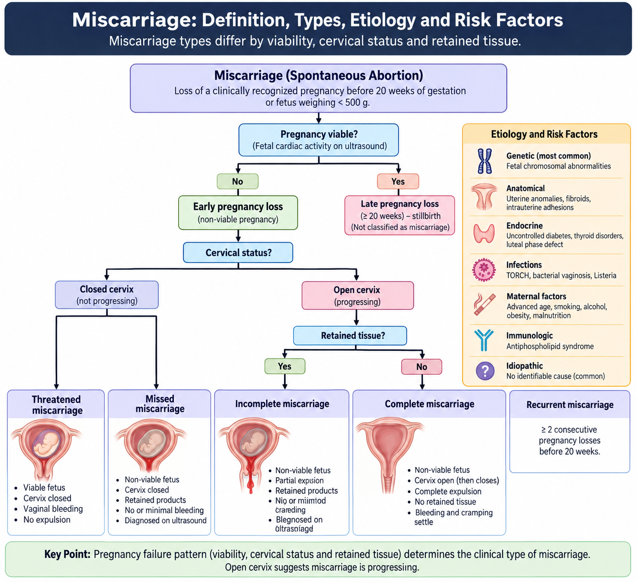
Miscarriage is the spontaneous loss of a pregnancy before the fetus has reached viability. It therefore refers to pregnancy loss without deliberate termination. The exact gestational threshold used to define viability may differ between health systems, so the central concept is more important than memorizing a locally variable numerical limit.
Types of miscarriage
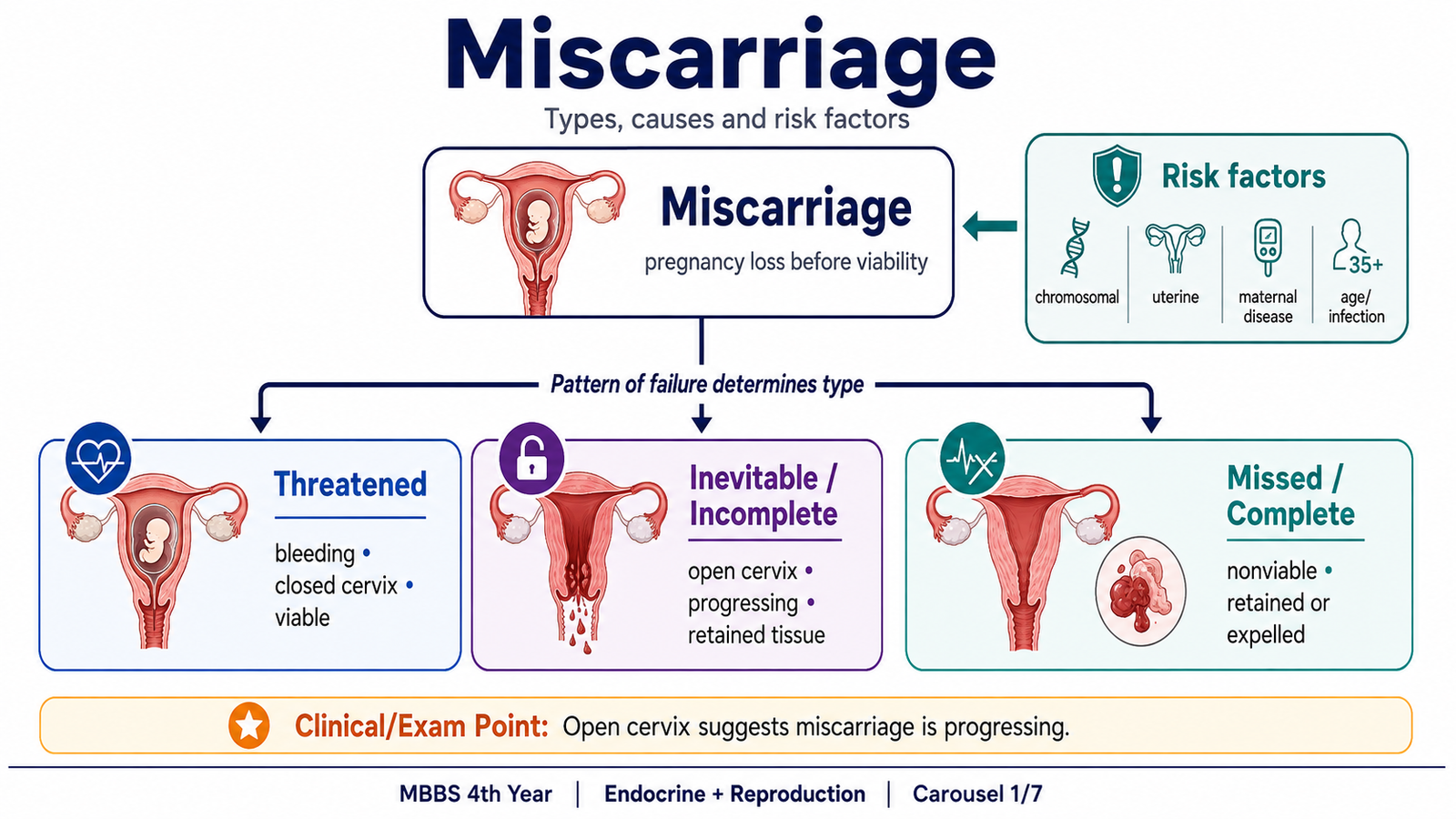
Different forms are distinguished mainly by whether the pregnancy is still viable, whether the cervix is open and whether products of conception remain inside the uterus. These distinctions help determine management.
| Type | Core idea | Typical cervical finding |
|---|---|---|
| Threatened | Bleeding occurs, but the pregnancy may remain viable. | Closed |
| Inevitable | Pregnancy loss is in progress and cannot be prevented. | Open |
| Incomplete | Some products have been expelled but some remain. | Often open |
| Complete | All products of conception have been expelled. | Closes after expulsion |
| Missed | The pregnancy is nonviable but remains within the uterus. | Closed |
| Recurrent | Repeated spontaneous pregnancy losses requiring evaluation for an underlying cause. | Depends on current pregnancy |
Etiology and risk factors
Early miscarriage often results from an abnormal embryo rather than from something the mother has done. Chromosomal abnormalities in the conceptus are an important cause of early spontaneous pregnancy loss. When development is genetically incompatible with continued pregnancy, embryonic growth stops and the pregnancy is eventually expelled or retained as a missed miscarriage.
Other important causes or predisposing factors include:
- Increasing maternal age: associated with a greater frequency of chromosomal errors in the oocyte and conceptus.
- Previous miscarriage: repeated previous losses increase the likelihood that an underlying maternal, genetic or anatomical factor may be present.
- Uterine abnormalities: congenital uterine anomalies or lesions that distort the uterine cavity may interfere with implantation and continuation of pregnancy.
- Endocrine or metabolic disease: poorly controlled maternal disorders such as diabetes or significant thyroid dysfunction may adversely affect pregnancy.
- Antiphospholipid syndrome: abnormal thrombosis and placental dysfunction may contribute to recurrent pregnancy loss.
- Cervical weakness: structural inability of the cervix to remain closed can contribute particularly to later spontaneous pregnancy loss.
- Smoking, alcohol and harmful drug exposure: these can adversely affect pregnancy and should be avoided.
- Some maternal infections or severe systemic illness: may cause pregnancy loss in selected circumstances.
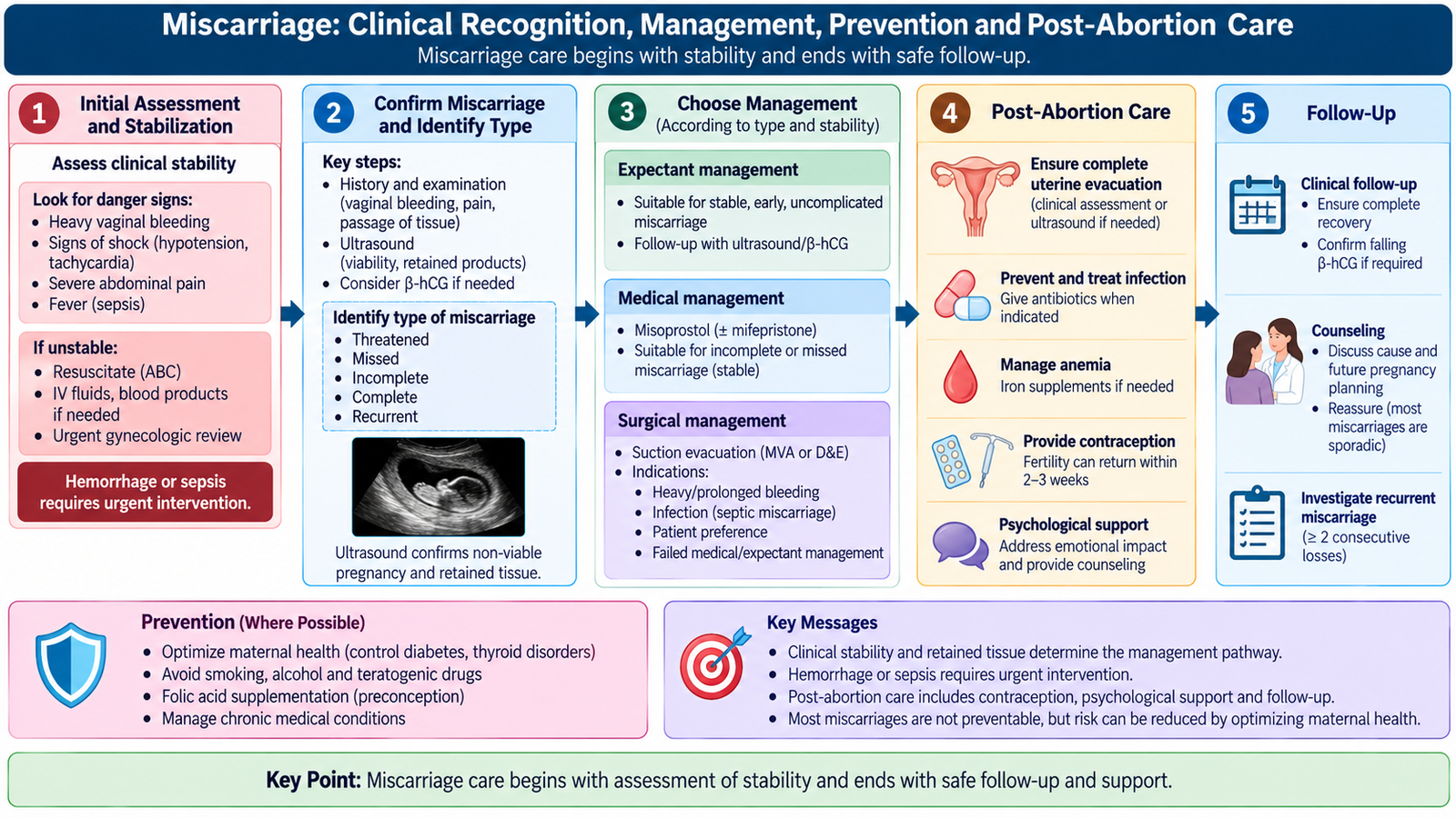
B. Miscarriage: Clinical Recognition, Management, Prevention and Post-Abortion Care
A patient with miscarriage commonly presents with vaginal bleeding, lower abdominal cramping or passage of tissue. The immediate clinical task is not simply to label the miscarriage. The clinician must first determine whether the patient is stable, whether significant hemorrhage or infection is present, whether the pregnancy is intrauterine, and whether products of conception remain within the uterus.
Clinical features and examination
- Vaginal bleeding ranging from light spotting to heavy bleeding.
- Lower abdominal or pelvic cramping caused by uterine contractions.
- Passage of tissue in incomplete or complete miscarriage.
- Reduction or disappearance of previously experienced pregnancy symptoms may occur in a missed miscarriage.
- Heavy bleeding may produce pallor, tachycardia, dizziness, hypotension or shock.
- Fever, uterine tenderness and offensive discharge suggest infection and possible septic miscarriage.
Speculum examination helps identify the source and amount of bleeding, whether tissue is visible at the cervical opening, and whether the cervix appears open. Abdominal and pelvic examination assess tenderness, uterine size and signs of peritoneal irritation. Severe pain, shock or marked abdominal tenderness should also raise concern for ectopic pregnancy, particularly if an intrauterine pregnancy has not been confirmed.
Diagnosis
Diagnosis combines clinical assessment with pregnancy testing and ultrasonography. Transvaginal ultrasound is particularly useful because it can establish whether the pregnancy is located inside the uterus, assess viability and identify retained products. Serum β-hCG may be useful when ultrasound findings are uncertain or when the pregnancy location is not yet established. A blood count helps assess blood loss when bleeding is significant.
A positive pregnancy test with no definite intrauterine or ectopic pregnancy on ultrasound may initially be described as a pregnancy of unknown location. Such a patient requires appropriate follow-up because an ectopic pregnancy cannot yet be excluded.
Management principles
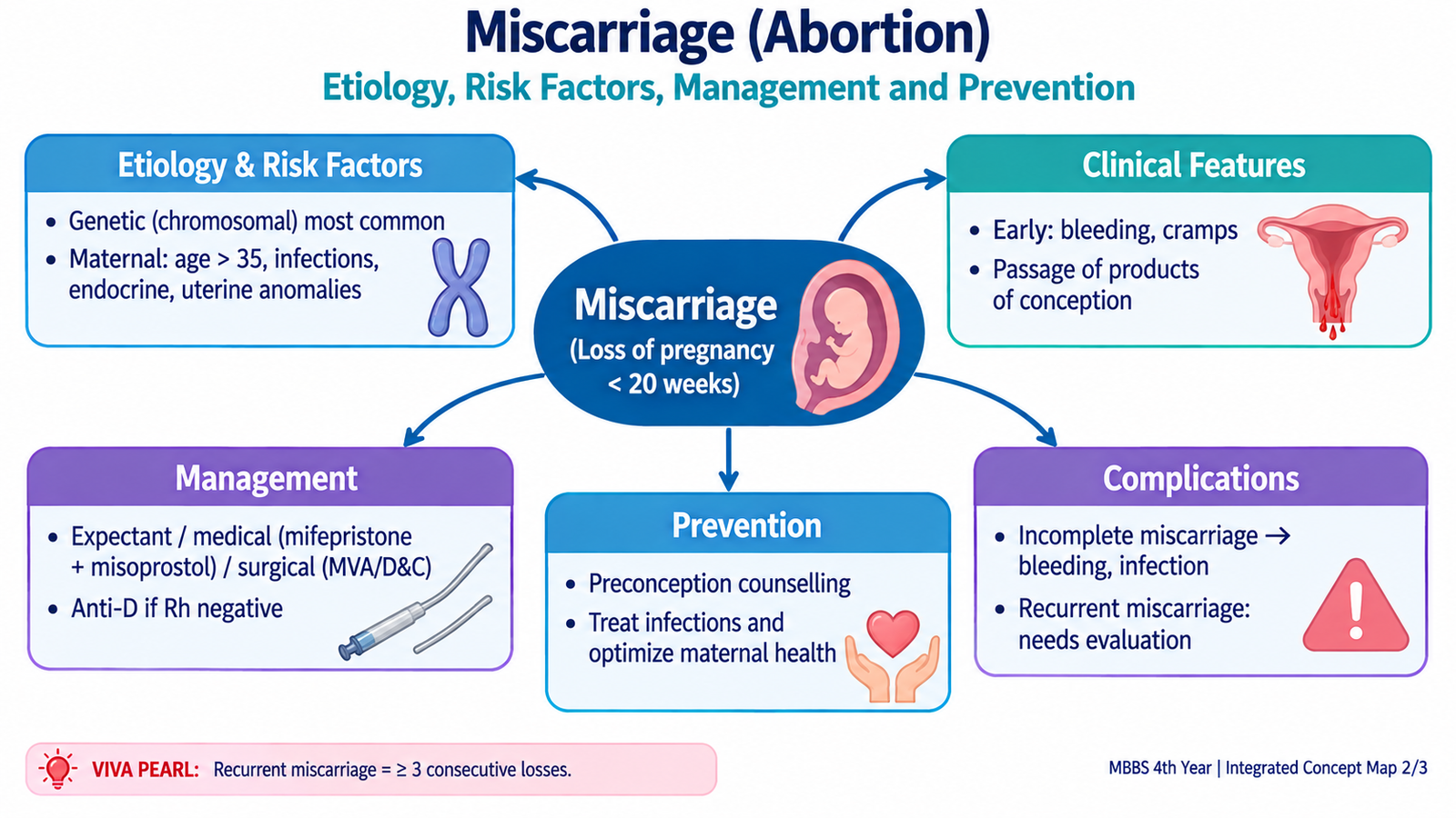
Management depends on the type of miscarriage, amount of bleeding, evidence of infection, ultrasound findings, clinical stability and the patient’s informed preference. Three broad approaches are expectant, medical and surgical management.
Allows spontaneous completion when the patient is stable and there is no urgent indication for intervention.
Uterotonic medication, commonly a prostaglandin such as misoprostol, promotes cervical opening and uterine evacuation. Mifepristone may be used in appropriate regimens.
Uterine evacuation by suction is used when rapid completion is required or when other approaches are unsuitable.
- Threatened miscarriage: establish pregnancy location and viability, assess bleeding and provide follow-up. The pregnancy may continue normally.
- Complete miscarriage: confirm that the uterus has emptied and that bleeding and symptoms are resolving.
- Inevitable, incomplete or missed miscarriage: expectant, medical or surgical management may be appropriate depending on the clinical situation.
- Heavy ongoing bleeding or hemodynamic instability: stabilize the patient and obtain urgent uterine evacuation when retained tissue is contributing to hemorrhage.
- Septic miscarriage: requires urgent stabilization, appropriate intravenous antimicrobial therapy and removal of infected retained products once the patient is stabilized.
Prevention of miscarriage
Many early miscarriages, particularly those due to chromosomal abnormalities, cannot be prevented. Prevention therefore focuses on modifiable risks and on identifying treatable causes when pregnancy loss is recurrent.
- Optimize control of important maternal medical and endocrine disorders before and during pregnancy.
- Avoid smoking, alcohol, harmful recreational drugs and unnecessary potentially harmful medication exposure.
- Evaluate recurrent miscarriage for specific anatomical, endocrine, immunological or genetic causes where appropriate.
- Treat an identified cause rather than giving nonspecific treatment to every patient.
- Provide preconception and early antenatal care for women with significant previous pregnancy loss or medical risk factors.
Principles of post-abortion care
Post-abortion care refers to the essential care given after spontaneous or induced pregnancy loss. Its purpose is to identify and treat immediate complications, support recovery and reduce preventable future reproductive morbidity. Care should be respectful, confidential and nonjudgmental.
Before discharge or completion of care, the patient should understand which symptoms require urgent review, particularly increasing bleeding, fever, worsening abdominal pain, fainting or persistent illness.
C. Ectopic Pregnancy: Etiology, Risk Factors and Clinical Features
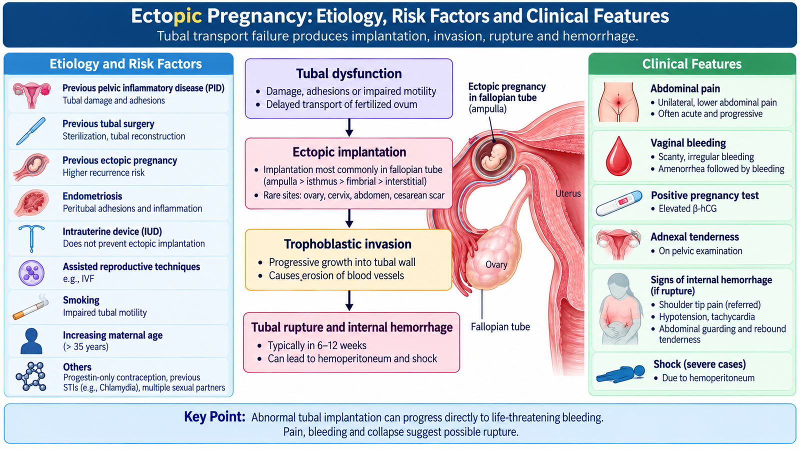
An ectopic pregnancy occurs when a fertilized ovum implants outside the normal endometrial cavity of the uterus. Most ectopic pregnancies are located in a fallopian tube, with the ampullary part being the commonest tubal site. The condition is clinically important because trophoblastic tissue can invade and erode the thin tubal wall and its vessels, producing internal hemorrhage.
How ectopic pregnancy develops
Etiology and risk factors
Anything that damages the fallopian tube or interferes with normal transport of the fertilized ovum can increase the risk. However, an ectopic pregnancy may occur even when no clear risk factor is found.
- Previous ectopic pregnancy: suggests persistent tubal disease and increases recurrence risk.
- Previous pelvic inflammatory disease: tubal inflammation and scarring can narrow the lumen and damage ciliary transport.
- Previous tubal or pelvic surgery: postoperative scarring or altered tubal anatomy may delay embryo transport.
- Infertility and assisted reproductive techniques: underlying tubal disease and altered embryo placement can contribute.
- Endometriosis or pelvic adhesions: may distort normal tubal anatomy.
- Smoking: is associated with impaired tubal function.
Clinical features
The classic presentation is amenorrhea followed by lower abdominal pain and vaginal bleeding, but patients do not always present with the complete triad. Symptoms depend partly on whether the ectopic pregnancy is still unruptured or has begun to bleed into the peritoneal cavity.
- Lower abdominal or pelvic pain: often unilateral initially because the abnormal pregnancy distends or irritates the affected tube.
- Vaginal bleeding: commonly light or irregular and results from inadequate hormonal support of the endometrium.
- Amenorrhea or delayed menstruation: reflects the presence of a pregnancy.
- Shoulder-tip pain: may occur when intraperitoneal blood irritates the diaphragm.
- Dizziness, fainting or collapse: may indicate major internal blood loss.
Examination may show lower abdominal tenderness, adnexal tenderness or cervical motion tenderness. With rupture and major hemorrhage the patient may become pale, tachycardic and hypotensive, with generalized abdominal tenderness, guarding or features of shock.
D. Ectopic Pregnancy: Diagnosis and Management
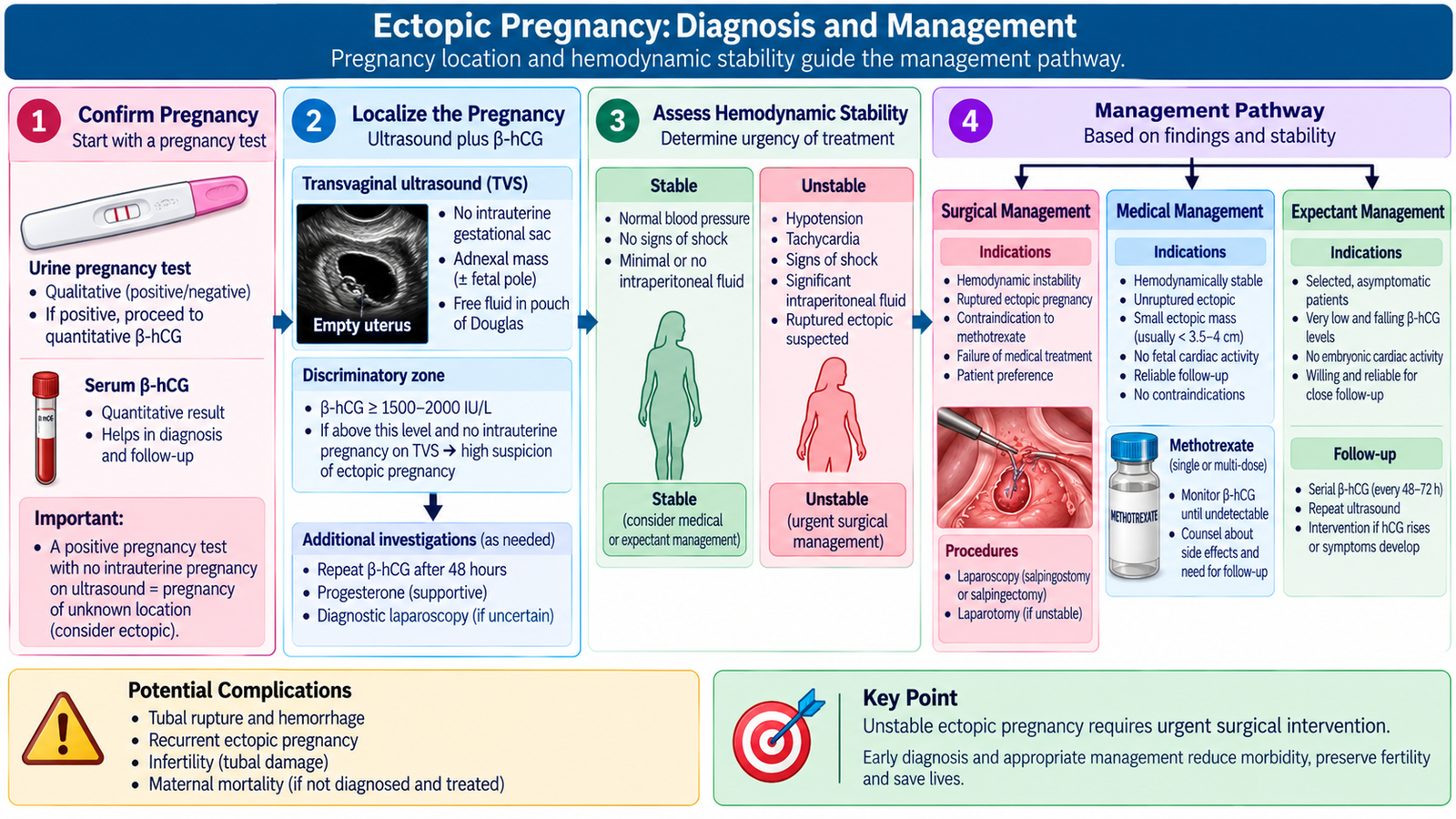
Diagnosis of ectopic pregnancy combines the clinical picture with evidence of pregnancy and determination of its location. No single symptom is sufficiently reliable by itself. The most useful approach connects pregnancy testing, transvaginal ultrasound and, when necessary, serial serum β-hCG measurements.
Diagnostic approach
Confirms that pregnancy tissue is present but does not determine its location.
Looks for an intrauterine pregnancy and for an adnexal gestational structure, mass or pelvic free fluid.
The trend helps interpret an uncertain early pregnancy when ultrasound does not yet establish the location or viability.
A clearly demonstrated extrauterine gestational sac containing a yolk sac or embryo is strong evidence of ectopic pregnancy. In contrast, an empty uterus alone is not enough to diagnose ectopic pregnancy because a very early intrauterine pregnancy may not yet be visible. This is why ultrasound findings must be interpreted together with the clinical situation and β-hCG trend.
Additional investigations are guided by the patient’s condition. A complete blood count helps assess anemia or blood loss. Blood grouping and compatibility testing are important when significant hemorrhage or transfusion may be anticipated.
Management
The first management decision is based on hemodynamic stability and the possibility of rupture. A ruptured ectopic pregnancy is a potentially fatal hemorrhagic emergency and must not be delayed for prolonged diagnostic observation.
In a clinically stable patient with an unruptured ectopic pregnancy, treatment may be expectant, medical or surgical depending on the clinical and ultrasound findings, β-hCG behavior, reliability of follow-up and suitability for each approach.
- Expectant management: may be appropriate in carefully selected stable patients when the ectopic pregnancy appears to be resolving spontaneously and reliable follow-up is possible.
- Medical management: methotrexate can be used in suitable unruptured ectopic pregnancies. It is a folate antagonist that suppresses rapidly proliferating trophoblastic tissue. Serial follow-up is required to ensure resolution.
- Surgical management: is required for rupture, significant bleeding, hemodynamic instability, unsuitable or failed medical treatment, or other circumstances in which definitive operative treatment is safer.
Surgical treatment generally removes the ectopic pregnancy through a laparoscopic approach when the patient is stable and appropriate facilities are available. Depending on tubal damage and fertility considerations, the affected tube may be opened to remove the pregnancy or removed when necessary. Detailed operative technique is beyond the required undergraduate level.
Complications and follow-up
- Tubal rupture and massive intraperitoneal hemorrhage.
- Hypovolemic shock and maternal death if severe hemorrhage is untreated.
- Loss or damage of a fallopian tube, which may affect future fertility.
- Increased risk of another ectopic pregnancy in a future conception.
After treatment, appropriate follow-up confirms resolution and provides counseling regarding future fertility. A woman with a previous ectopic pregnancy should seek assessment early in a future pregnancy so that the location can be established.
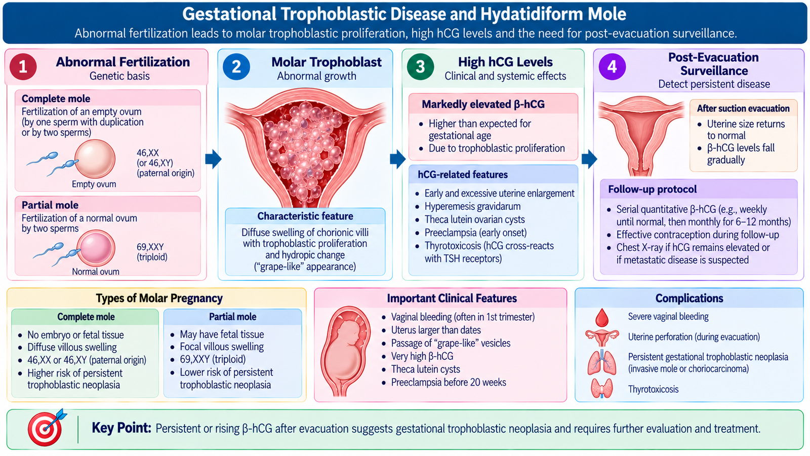
E. Gestational Trophoblastic Disease and Hydatidiform Mole
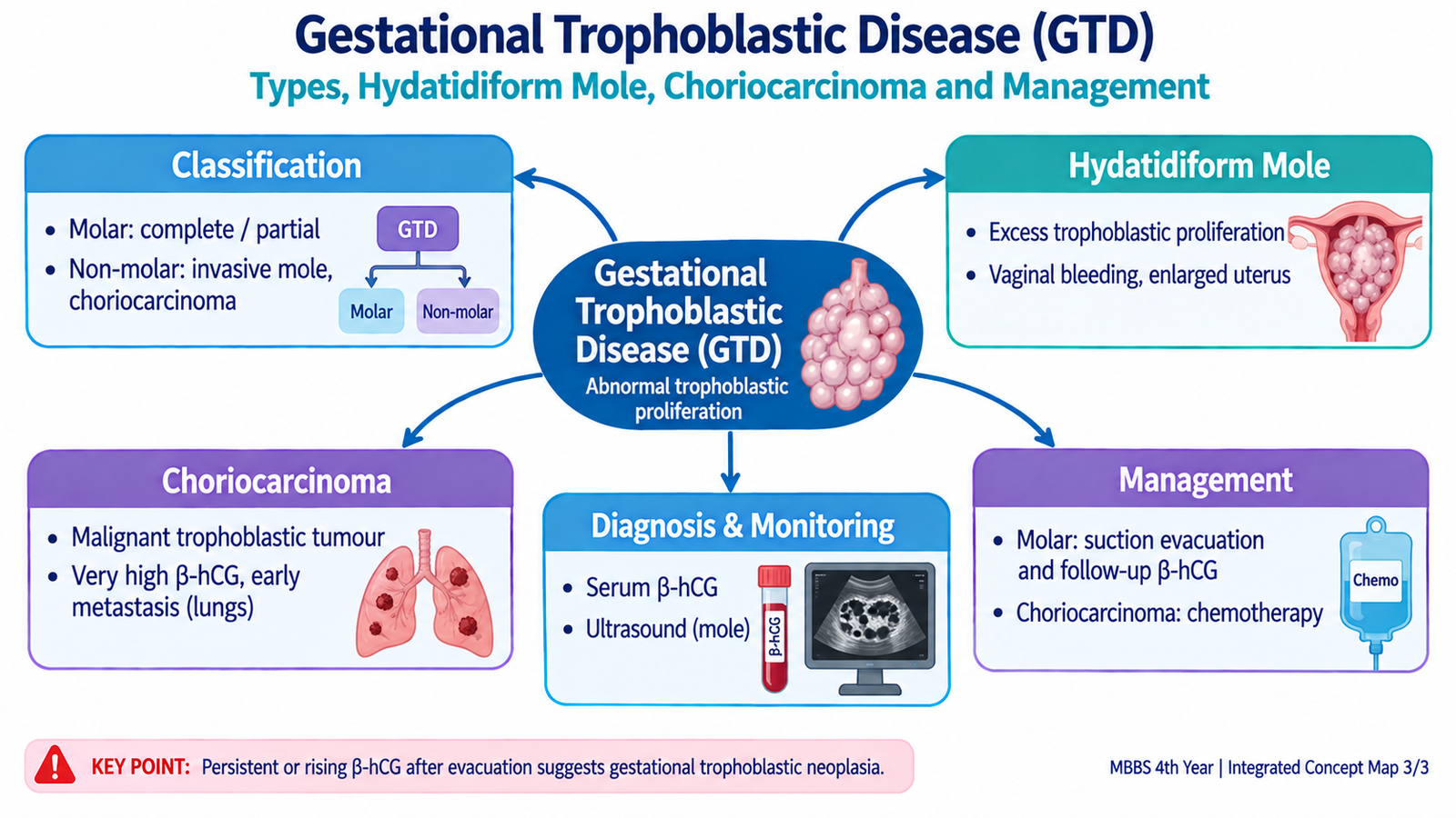
Gestational trophoblastic disease (GTD) is a group of pregnancy-related disorders caused by abnormal proliferation of placental trophoblastic tissue. Trophoblast normally forms an important part of the placenta and produces human chorionic gonadotropin (hCG). When trophoblastic growth becomes abnormal, hCG production may become excessive or persist after the pregnancy has ended.
Classification of gestational trophoblastic disease
- Hydatidiform mole
- Complete hydatidiform mole
- Partial hydatidiform mole
- Gestational trophoblastic neoplasia
- Invasive mole
- Gestational choriocarcinoma
- Placental-site trophoblastic tumor
- Epithelioid trophoblastic tumor
Hydatidiform mole: etiology and pathogenesis
A hydatidiform mole results from abnormal fertilization and produces abnormal chorionic villi together with excessive trophoblastic proliferation. The genetic mechanism differs between complete and partial moles.
| Feature | Complete mole | Partial mole |
|---|---|---|
| Fertilization concept | An ovum lacking functional maternal nuclear chromosomes is fertilized; the nuclear genome is essentially paternal in origin. | A normal ovum receives an extra paternal chromosome set, commonly producing triploidy. |
| Embryo / fetus | Absent | May be present but is abnormal and nonviable |
| Villous change | Diffuse hydropic swelling | Focal hydropic change |
| Trophoblastic proliferation | More diffuse and prominent | Usually less extensive and more focal |
| Persistent neoplasia risk | Higher | Lower |
Clinical features of hydatidiform mole
Vaginal bleeding is the most frequent presentation. Features related to excessive hCG or abnormal placental tissue are particularly suggestive of a complete mole.
- Vaginal bleeding: results from separation and disruption of abnormal molar tissue.
- Uterus larger than expected for the pregnancy: may result from abundant molar tissue, although uterine size is not always increased.
- Marked nausea and vomiting: may occur because of high circulating hCG.
- Unusually early development of preeclampsia: is an important clue to abnormal placental disease.
- Clinical hyperthyroidism: may occur because very high hCG can stimulate thyroid receptors.
- Absence of fetal parts or fetal cardiac activity in a complete mole: reflects absence of an embryo.
- Passage of translucent vesicle-like tissue may occur but is not required for diagnosis.
Diagnosis
Diagnosis is based on the clinical presentation, quantitative serum hCG, ultrasonography and histopathological examination of evacuated tissue. Ultrasound of a complete mole may show a heterogeneous intrauterine mass with numerous small cystic spaces and no normal fetus. This produces the classic vesicular or “snowstorm-like” appearance. Partial moles may be less obvious and can resemble a failed pregnancy with an abnormal fetus and abnormal placenta.
Histopathological examination confirms the nature of the molar tissue and helps distinguish complete from partial disease.
Management
The main treatment is evacuation of the molar pregnancy, usually by suction evacuation. Significant hemorrhage or other complications should be stabilized before or during definitive treatment as required. Evacuated tissue should be examined histologically.
After evacuation, serial quantitative hCG surveillance is essential. Falling hCG indicates regression of trophoblastic tissue. Persistent elevation or a renewed rise suggests persistent gestational trophoblastic neoplasia and requires specialist assessment and treatment. A new pregnancy during surveillance can itself raise hCG and make interpretation difficult, so reliable contraception is used during the required follow-up period according to specialist advice.
Complications
- Heavy uterine bleeding and anemia.
- Severe nausea and vomiting related to high hCG.
- Hyperthyroid manifestations from hCG-mediated thyroid stimulation.
- Unusually early preeclampsia.
- Persistent gestational trophoblastic neoplasia, particularly after a complete mole.
F. Choriocarcinoma
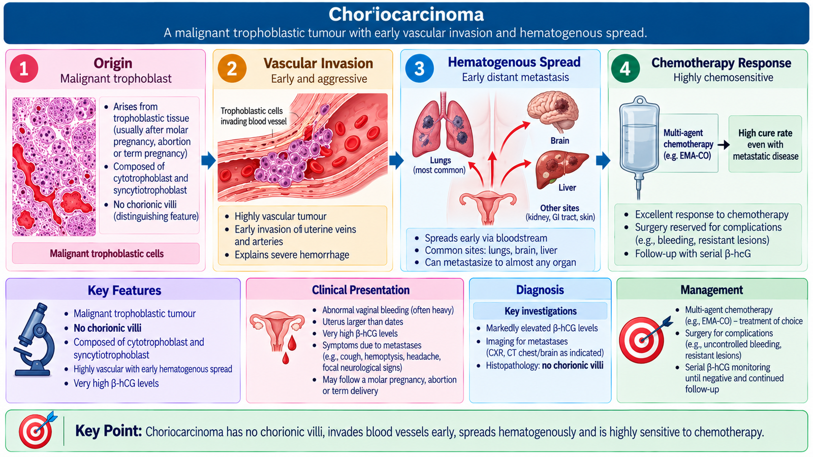
Gestational choriocarcinoma is a highly malignant trophoblastic tumor composed of proliferating trophoblastic cells. It can develop after a hydatidiform mole, miscarriage, ectopic pregnancy or a previous viable pregnancy. Unlike a hydatidiform mole, choriocarcinoma does not form chorionic villi. Its trophoblastic cells directly invade blood vessels, which explains its tendency to produce hemorrhage and to spread early through the bloodstream.
Etiology and disease mechanism
Microscopically, the tumor contains malignant cytotrophoblast and syncytiotrophoblast without chorionic villi. Extensive hemorrhage and necrosis are characteristic because the tumor is highly vascular and destroys vessel walls.
Clinical features
- Persistent or irregular vaginal bleeding after a pregnancy: may be the first clue.
- Persistently elevated or rising hCG: indicates continuing active trophoblastic tissue.
- Pelvic or uterine enlargement may be present.
- Pulmonary metastases: may produce cough, chest symptoms or hemoptysis.
- Brain or other organ metastases: may produce neurological or organ-specific symptoms in advanced disease.
The lungs are a common site of hematogenous spread. Other important metastatic sites include the vagina, brain and liver. Because metastases can also be very vascular, they may themselves bleed.
Diagnosis
Diagnosis is strongly suggested by the combination of a recent or previous pregnancy, persistent abnormal bleeding, high or rising serum hCG and imaging evidence of pelvic disease or metastases. Pelvic ultrasonography assesses uterine disease, while additional imaging is used to determine metastatic spread when indicated. The pattern of hCG after a molar pregnancy is particularly important because failure of hCG to regress normally suggests persistent neoplasia.
Management
Choriocarcinoma is highly sensitive to chemotherapy. Treatment is therefore primarily systemic chemotherapy, selected according to the extent and risk category of disease. Lower-risk disease can often be treated with single-agent chemotherapy, whereas higher-risk or extensive disease requires combination chemotherapy. Surgery is reserved for selected circumstances rather than being the routine primary treatment.
Complications
- Severe uterine or metastatic hemorrhage.
- Pulmonary compromise from lung metastases.
- Neurological deterioration or intracranial bleeding from brain metastases.
- Other organ damage from widespread hematogenous disease.
G. Mifepristone in Reproductive Care
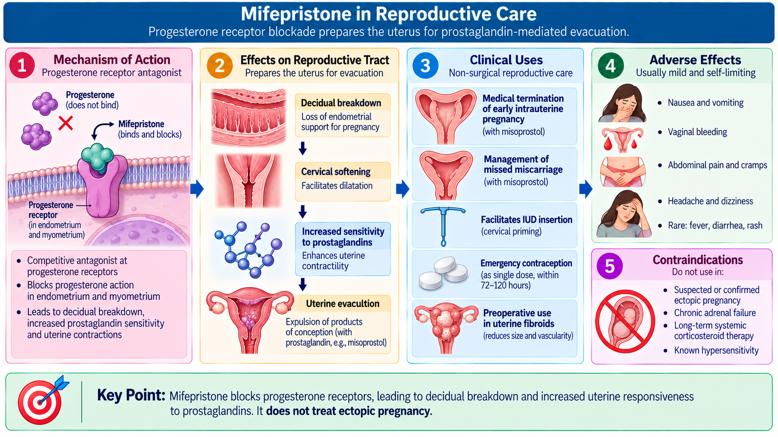
Mifepristone is a synthetic steroid that acts mainly as a progesterone-receptor antagonist in its reproductive use. Progesterone is essential for maintaining the decidua and supporting an early intrauterine pregnancy. Blocking progesterone therefore destabilizes the endometrium and prepares the uterus and cervix for expulsion of the pregnancy.
Mechanism of action
This explains why mifepristone is commonly combined with misoprostol, a prostaglandin analogue. Mifepristone removes progesterone support and makes the uterus more responsive, while misoprostol stimulates uterine contraction and cervical change.
Clinical uses in the reproductive context
- Medical termination of an intrauterine pregnancy, usually in combination with a prostaglandin such as misoprostol.
- Medical management of selected early pregnancy loss or missed miscarriage, where an antiprogestin followed by a prostaglandin improves uterine evacuation in appropriate clinical settings.
Adverse effects
Most reproductive adverse effects follow from the intended disruption of pregnancy and uterine evacuation rather than from unrelated organ toxicity.
- Vaginal bleeding.
- Lower abdominal cramping or pain.
- Nausea and vomiting.
- Diarrhea, particularly when used with a prostaglandin.
- Headache, dizziness or weakness.
- Heavy or prolonged bleeding requiring medical or surgical intervention in a small proportion of patients.
- Incomplete expulsion requiring further treatment.
- Infection, which is uncommon but potentially serious.
Mifepristone also has antiglucocorticoid activity, which is relevant when considering significant adrenal disease or long-term systemic corticosteroid therapy.
Important Comparison — Early Pregnancy Bleeding
Miscarriage, ectopic pregnancy and molar pregnancy can all present with vaginal bleeding. The following distinctions help organize the diagnostic approach.
| Feature | Miscarriage | Ectopic pregnancy | Hydatidiform mole |
|---|---|---|---|
| Basic process | Loss of intrauterine pregnancy | Implantation outside uterine cavity | Abnormal trophoblastic pregnancy |
| Pain | Cramping common | Often unilateral; severe if rupture | Not usually dominant feature |
| Important clue | Cervical status and retained tissue | Adnexal findings or internal bleeding | Excessive hCG-related manifestations |
| Ultrasound focus | Viability and retained products | Pregnancy location and adnexa | Abnormal vesicular placental tissue |
| Major danger | Hemorrhage or infection | Tubal rupture and internal hemorrhage | Persistent trophoblastic neoplasia |
| Management principle | Expectant, medical or uterine evacuation | Expectant, methotrexate or surgery according to stability | Evacuation followed by hCG surveillance |
⭐ AIM High-Yield Review
Revise these points after understanding the full chapter.
Early Pregnancy Disorders
Review the clinical differentiation of miscarriage, ectopic pregnancy and gestational trophoblastic disease in early pregnancy.