
Topic 15 — Female Infertility and Benign Gynecologic Disorders: PCOS, Fibroids, Endometriosis, Menstrual and Pelvic-Floor Disorders
Topic Introduction
Female infertility and benign gynecological disorders are closely connected because normal fertility depends on normal ovarian function, patent fallopian tubes, a receptive uterus, an intact pelvic anatomy and coordinated hormonal control. Disorders such as polycystic ovary syndrome, fibroids and endometriosis can interfere with menstruation, ovulation, implantation or pelvic function. Other common problems, including abnormal uterine bleeding, uterovaginal prolapse and urinary incontinence, may markedly affect quality of life even when fertility is not the main concern. This chapter first establishes the relevant anatomy, then explains infertility and reproductive pharmacology, followed by the major benign gynecological disorders and their investigation and management.
A. Applied Anatomy of the Female Reproductive Tract
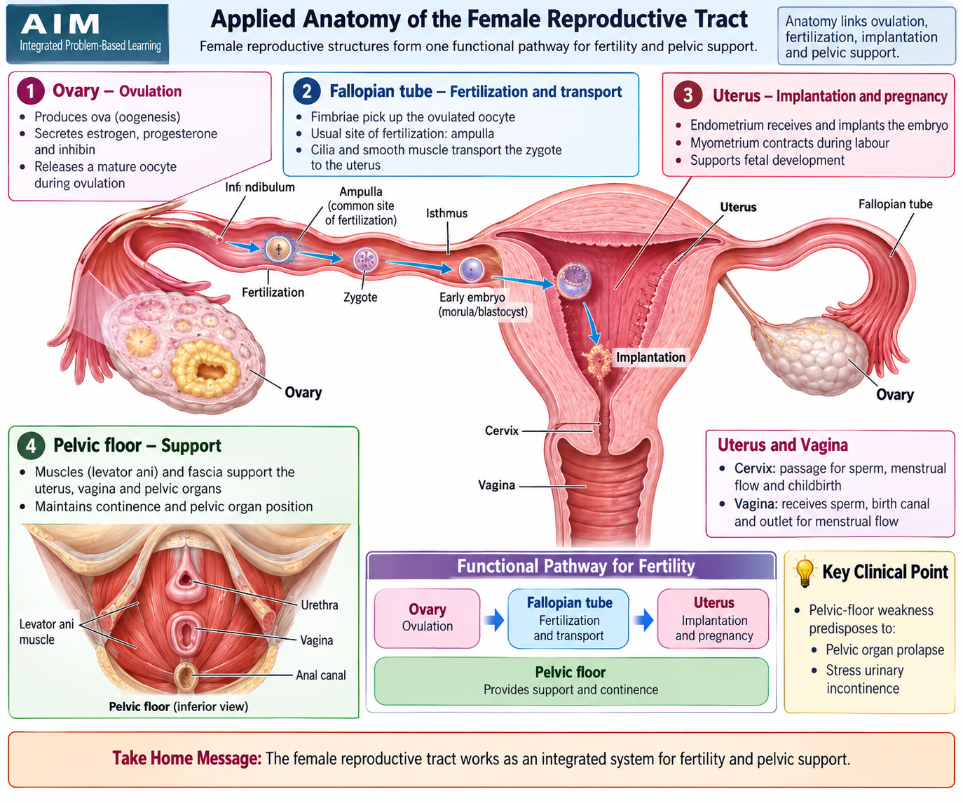
Understanding the anatomy of the perineum, vagina, cervix, uterus, adnexa and ovaries makes the later disorders much easier to understand. These structures form a continuous reproductive pathway: the ovaries release the oocyte, the fallopian tube receives it and is the usual site of fertilization, the uterus provides the site for implantation, and the cervix and vagina form the lower reproductive tract. The pelvic floor and connective-tissue supports maintain the normal position of the pelvic organs.
Perineum and pelvic floor
The perineum is the diamond-shaped region below the pelvic diaphragm. Its boundaries are formed by the pubic symphysis anteriorly, coccyx posteriorly and ischial tuberosities laterally. A line joining the ischial tuberosities divides it into an anterior urogenital triangle and a posterior anal triangle.
The perineal body is a central fibromuscular structure into which several pelvic-floor muscles attach. Together with the levator ani and connective-tissue supports, it helps maintain the position of the vagina, uterus, bladder and rectum. Damage during childbirth can weaken this support and contribute to uterovaginal prolapse or stress urinary incontinence.
Vagina and cervix
The vagina is a distensible fibromuscular canal extending from the vulva to the cervix. The bladder and urethra lie anterior to it, while the rectum lies posteriorly. These close relationships explain why weakness of the anterior vaginal support can allow the bladder to descend, while posterior weakness can permit rectal bulging.
The cervix forms the lower part of the uterus and projects into the upper vagina. Cervical mucus changes during the menstrual cycle and helps sperm passage around ovulation. Structural or inflammatory abnormalities involving the cervix may therefore contribute to infertility, although cervical causes are less common than ovulatory or tubal causes.
Uterus and its supports
The uterus consists of the fundus, body, isthmus and cervix. Its wall has an inner endometrium, a thick smooth-muscle myometrium and an outer serosal covering where present. The endometrium undergoes hormone-dependent cyclical changes, while the myometrium is the tissue from which uterine leiomyomas arise.
Uterine support depends on the pelvic floor together with the uterosacral, cardinal and other fascial supports. Failure of these structures allows downward displacement of the uterus and vaginal walls.
Adnexa, fallopian tubes and ovaries
The term adnexa mainly refers to the ovaries and fallopian tubes. The tube contains fimbrial, ampullary, isthmic and intramural portions. Fertilization most commonly takes place within the ampullary region. Tubal obstruction or distortion can prevent sperm and oocyte transport and is therefore an important cause of infertility.
The ovaries contain follicles at different stages of development and produce estrogen, progesterone and androgens. Normal cyclic follicular development and ovulation depend on coordinated hypothalamic, pituitary and ovarian function. Disturbance of this coordination is central to PCOS and many cases of anovulatory infertility.
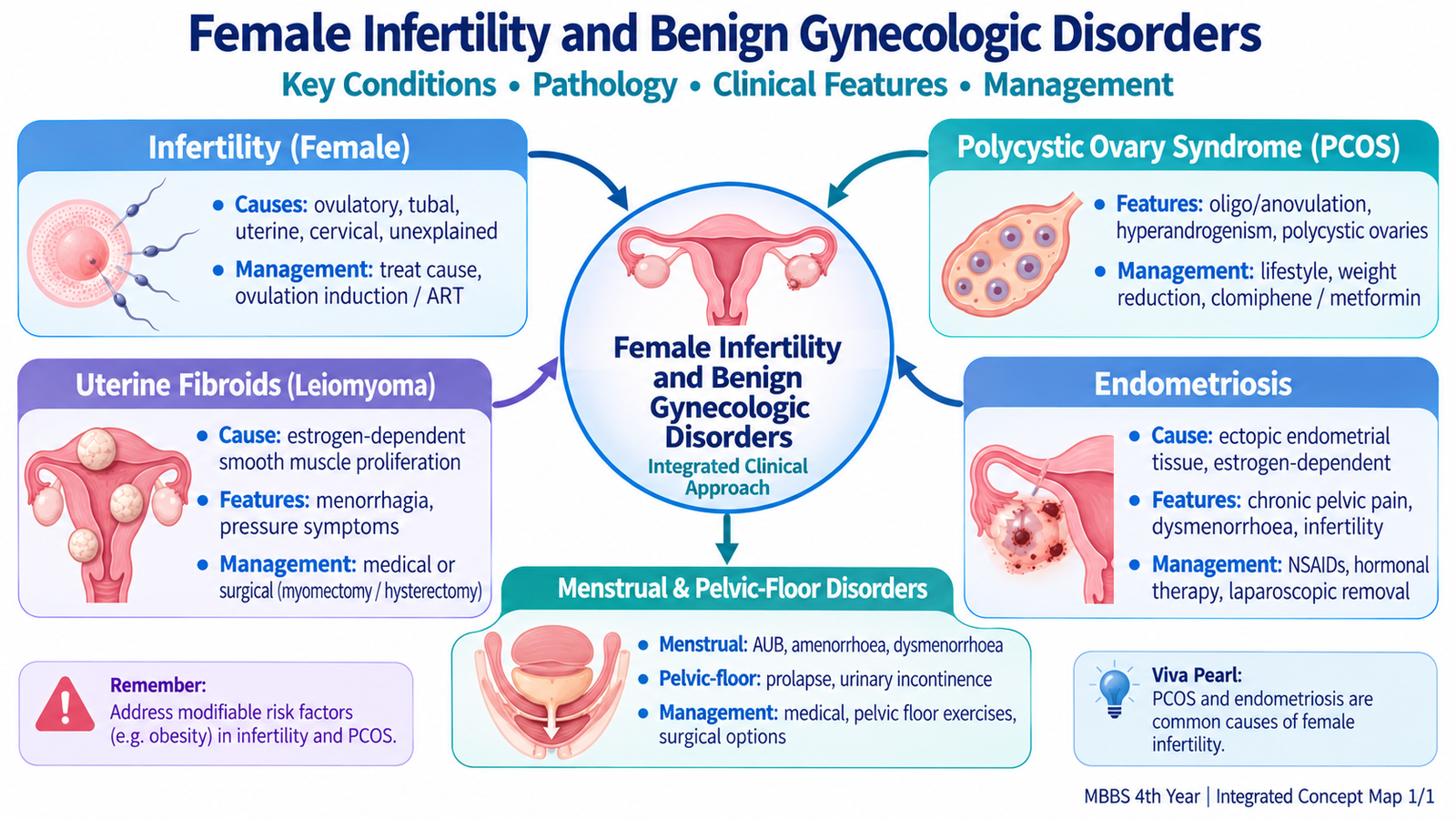
B. Female Infertility: Causes, Evaluation and Management
Definition and core concept
Infertility is failure to achieve pregnancy after about 12 months of regular unprotected sexual intercourse. Evaluation may be started earlier when there is an obvious reproductive disorder or when delay could significantly reduce the chance of conception. Infertility is best approached as a problem involving the couple rather than the woman alone, although this topic focuses on female causes.
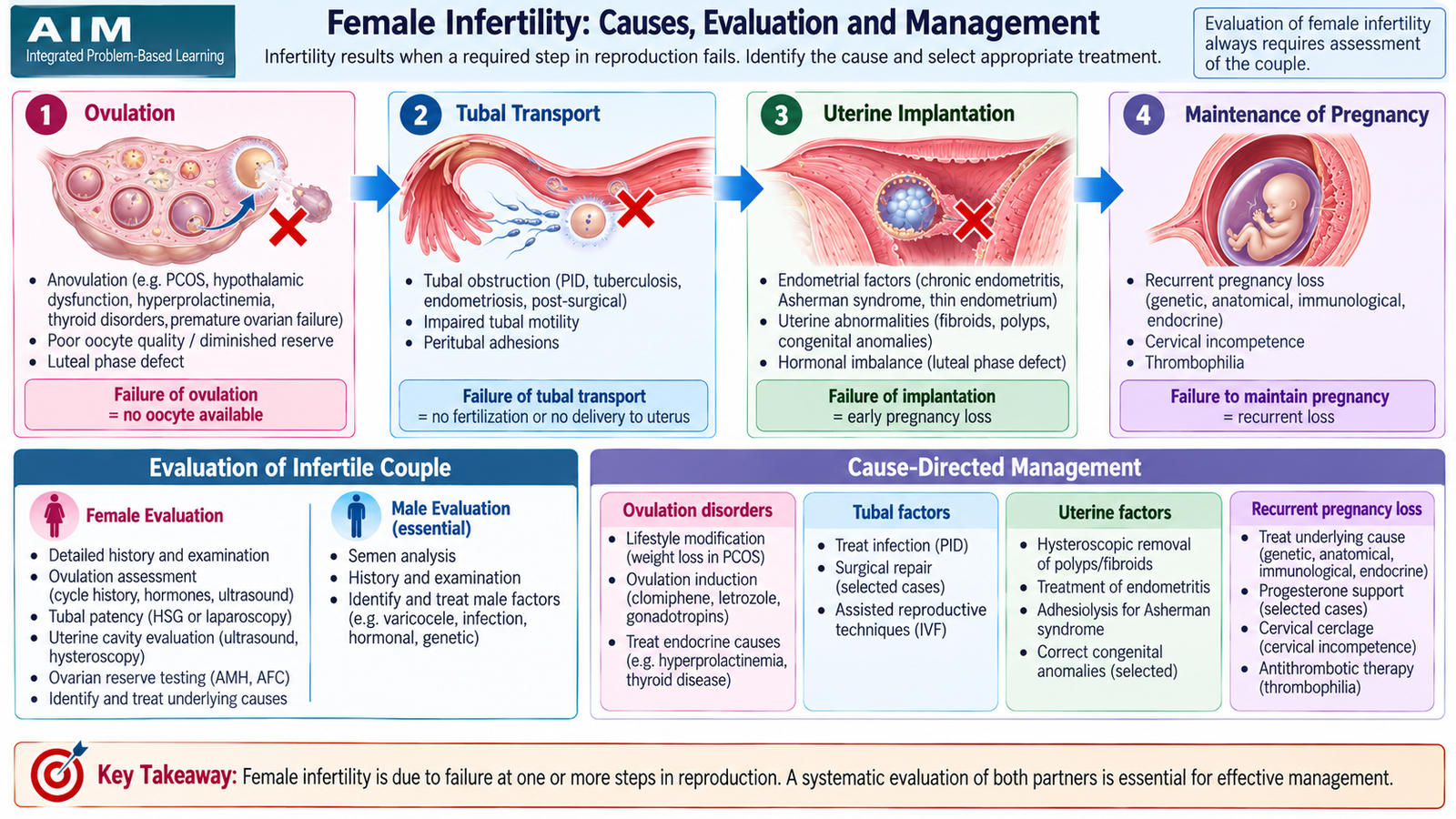
Pregnancy requires successful completion of several steps: development and release of an oocyte, capture by a patent fallopian tube, fertilization, transport of the embryo into the uterus and implantation in a suitable endometrium. A defect at any of these points can produce infertility.
Follicular development → ovulation → tubal pickup → fertilization → embryo transport → implantation
Major causes of female infertility
The cause can usually be understood by asking which part of the reproductive sequence has failed.
- Ovulatory dysfunction: PCOS is an important cause. Hypothalamic, pituitary, thyroid and other endocrine disturbances may also impair ovulation.
- Tubal disease: previous pelvic infection, surgery or other tubal damage may obstruct or distort the tubes.
- Peritoneal factors: endometriosis and pelvic adhesions can interfere with tubo-ovarian anatomy and fertilization.
- Uterine factors: submucosal fibroids, congenital abnormalities, intrauterine adhesions or important endometrial disease may interfere with implantation.
- Cervical factors: abnormalities that impair sperm passage are less common but may contribute.
- Reduced ovarian reserve or reproductive aging: the quantity and quality of available oocytes decline with advancing reproductive age.
- Unexplained infertility: routine assessment occasionally shows no clear abnormality despite failure to conceive.
Clinical evaluation
The history should determine whether ovulation is likely to be occurring and whether there are clues to tubal, uterine or endocrine disease. Menstrual regularity, previous pregnancies, pelvic infection, pelvic surgery, endometriosis symptoms and exposure to treatments affecting ovarian function are particularly relevant.
Examination looks for features such as obesity, hirsutism, acne or acanthosis suggesting PCOS, thyroid abnormalities, galactorrhea, pelvic masses and signs of endometriosis or other pelvic pathology.
Investigations
- Assessment of ovulation: menstrual history and appropriate hormonal assessment help determine whether regular ovulation is occurring.
- Ovarian reserve assessment: may be useful when reduced reserve is suspected or assisted reproduction is being considered.
- Pelvic ultrasonography: assesses ovaries and uterus and may identify polycystic ovarian morphology, fibroids, endometrioma or other structural abnormalities.
- Tubal patency assessment: contrast-based or other appropriate techniques determine whether the fallopian tubes are open.
- Endocrine testing: thyroid, prolactin and other hormonal investigations are selected according to the clinical pattern.
- Partner assessment: semen analysis remains essential because infertility may have male, female or combined causes.
Management principles
Treatment is directed at the identified cause and the reproductive goals of the couple. Ovulatory disorders may require correction of the underlying endocrine problem and ovulation induction. Tubal or uterine structural disease may require appropriate gynecological intervention. Endometriosis and fibroids are managed according to their contribution to symptoms and infertility.
Assisted reproductive techniques can bypass some reproductive barriers when simpler measures are unsuccessful or when significant tubal or other factors make spontaneous conception unlikely. Management should therefore progress from identification of the cause to the least invasive appropriate intervention.
C. Hormonal Pharmacology in Female Infertility and Benign Gynecology
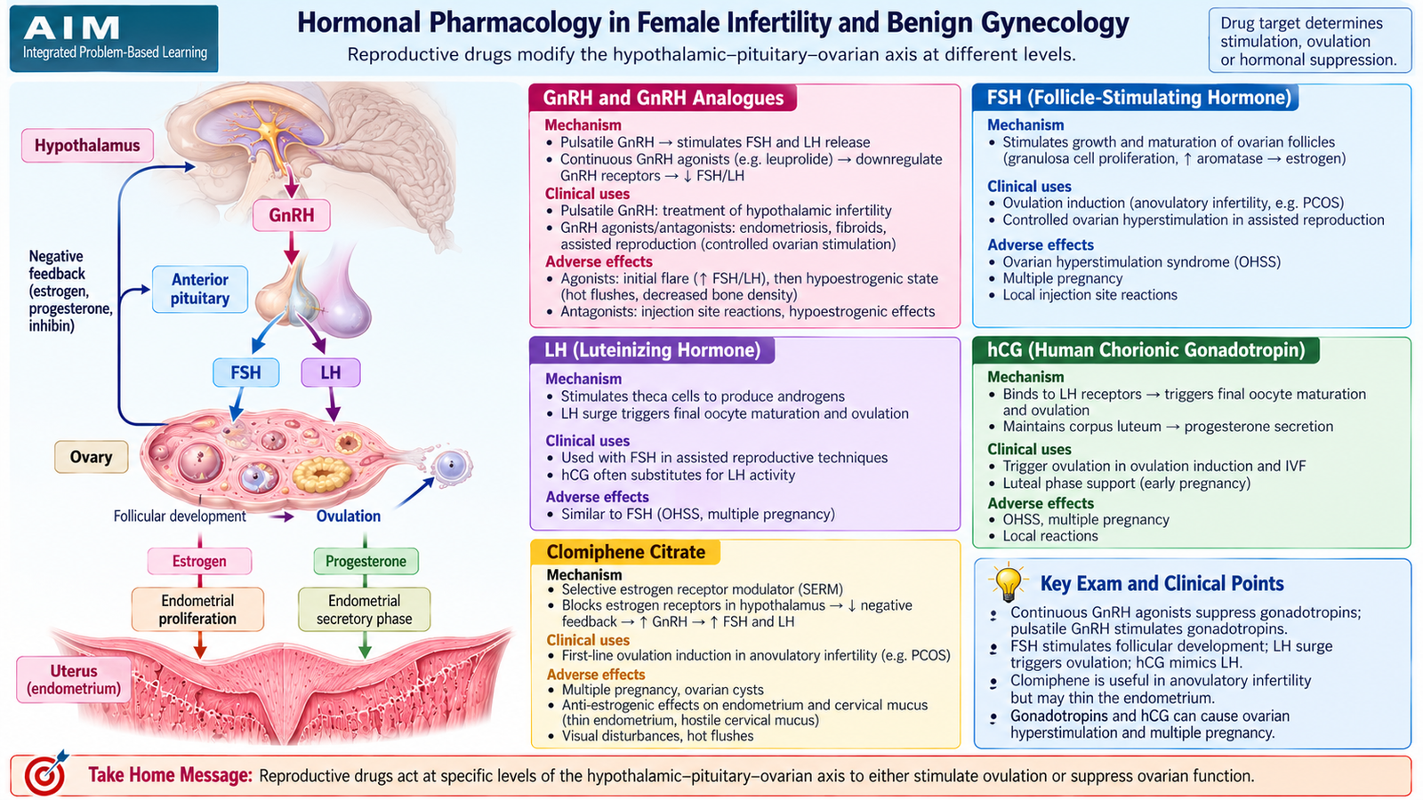
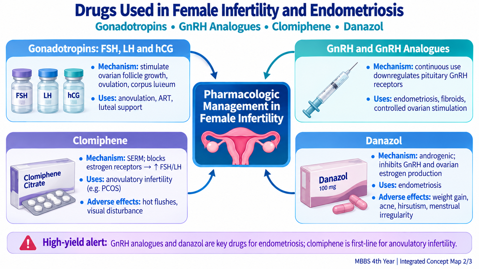
Several drugs used in infertility and benign gynecological disorders act on the hypothalamic–pituitary–ovarian axis. The key principle is that the same hormonal pathway can produce opposite effects depending on how it is stimulated. Physiological pulsatile GnRH stimulates pituitary gonadotropin release, whereas continuous exposure to a GnRH agonist eventually suppresses gonadotropin secretion.
FSH, LH and hCG
Follicle-stimulating hormone and luteinizing hormone act through membrane G-protein-coupled receptors in the ovary. FSH primarily promotes granulosa-cell function and follicular development. LH acts mainly on theca cells to support androgen production and, later in the cycle, participates in final follicular maturation and ovulation.
LH/hCG: LH-receptor activation → final follicular maturation and luteinization → ovulation and corpus-luteum support.
Human chorionic gonadotropin has substantial LH-like activity because it activates the LH receptor. It can therefore be used to trigger final follicular maturation after follicles have been stimulated to develop.
Pharmacokinetic relevance
Gonadotropins are peptide or glycoprotein hormones and are not suitable for routine oral administration because they would be degraded in the gastrointestinal tract. They are therefore given parenterally. Recombinant and purified preparations are available. hCG has a relatively prolonged biological effect compared with LH because of differences in its glycoprotein structure.
Clinical uses
- FSH: stimulation of follicular development in selected women with anovulatory infertility and during controlled ovarian stimulation.
- LH: may be combined with FSH when additional LH activity is required.
- hCG: mimics the LH surge and is used to induce final follicular maturation and ovulation.
Important adverse effects
Because gonadotropins can stimulate several ovarian follicles simultaneously, treatment can produce ovarian enlargement, multiple pregnancy and ovarian hyperstimulation syndrome. Careful monitoring of ovarian response is therefore important.
GnRH and GnRH analogues
Native gonadotropin-releasing hormone is a peptide with a very short duration of action. Its physiological secretion is pulsatile. Pulsatile exposure maintains pituitary sensitivity and releases FSH and LH. Continuous stimulation, however, causes receptor desensitization and down-regulation.
Continuous GnRH agonist → brief initial stimulation → receptor down-regulation → fall in FSH/LH → reduced ovarian estrogen production.
Synthetic GnRH agonists are more resistant to degradation and have a longer duration of action than native GnRH. Long-acting or depot preparations are therefore useful when prolonged suppression is desired. GnRH antagonists occupy the receptor without first stimulating it, so suppression of gonadotropins occurs rapidly and without an initial flare.
Clinical uses
- Pulsatile GnRH can stimulate gonadotropin secretion in selected hypothalamic causes of infertility.
- GnRH agonists may suppress ovarian hormone production in endometriosis and temporarily reduce fibroid size.
- GnRH agonists and antagonists are used in controlled reproductive protocols to regulate or prevent an unwanted endogenous LH surge.
Adverse effects
Prolonged suppression produces a hypoestrogenic state. Hot flushes, vaginal dryness and loss of bone mineral density may therefore occur. Continuous GnRH agonists can also cause an initial temporary flare because gonadotropin release increases before receptor down-regulation develops.
Clomiphene
Clomiphene is a selective estrogen receptor modulator with predominantly anti-estrogenic action at the hypothalamus. By reducing the hypothalamic perception of estrogen feedback, it increases GnRH output and subsequently raises pituitary FSH and LH secretion.
Clomiphene is given orally and undergoes hepatic metabolism. Some of its components persist for a relatively long period. It is used for ovulation induction in selected women with anovulatory infertility, including appropriate patients with PCOS.
Adverse effects include hot flushes, ovarian enlargement, multiple pregnancy and visual disturbances. Anti-estrogenic effects on cervical mucus and the endometrium can also occur.
Danazol
Danazol is a synthetic steroid with androgenic properties. It suppresses pituitary gonadotropin secretion and inhibits ovarian steroidogenesis, producing a low-estrogen environment that causes endometriotic tissue to become less active.
Danazol is orally active and is metabolized in the liver. It can suppress endometriosis, but its use is limited by androgenic and metabolic adverse effects.
- Weight gain and edema
- Acne and hirsutism
- Deepening of the voice
- Menstrual disturbance or amenorrhea
- Adverse lipid changes
- Potential hepatic adverse effects
Because of its androgenic actions, danazol must be avoided during pregnancy.
| Drug/Class | Main Action | Major Use in This Topic | Important Adverse Effect |
|---|---|---|---|
| FSH | Stimulates follicular growth | Ovarian stimulation | Ovarian hyperstimulation |
| hCG | LH-receptor agonist | Triggers final follicular maturation | Ovarian hyperstimulation |
| Continuous GnRH agonist | Down-regulates pituitary GnRH receptors | Endometriosis/fibroid suppression | Hypoestrogenism |
| GnRH antagonist | Immediate receptor blockade | Controlled ovarian stimulation | Hypoestrogenic effects |
| Clomiphene | Reduces estrogen negative feedback | Ovulation induction | Visual symptoms/multiple pregnancy |
| Danazol | Suppresses gonadotropins and ovarian steroids | Endometriosis | Androgenic effects |
D. Polycystic Ovary Syndrome
Definition and core concept
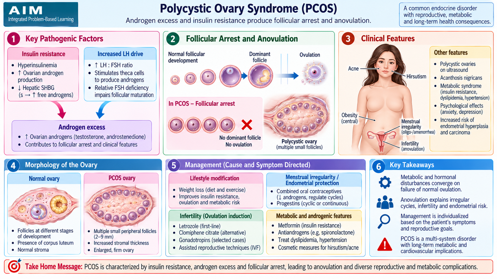
Polycystic ovary syndrome (PCOS) is a common endocrine disorder characterized by varying combinations of ovulatory dysfunction, androgen excess and polycystic ovarian morphology. The key problem is abnormal follicular development. Many follicles begin developing, but normal selection and maturation of a dominant follicle is disturbed, producing chronic or recurrent anovulation.
Etiology and risk factors
PCOS is multifactorial rather than being caused by one isolated hormone abnormality. Genetic susceptibility interacts with abnormalities of ovarian androgen production, hypothalamic–pituitary signalling and insulin action.
- Family or genetic predisposition
- Insulin resistance and compensatory hyperinsulinemia
- Overweight or obesity, which can intensify insulin resistance and hyperandrogenism
- Abnormal regulation of ovarian androgen production
Pathogenesis
Increased ovarian androgen production is central to PCOS. Abnormal gonadotropin signalling can favor LH-driven theca-cell androgen synthesis. Hyperinsulinemia can further stimulate ovarian androgen production and reduce hepatic production of sex hormone-binding globulin, increasing biologically active free androgen.
Persistent anovulation also means that regular corpus-luteum formation and progesterone exposure do not occur. The endometrium may therefore experience prolonged estrogenic stimulation without adequate cyclical progesterone opposition.
Morphology
Gross morphology
- Ovaries may be enlarged, often bilaterally.
- Multiple small cystic follicles are commonly arranged beneath the ovarian surface.
- The outer cortex may appear thickened.
Microscopic morphology
- Multiple follicular cysts or arrested follicles
- Hyperplasia and luteinization of theca interna cells
- Increased ovarian stromal tissue
- Relative absence of normal features indicating regular ovulation, such as sequential corpora lutea
Clinical features
Clinical features follow directly from anovulation, androgen excess and metabolic disturbance.
- Oligomenorrhea or amenorrhea: due to infrequent or absent ovulation.
- Infertility: due mainly to anovulation.
- Hirsutism and acne: caused by androgen excess.
- Weight gain or obesity: commonly accompanies insulin resistance but is not present in every patient.
- Acanthosis nigricans: may indicate significant insulin resistance.
Investigations and diagnosis
Diagnosis is based on the overall pattern rather than a single laboratory test. Commonly used diagnostic frameworks identify a combination of ovulatory dysfunction, clinical or biochemical hyperandrogenism and polycystic ovarian morphology after important alternative causes have been excluded.
- Pregnancy testing where appropriate
- Assessment of androgen excess
- Thyroid and prolactin assessment when clinically indicated
- Evaluation for other endocrine causes of androgen excess when the presentation is atypical
- Pelvic ultrasonography for ovarian morphology and uterine assessment
- Assessment of glucose and other metabolic risk factors where appropriate
Management
Management is individualized according to whether the main concern is menstrual irregularity, hyperandrogenic symptoms, infertility or metabolic risk.
- Lifestyle intervention: weight reduction and physical activity can improve insulin sensitivity and may restore more regular ovulation in overweight patients.
- Cycle control: hormonal therapy or periodic progestogen exposure can regulate bleeding and protect the endometrium when pregnancy is not desired.
- Hyperandrogenic symptoms: hormonal suppression and selected antiandrogen therapy may be considered with appropriate pregnancy prevention.
- Infertility: ovulation induction is used when anovulation is the principal factor. Clomiphene is an established pharmacological ovulation-inducing agent included in this curriculum.
- Metabolic abnormalities: insulin resistance and associated metabolic problems should be addressed; metformin may be useful in selected women, particularly when metabolic indications are present.
Complications and prognosis
Persistent anovulation can cause infertility and prolonged unopposed estrogen exposure, increasing the risk of endometrial hyperplasia and eventually endometrial carcinoma in susceptible patients. Insulin resistance also increases long-term metabolic risk. Despite these issues, reproductive and menstrual outcomes can often be substantially improved when treatment is matched to the patient’s main problem.
E. Uterine Fibroids
Definition and core concept
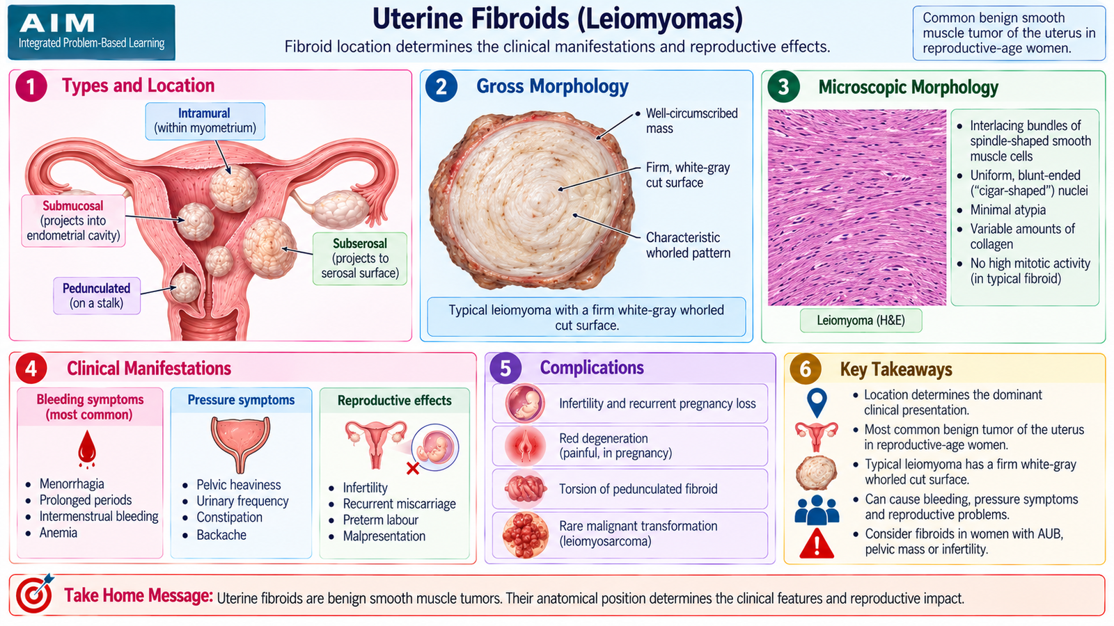
Uterine fibroids, or leiomyomas, are benign neoplasms of uterine smooth muscle. They arise within the myometrium and are strongly influenced by ovarian steroid hormones. Their clinical effects depend less on their absolute number than on their size and anatomical location.
Etiology and risk factors
Leiomyomas are clonal smooth-muscle tumors. Somatic molecular abnormalities are found in many tumors, while estrogen and progesterone promote their growth. This hormonal dependence explains why fibroids are predominantly disorders of reproductive-age women, may enlarge during periods of strong hormonal stimulation and commonly shrink after menopause.
Important associations and risk factors include:
- Reproductive age
- Family predisposition
- Greater lifetime exposure to ovarian steroid hormones
- Obesity, which may increase estrogenic influence
- Nulliparity in some patients
Location
- Intramural: located within the myometrium.
- Submucosal: project toward the endometrial cavity and are particularly likely to produce abnormal bleeding or interfere with implantation.
- Subserosal: project outward toward the serosal surface and more often produce pressure symptoms when large.
Morphology
Gross morphology
Fibroids are typically sharply circumscribed, firm, round masses. On cut section they have a characteristic white-gray whorled appearance produced by interlacing bundles of smooth muscle.
Larger fibroids may undergo secondary changes when their blood supply becomes inadequate. Hyaline degeneration is common; cystic change, calcification or hemorrhagic red degeneration may also occur.
Microscopic morphology
The tumor consists of intersecting fascicles of uniform smooth-muscle cells. The nuclei are elongated and blunt-ended, often described as cigar-shaped. Typical leiomyomas show little cytological atypia and low mitotic activity.
Clinical features
Many fibroids are asymptomatic. Symptoms arise when a tumor distorts the endometrial cavity, increases the effective uterine surface area or compresses neighboring structures.
- Heavy menstrual bleeding: particularly important with submucosal or cavity-distorting fibroids.
- Pelvic pressure or abdominal enlargement: due to increasing uterine size.
- Urinary frequency or pressure symptoms: when the enlarged uterus compresses the bladder.
- Pain: may occur with degeneration or torsion of a pedunculated lesion.
- Infertility: particularly when the uterine cavity is distorted or tubal relationships are affected.
Assessment and management
Pelvic examination may identify an enlarged, irregular, firm uterus. Ultrasonography is the usual first imaging investigation because it demonstrates the size, number and location of fibroids and their relationship to the uterine cavity.
Management depends on symptoms, severity of bleeding, fibroid location, age and fertility wishes.
- Asymptomatic fibroids may be observed.
- Medical treatment can reduce heavy menstrual bleeding.
- GnRH analogues can temporarily suppress ovarian hormone production and reduce fibroid volume, but prolonged use is limited by hypoestrogenic adverse effects.
- Myomectomy removes fibroids while preserving the uterus and may be appropriate when future fertility is important.
- Hysterectomy provides definitive treatment when symptoms are significant and uterine preservation is not required.
- Other uterus-preserving interventions may be considered in appropriately selected patients.
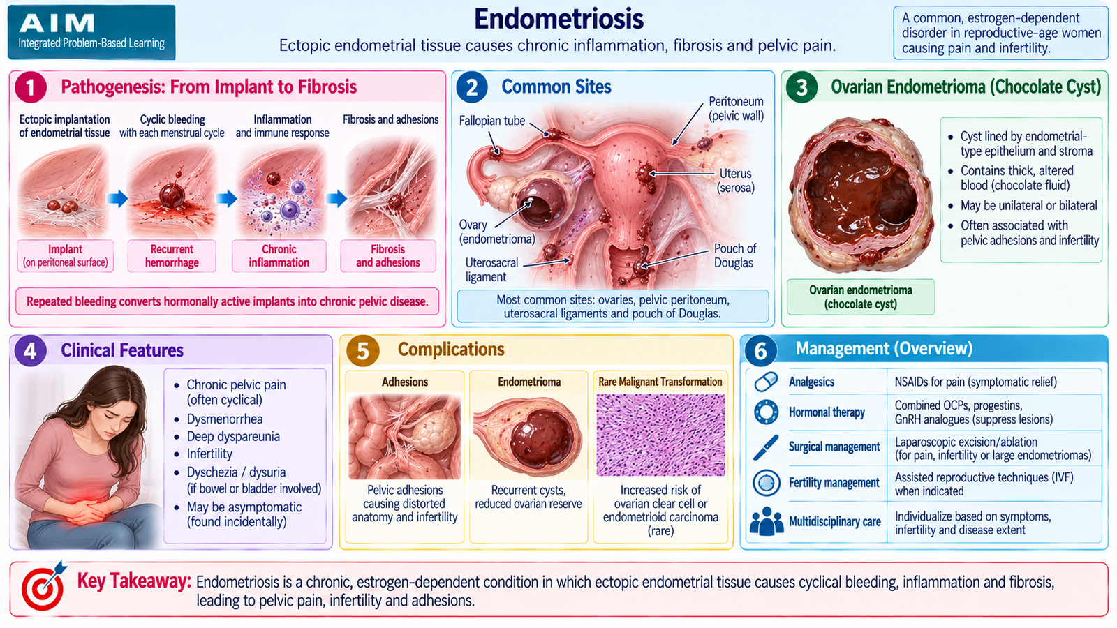
F. Endometriosis
Definition
Endometriosis is the presence of functional endometrial glands and stroma outside the normal endometrial cavity. The ectopic tissue remains hormonally responsive and can undergo cyclical bleeding, which produces inflammation, fibrosis and adhesions.
Common sites
The ovaries are a particularly important site. Other common pelvic locations include pelvic peritoneum, uterosacral ligaments and the region behind the uterus. Disease at different sites explains why the clinical presentation varies between patients.
Etiology and pathogenesis
No single theory explains every case. Several mechanisms are accepted as contributors.
- Retrograde menstruation: viable endometrial cells pass through the fallopian tubes into the peritoneal cavity, implant and grow at ectopic sites.
- Coelomic metaplasia: peritoneal cells may transform into endometrial-type tissue.
- Lymphatic or vascular dissemination: helps explain endometriosis at unusual distant sites.
Once implanted, the ectopic tissue is stimulated by estrogen. Cyclical tissue breakdown and bleeding trigger repeated inflammation. Healing then produces fibrosis and adhesions, which distort pelvic anatomy.
Morphology
Gross morphology
- Small red, blue, brown or black implants may occur on pelvic surfaces.
- Old hemorrhage produces dark lesions sometimes described as powder-burn lesions.
- Repeated ovarian hemorrhage can produce an endometriotic cyst containing altered brown blood, classically called a chocolate cyst.
- Chronic inflammation produces fibrous adhesions and distortion of pelvic structures.
Microscopic morphology
Typical lesions contain ectopic endometrial glands and endometrial-type stroma. Repeated hemorrhage frequently results in hemosiderin deposition and hemosiderin-laden macrophages.
Clinical features
- Progressive dysmenorrhea: pain results from cyclical bleeding and inflammatory mediator release.
- Chronic pelvic pain: related to chronic inflammation and adhesions.
- Deep dyspareunia: particularly with posterior pelvic disease.
- Infertility: inflammation, adhesions and distortion of tubo-ovarian anatomy may interfere with oocyte pickup and fertilization.
- Site-specific bowel or urinary symptoms may occur when adjacent organs are involved.
Investigations
Diagnosis begins with history and pelvic examination. Ultrasonography is useful for identifying ovarian endometriomas and other pelvic pathology. MRI may help characterize more complex or deep pelvic disease when required. Laparoscopy provides direct visualization of pelvic lesions and allows biopsy or treatment when operative assessment is appropriate.
Management
Treatment is determined by the severity of pain, extent of disease, age and fertility wishes. Medical treatment suppresses ovarian hormonal stimulation but does not mechanically remove existing fibrosis and adhesions.
- NSAIDs may reduce pain associated with menstruation.
- Combined hormonal methods or progestogens can suppress cyclical activity of endometriotic tissue.
- GnRH analogues produce a hypoestrogenic state and can suppress active lesions.
- Danazol suppresses gonadotropins and ovarian steroid production but is now limited by androgenic adverse effects.
- Surgical treatment may involve excision or ablation of lesions, treatment of ovarian endometriomas and division of significant adhesions.
- Infertility management is individualized according to age, anatomical distortion, ovarian factors and other causes of infertility.
G. Endometritis
Core concept
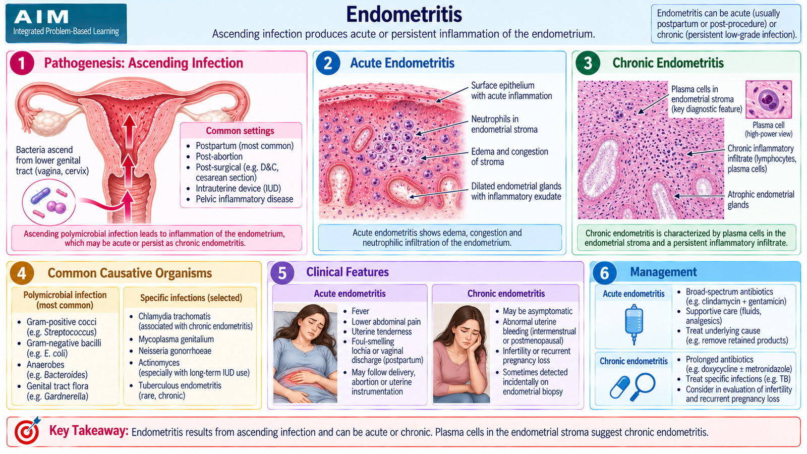
Endometritis is inflammation of the endometrial lining of the uterus. It can be acute or chronic. The underlying mechanism is usually entry or persistence of infectious or inflammatory material within the endometrial cavity, followed by recruitment of inflammatory cells.
Acute endometritis
Acute endometritis is most often associated with circumstances that allow organisms to ascend from the lower genital tract, particularly after childbirth, miscarriage, retained products of conception or uterine instrumentation. The infection is commonly polymicrobial rather than being caused by one unique organism.
Chronic endometritis
Chronic endometritis develops when inflammation persists. Important settings include chronic pelvic infection, retained intrauterine material and some specific infections. Genital tuberculosis is an important cause of chronic granulomatous endometritis in regions where tuberculosis occurs.
The characteristic histological clue to chronic endometritis is the presence of plasma cells within the endometrial stroma. Persistent inflammation can alter normal endometrial function and may contribute to abnormal bleeding or impaired fertility in some patients.
Why the pathology matters
The endometrium must undergo controlled cyclical change and provide a receptive surface for implantation. Persistent inflammation can disturb this environment. The clinical significance therefore extends beyond infection itself and may include abnormal bleeding or reproductive difficulty depending on cause and severity.
H. Abnormal Uterine Bleeding and Important Menstrual Disorders
Core concept
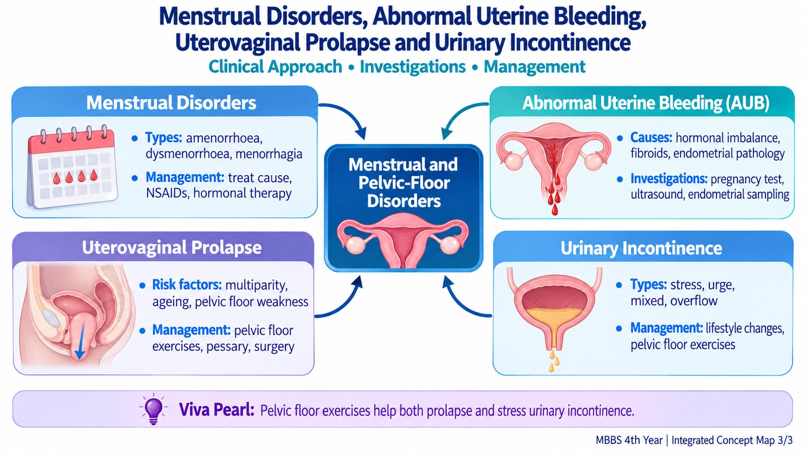
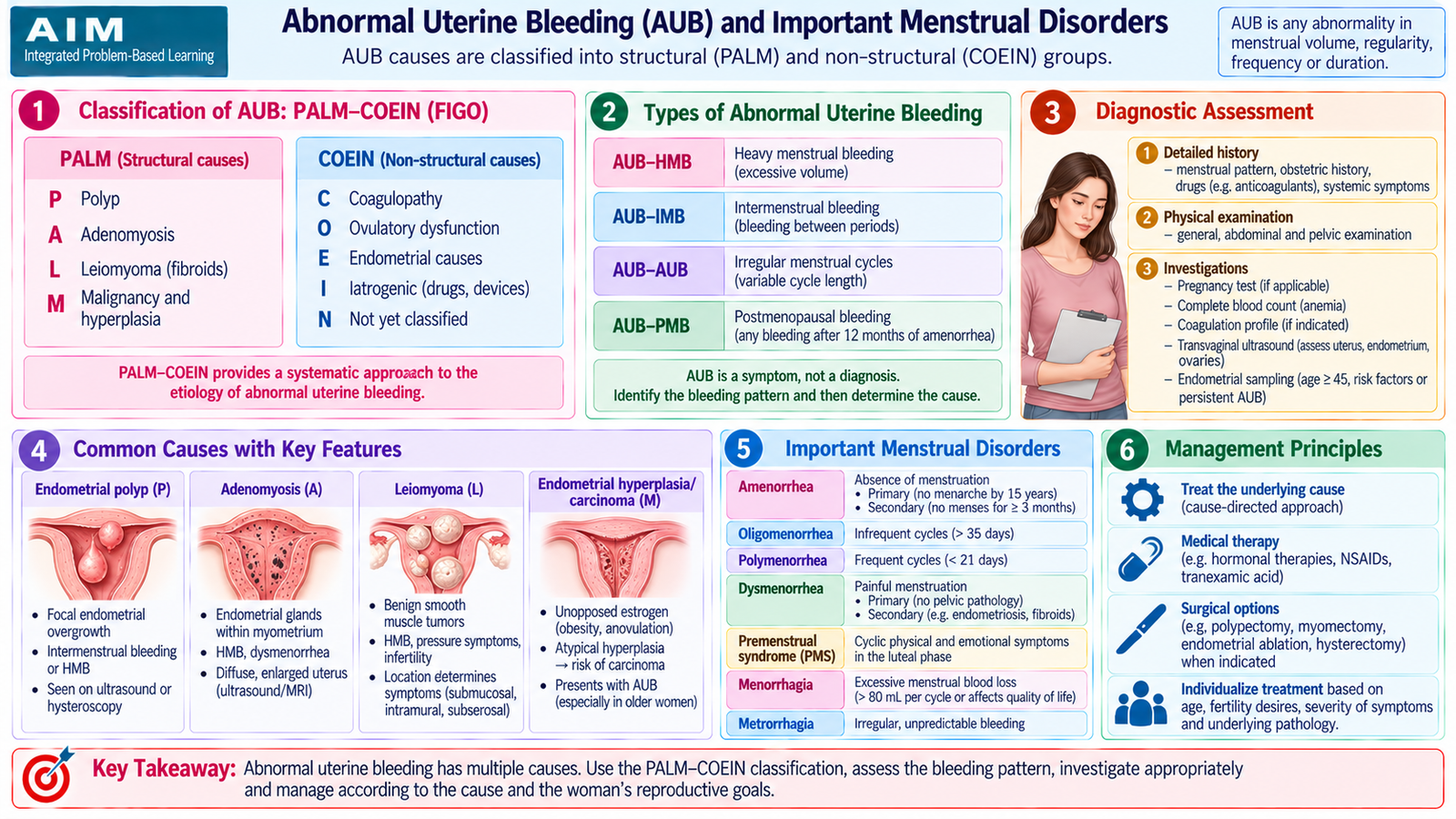
Abnormal uterine bleeding (AUB) describes uterine bleeding that is abnormal in its regularity, timing, duration or amount. AUB is a clinical presentation, not a final diagnosis. The aim of assessment is therefore to identify whether the cause is structural, hormonal, systemic, medication-related or due to an endometrial functional disturbance.
Etiological approach to AUB
A useful framework is the PALM–COEIN classification. PALM contains structural abnormalities that can often be demonstrated by imaging or pathology, whereas COEIN contains mainly non-structural causes.
| Group | Cause | Clinical Idea |
|---|---|---|
| P | Polyp | Focal endometrial lesion |
| A | Adenomyosis | Endometrial tissue within myometrium |
| L | Leiomyoma | Fibroid, especially cavity-distorting lesions |
| M | Malignancy/hyperplasia | Important pathology requiring exclusion |
| C | Coagulopathy | Systemic bleeding disorder |
| O | Ovulatory dysfunction | Irregular endometrial exposure to hormones |
| E | Endometrial | Local functional/hemostatic disturbance |
| I | Iatrogenic | Drug or device related |
| N | Not otherwise classified | Other recognized causes |
Important menstrual disorders
Amenorrhea
Amenorrhea means absence of menstruation. It may reflect failure of reproductive tract development or outflow, ovarian failure, hypothalamic–pituitary dysfunction, endocrine disease or physiological states such as pregnancy. In any reproductive-age woman with previously established menstruation, pregnancy should be considered early in the assessment.
Oligomenorrhea or infrequent menstruation
Infrequent menstruation commonly reflects infrequent ovulation. PCOS is an important cause, but thyroid disease, prolactin disturbance, marked weight change and hypothalamic dysfunction may produce a similar pattern.
Heavy menstrual bleeding
Excessive menstrual bleeding may arise from fibroids, adenomyosis, endometrial dysfunction, ovulatory abnormalities or systemic bleeding disorders. Chronic blood loss can lead to iron-deficiency anemia.
Dysmenorrhea
Dysmenorrhea means painful menstruation. Primary dysmenorrhea occurs without identifiable pelvic pathology and is strongly related to prostaglandin-mediated uterine contractions. Secondary dysmenorrhea results from an underlying condition such as endometriosis or adenomyosis.
Diagnostic approach
Evaluation should begin with the bleeding pattern, reproductive history, pregnancy possibility, medication use, symptoms suggesting anemia, endocrine disease or bleeding disorder, and features suggesting pelvic pathology.
- Pregnancy testing: essential when pregnancy is possible because pregnancy-related bleeding requires a different approach.
- Complete blood count: assesses anemia when bleeding is significant.
- Thyroid or prolactin testing: selected when the menstrual pattern suggests endocrine dysfunction.
- Coagulation assessment: considered when the history suggests an inherited or acquired bleeding tendency.
- Pelvic ultrasonography: looks for fibroids, ovarian pathology and other structural causes.
- Endometrial assessment: may be required when persistent bleeding or clinical risk raises concern for endometrial pathology.
Primary-care management
Management should address the cause while controlling symptoms and preventing complications. Iron deficiency should be identified and corrected. NSAIDs can reduce prostaglandin-mediated dysmenorrhea and may reduce menstrual blood loss. Tranexamic acid can reduce heavy menstrual bleeding in suitable patients. Hormonal methods can regulate cycles and reduce bleeding when appropriate.
Amenorrhea and oligomenorrhea require cause-directed management rather than simply inducing bleeding. Patients with severe ongoing bleeding, hemodynamic compromise, marked anemia, pregnancy-related bleeding, a significant pelvic mass or concern for malignancy require urgent or specialist assessment.
I. Uterovaginal Prolapse
Definition and core concept
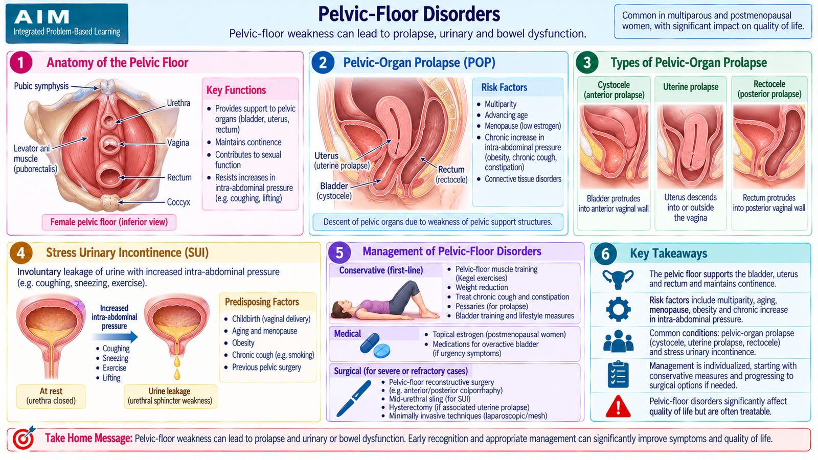
Uterovaginal prolapse is downward displacement of the uterus, vaginal apex or vaginal walls because the normal pelvic-floor and connective-tissue supports have weakened. The disorder is therefore best understood as a failure of support rather than simply a problem of the uterus itself.
Etiology and risk factors
The levator ani, perineal body and endopelvic fascial and ligamentous supports normally resist downward forces. Childbirth may stretch or injure these structures, while aging and reduced tissue strength can progressively worsen support.
- Pregnancy and vaginal childbirth, particularly repeated childbirth
- Pelvic-floor muscle or connective-tissue injury
- Advancing age and postmenopausal tissue changes
- Chronic elevation of intra-abdominal pressure from cough, constipation or heavy physical strain
- Obesity
- Previous pelvic surgery in selected cases
Anatomical patterns
Different components of the pelvic support system may fail. Anterior vaginal wall descent may be associated with bladder prolapse, posterior wall weakness may be associated with rectal bulging, and loss of apical support permits descent of the uterus or upper vagina.
Clinical features
- Sensation of something coming down through the vagina
- Visible or palpable vaginal bulge
- Pelvic dragging or pressure
- Urinary frequency, difficulty voiding or urinary incontinence
- Difficulty with defecation in some posterior-compartment defects
- Sexual discomfort in some patients
Complications
Exposed prolapsed tissue may become dry, irritated and ulcerated. Distortion of the urinary tract can cause incomplete bladder emptying and predispose to recurrent urinary problems. Severe longstanding prolapse may considerably impair mobility and daily activity.
Management
Treatment depends on symptom severity, degree of prolapse, general health, sexual activity and whether the woman wishes to preserve her uterus or future fertility.
- Pelvic-floor muscle training: strengthens remaining muscular support and is particularly useful in mild disease.
- Correction of aggravating factors: chronic constipation, chronic cough and excessive strain should be addressed.
- Vaginal pessary: provides mechanical support and is useful when conservative management is preferred or surgery is unsuitable.
- Surgery: reconstructive procedures restore support when symptoms are significant and conservative measures are insufficient. The operation selected depends on the compartment involved and the patient’s reproductive preferences.
Prevention principles
Pelvic-floor rehabilitation, appropriate postpartum recovery and reduction of chronic straining may help preserve pelvic support. Preventive measures are particularly relevant because prolapse commonly develops gradually after cumulative injury rather than following one isolated event.
J. Urinary Incontinence
Definition and classification
Urinary incontinence is involuntary leakage of urine. Different types arise from different failures of storage, sphincter support, bladder contraction or the ability to reach and use a toilet. Correct classification is important because treatment differs substantially between types.
Stress urinary incontinence
Urine leaks when intra-abdominal pressure rises during coughing, sneezing, laughing or physical activity. The underlying problem is inadequate urethral support or sphincter competence, often related to pelvic-floor damage.
Urge urinary incontinence
Urine leakage is preceded by a sudden compelling desire to void. It is usually associated with involuntary detrusor contractions as part of an overactive bladder syndrome.
Mixed urinary incontinence
Mixed incontinence contains clinically important features of both stress and urge incontinence.
Overflow incontinence
The bladder fails to empty adequately and becomes chronically overdistended. Urine then leaks from the overly full bladder. Causes include outlet obstruction and impaired detrusor contractility.
Functional incontinence
The urinary tract may function normally, but physical, cognitive or environmental limitations prevent the patient from reaching or using a toilet in time.
Etiology and risk factors
- Pregnancy and childbirth-related pelvic-floor injury
- Aging and loss of pelvic tissue support
- Pelvic organ prolapse
- Obesity and chronic raised intra-abdominal pressure
- Neurological disease affecting bladder control
- Urinary infection or bladder irritation causing transient urgency
- Urinary retention or impaired bladder contractility
- Mobility or cognitive impairment in functional incontinence
Clinical assessment
History should determine whether leakage occurs with physical stress, follows urgency, is associated with incomplete emptying or results from functional limitations. The amount and timing of leakage, urinary frequency, nocturia, dysuria, obstetric history and pelvic surgery are useful clues.
Examination includes abdominal and pelvic assessment, evaluation for prolapse and assessment of pelvic-floor function. A cough stress test may demonstrate stress leakage when appropriate.
Investigations
- Urinalysis: helps identify infection, blood or other urinary abnormalities.
- Bladder diary: can show the relationship between fluid intake, voiding, urgency and leakage.
- Post-void residual assessment: is especially helpful when incomplete bladder emptying or overflow is suspected.
- Further urodynamic or specialist testing: is reserved for selected patients when the diagnosis remains uncertain or intervention is being planned.
Management
Conservative measures are important because many patients improve without immediate invasive treatment.
- Stress incontinence: pelvic-floor muscle training is first-line conservative therapy. Persistent troublesome cases may require specialist procedures that improve urethral support.
- Urge incontinence: bladder training and modification of aggravating factors are important. Antimuscarinic drugs or beta-3 adrenergic agonists may be considered when conservative treatment is insufficient.
- Mixed incontinence: treatment is directed at the component producing the greatest symptoms.
- Overflow incontinence: identify and correct urinary retention or the underlying obstruction or bladder-emptying problem.
- Functional incontinence: improve mobility, caregiver support and access to toilet facilities.
Prevention
Pelvic-floor muscle training during appropriate periods of pregnancy and postpartum recovery, avoidance of chronic straining, treatment of persistent cough and weight management can reduce factors that contribute to pelvic-floor dysfunction. Early assessment of prolapse and urinary symptoms can also prevent progressive functional impairment.
Integrated Mechanism Flow — PCOS to Infertility and Endometrial Risk
Genetic/metabolic susceptibility
Insulin resistance + altered gonadotropin signalling
Ovarian androgen excess
Follicular arrest and anovulation
Irregular cycles + infertility
Prolonged unopposed estrogenic effect on endometrium
The same underlying anovulation therefore explains both the reproductive problem and an important long-term endometrial complication of PCOS.
Important Comparison — Major Types of Urinary Incontinence
| Type | Typical Clue | Main Mechanism | Main Management Principle |
|---|---|---|---|
| Stress | Leakage with cough/sneeze/exertion | Poor urethral/pelvic support | Pelvic-floor strengthening |
| Urge | Urgency followed by leakage | Detrusor overactivity | Bladder training ± medication |
| Mixed | Stress + urgency symptoms | Combined mechanisms | Treat predominant component |
| Overflow | Incomplete emptying, dribbling | Overdistended retained bladder | Correct retention/cause |
| Functional | Cannot reach/use toilet | Mobility/cognitive/environmental limitation | Improve access and support |
⭐ AIM High-Yield Review
Female Infertility and Benign Gynecologic Disorders
Selected videos reinforce the major concepts in female infertility, PCOS, fibroids, endometriosis, pelvic organ prolapse and urinary incontinence.
Best for reproductive physiology, major female causes of infertility and the clinical evaluation pathway.
Covers pathophysiology, insulin resistance, hyperandrogenism, clinical features, infertility and management.
Direct 4th-year coverage of fibroid location, morphology, symptoms, endometriosis, infertility and management principles.
Reinforces pelvic support failure, prolapse patterns, clinical recognition and management principles.
Short visual review of stress, urge and overflow incontinence with causes and treatment principles.