
This chapter follows the KMU learning outcomes and connects pathology, prevention and clinical gynecology in a logical sequence. First understand how each malignancy develops and presents; then use the final AIM High-Yield Review for rapid revision.
Topic 16 — Gynecologic Malignancies: Cervical, Endometrial and Ovarian Cancer
Understand the pathology, clinical presentation, diagnosis, staging, prevention and management principles of the three major gynecologic malignancies emphasized in this topic.
Topic Introduction
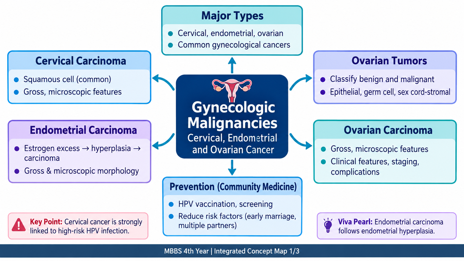
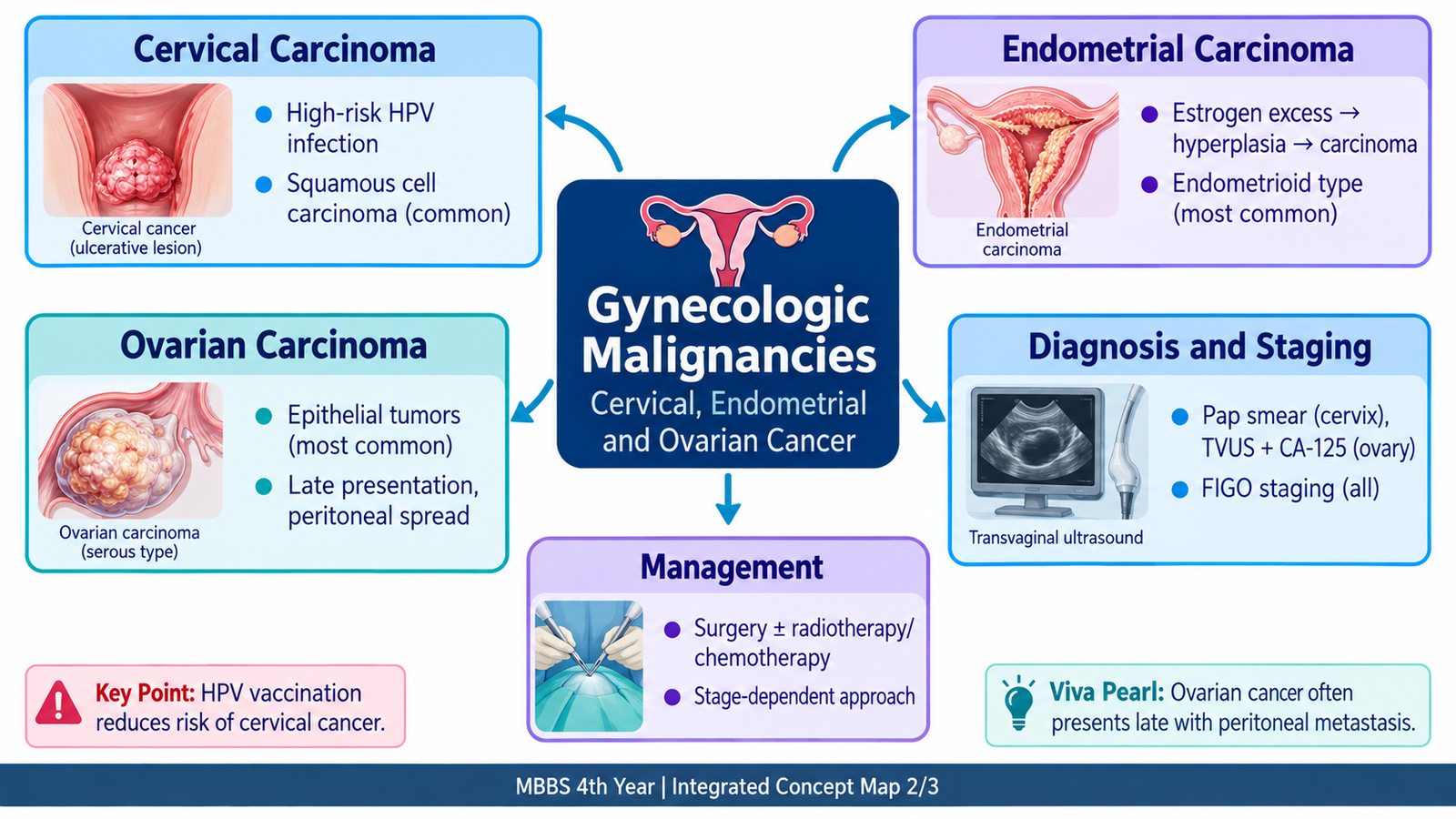
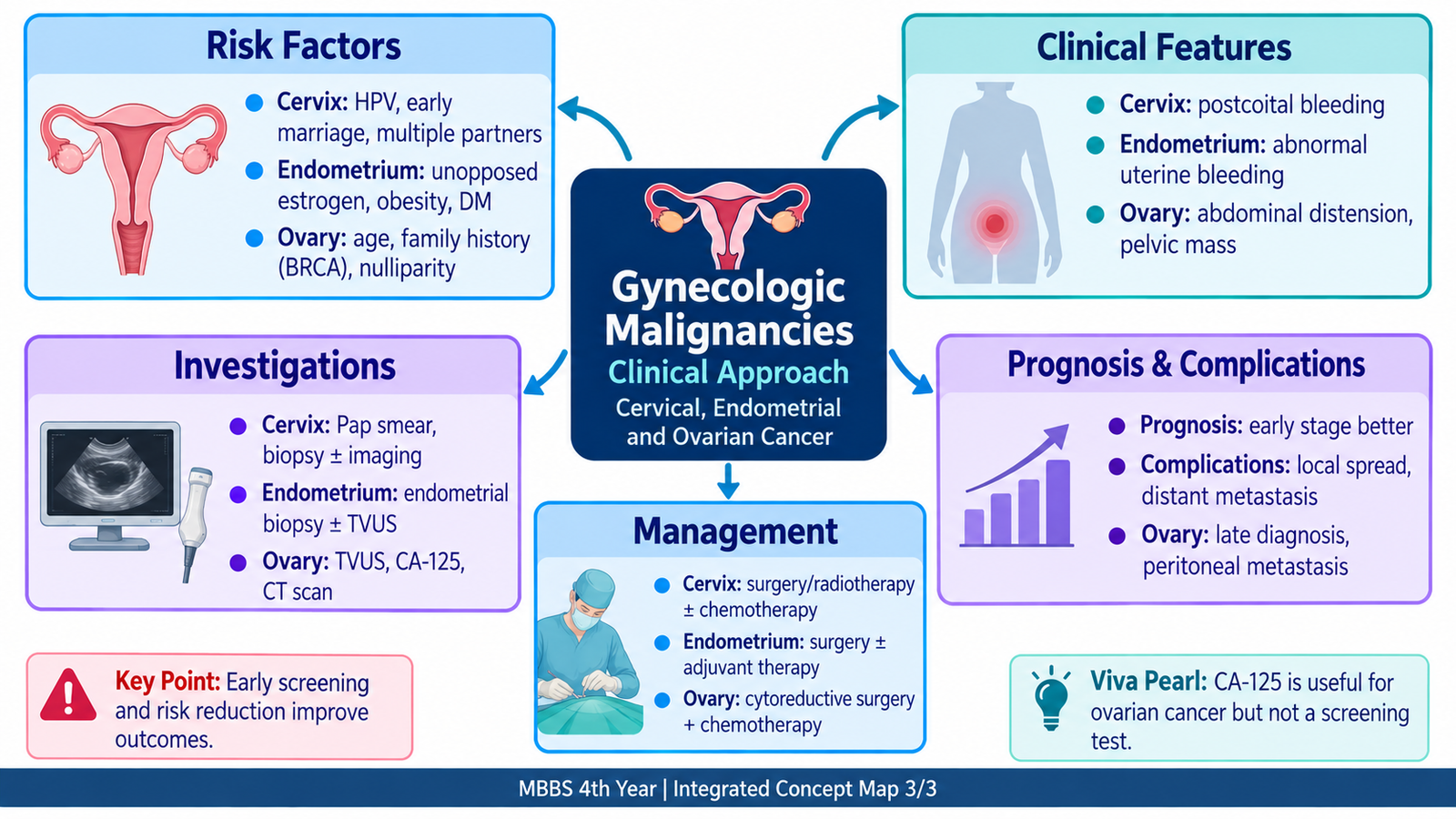
Gynecologic malignancies are cancers arising from the female reproductive tract. This chapter focuses on carcinoma of the cervix, endometrial carcinoma and ovarian carcinoma because each has a different pattern of causation, presentation and spread. Cervical cancer is strongly linked to persistent high-risk human papillomavirus infection and is particularly important because much of its burden is preventable. Endometrial carcinoma commonly produces abnormal uterine bleeding and is closely related to hormonal and precursor endometrial changes. Ovarian malignancy often remains clinically silent until disease has spread within the peritoneal cavity. By linking risk factors, pathogenesis, morphology, clinical features, investigations, staging and treatment principles, these diseases become much easier to distinguish and understand.
A. Major Types of Gynecologic Malignancy
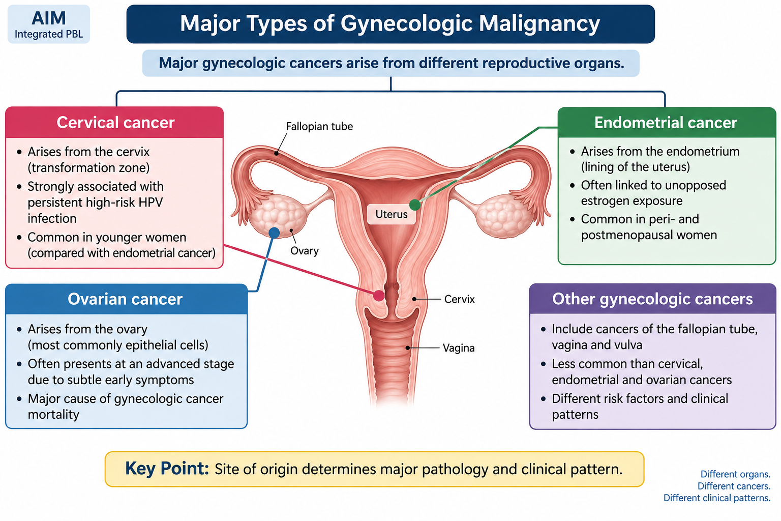
Gynecologic cancers are classified mainly according to the organ or tissue from which they arise. For this topic, the key cancers are cervical, endometrial and ovarian malignancies. Recognizing their site of origin provides the first framework for understanding their different risk factors, clinical presentations and patterns of spread.
Major groups
- Cervical carcinoma — most commonly squamous cell carcinoma; adenocarcinoma is another important type.
- Endometrial carcinoma — arises from the endometrial lining of the uterine body; endometrioid carcinoma is the common major pattern.
- Ovarian malignancies — include epithelial, germ-cell and sex-cord stromal tumors; epithelial carcinomas account for most malignant ovarian tumors in adults.
- Vulvar carcinoma and vaginal carcinoma are additional gynecologic malignancies.
- Gestational trophoblastic neoplasia represents malignant proliferation of trophoblastic tissue related to pregnancy.
The three major cancers emphasized here should not be viewed as one disease group with identical behavior. Cervical carcinoma is strongly infection-related, endometrial carcinoma is frequently linked to abnormal estrogenic stimulation or specific molecular pathways, while ovarian carcinoma commonly spreads across peritoneal surfaces before producing obvious symptoms.
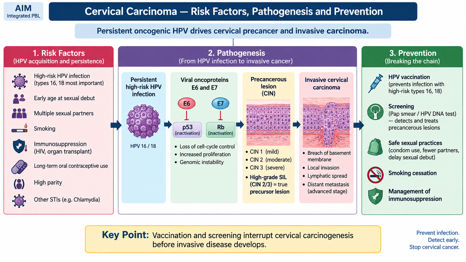
B. Cervical Carcinoma — Risk Factors, Pathogenesis and Prevention
Cervical carcinoma usually develops through a long sequence beginning with infection of cervical squamous epithelium by a high-risk human papillomavirus (HPV). Most HPV infections resolve, but persistent high-risk infection can produce precancerous epithelial abnormalities and eventually invasive carcinoma. This long precursor phase explains why vaccination and screening can prevent many cases.
Important risk factors
Persistent infection with an oncogenic HPV type is the central causal factor. Other factors increase the probability of acquiring HPV, failing to clear the infection or progressing from precancer to invasive malignancy.
- Persistent infection with high-risk HPV, particularly oncogenic types such as HPV 16 and 18.
- Early onset of sexual activity and greater lifetime exposure to sexual partners, because these increase opportunity for HPV transmission.
- Smoking, which promotes local carcinogenic effects and reduces effective immune clearance.
- Immunosuppression, which reduces the ability to control persistent HPV infection.
- High parity and prolonged exposure to other cofactors may contribute to progression in an HPV-infected cervix.
- Failure to participate in appropriate cervical screening increases the chance that a precursor lesion will remain undetected.
Pathogenesis
The most important changes occur near the transformation zone, where endocervical columnar epithelium meets squamous epithelium. This actively changing epithelial region is particularly susceptible to oncogenic HPV infection.
Two viral proteins are particularly important. E6 promotes loss of normal p53 tumor-suppressor activity, while E7 interferes with the retinoblastoma (RB) pathway. Loss of these cell-cycle checkpoints allows damaged epithelial cells to proliferate rather than undergo normal growth arrest or apoptosis.
Low-grade squamous intraepithelial lesions may regress, especially when HPV infection is cleared. Persistent high-grade lesions have a much greater risk of progressing to invasive carcinoma because abnormal cells occupy a greater thickness of the epithelium and accumulate further genetic damage.
Prevention
Cervical carcinoma is particularly suitable for prevention because its major causal infection can be reduced and because a recognizable precancerous phase usually precedes invasive disease.
- HPV vaccination reduces infection with important oncogenic HPV types and therefore represents primary prevention.
- Cervical screening using accepted cytology- and/or HPV-based methods detects women at risk before invasive cancer develops.
- Evaluation and treatment of precancerous lesions interrupts progression to invasive carcinoma.
- Smoking cessation removes an important modifiable cofactor.
- Education regarding HPV transmission and safer sexual practices can reduce exposure.
C. Cervical Carcinoma — Morphology, Clinical Features and Staging
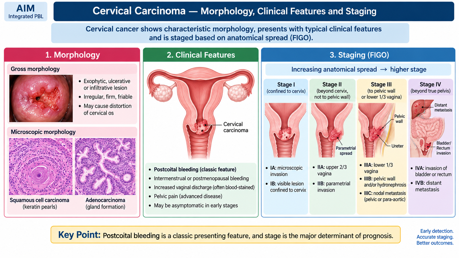
Once neoplastic cervical epithelial cells cross the basement membrane and invade the underlying stroma, the lesion becomes invasive cervical carcinoma. Its gross appearance, microscopic pattern and direction of spread explain many of the symptoms and staging findings seen clinically.
Gross morphology
Most tumors arise near the transformation zone. As the carcinoma enlarges it may project outward, ulcerate the cervical surface or infiltrate deeply into the cervix and adjacent tissues.
- Exophytic or fungating mass: projects from the cervical surface and may bleed easily because malignant tissue is friable.
- Ulcerative lesion: produces irregular tissue destruction and necrosis.
- Infiltrative lesion: spreads into the cervical stroma and may eventually extend into the parametrium.
Microscopic morphology
Squamous cell carcinoma is the classic major histological type. Malignant squamous cells invade the cervical stroma in nests, cords or irregular sheets. Well-differentiated tumors may form keratin pearls and show intercellular bridges. Poorly differentiated tumors show greater atypia and less obvious squamous maturation.
Adenocarcinoma arises from glandular epithelium and forms malignant glands containing atypical epithelial cells. Its increasing clinical importance makes recognition of both squamous and glandular cervical malignancy essential.
Clinical features
Early cervical carcinoma can be asymptomatic. When symptoms develop, bleeding is common because the tumor disrupts the cervical surface and forms fragile abnormal vessels.
- Postcoital bleeding is a classic presentation because contact traumatizes the friable cervical lesion.
- Intermenstrual or irregular vaginal bleeding.
- Postmenopausal bleeding.
- Persistent watery, blood-stained or offensive vaginal discharge.
- Pelvic or back pain in more advanced disease.
- Urinary or bowel symptoms when adjacent structures become involved.
On speculum examination, the cervix may appear irregular, ulcerated, fungating or friable and may bleed on contact. Advanced parametrial extension can reduce cervical mobility. Obstruction of the ureters by advanced pelvic disease may produce hydroureter and hydronephrosis.
Principles of cervical cancer staging
Cervical carcinoma is staged according to the anatomical extent of disease. The important undergraduate principle is to understand how progressively greater local extension, nodal disease and distant spread move the tumor from Stage I toward Stage IV.
| Stage | Essential anatomical concept |
|---|---|
| I | Carcinoma confined to the cervix. Stage IA is microscopic invasion; Stage IB represents larger invasive disease still confined to the cervix. |
| II | Tumor extends beyond the uterus but has not reached the pelvic wall or lower third of the vagina. Parametrial invasion places disease in Stage IIB. |
| III | Involvement of the lower third of the vagina, pelvic wall, obstructive hydronephrosis/non-functioning kidney, and/or regional nodal disease. |
| IV | Tumor invades adjacent pelvic organs such as bladder or rectal mucosa, or spreads beyond the pelvis to distant sites. |
Increasing stage reflects increasing tumor spread and therefore generally indicates a less favorable prognosis and a greater need for multimodal treatment.
D. Cervical Carcinoma — Investigations, Management and Prognosis
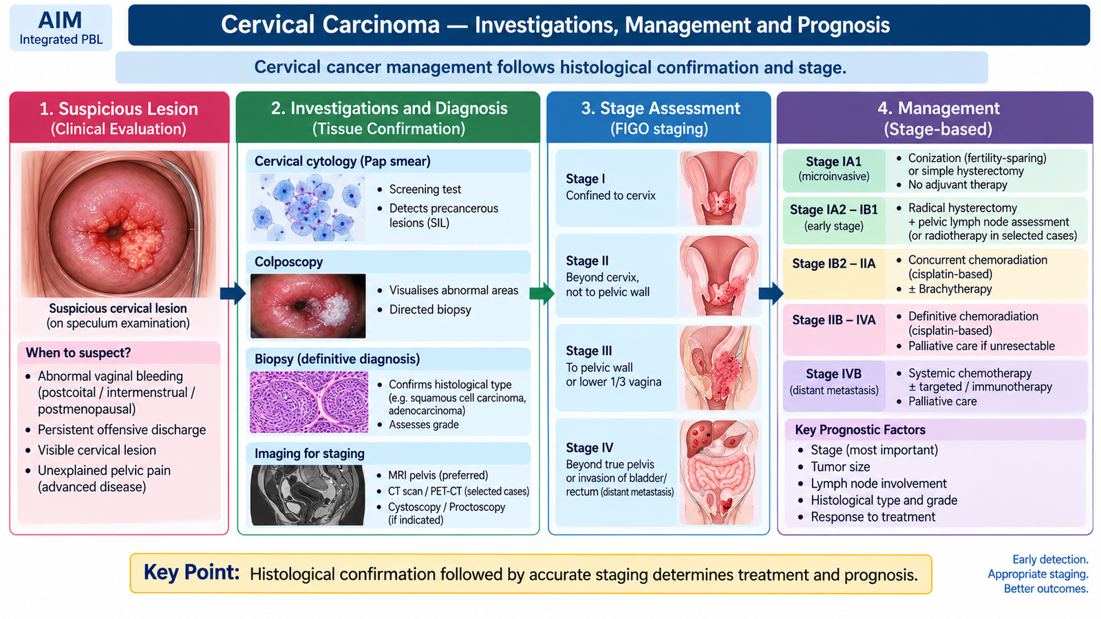
Clinical evaluation of suspected cervical carcinoma aims first to confirm malignancy histologically and then determine the extent of disease. Management is therefore based not simply on the presence of cancer, but on tumor stage, tumor size, nodal involvement, general health and, in carefully selected early disease, fertility considerations.
Diagnostic approach
- History: characterize postcoital, intermenstrual or postmenopausal bleeding, vaginal discharge, pelvic pain and symptoms of advanced disease.
- Pelvic and speculum examination: identifies a visible cervical lesion and assesses local extension.
- Biopsy: histological demonstration of invasive carcinoma provides definitive diagnosis.
- Colposcopy-directed biopsy is useful when an abnormal epithelial area requires targeted assessment.
- Imaging assists assessment of local tumor extent, lymph nodes, urinary obstruction and distant disease when clinically appropriate.
Cervical cytology and HPV testing are highly valuable in screening and precursor detection, but a suspicious invasive lesion requires tissue diagnosis.
Management principles
Management becomes more intensive as disease extends beyond the cervix. Detailed operative technique is not required at undergraduate level; the key concept is the relationship between stage and treatment modality.
- Preinvasive disease: appropriately selected cervical precursor lesions are treated locally to prevent invasion.
- Very early invasive disease: selected patients may be managed with conservative excisional or fertility-preserving procedures when oncologically appropriate.
- Early invasive carcinoma: surgery, including hysterectomy-based treatment with appropriate nodal assessment, is an important option in suitable patients. Radiotherapy is an alternative in selected situations.
- Locally advanced carcinoma: concurrent chemoradiation with brachytherapy is a major treatment principle.
- Recurrent or metastatic disease: systemic treatment and symptom-directed palliative measures are considered according to disease extent and patient condition.
Prognosis
The strongest prognostic factor is the stage at diagnosis. A small tumor confined to the cervix has a much better outcome than a tumor involving pelvic structures or distant organs. Lymph-node involvement, increasing tumor size and unfavorable pathological features are also associated with poorer prognosis.
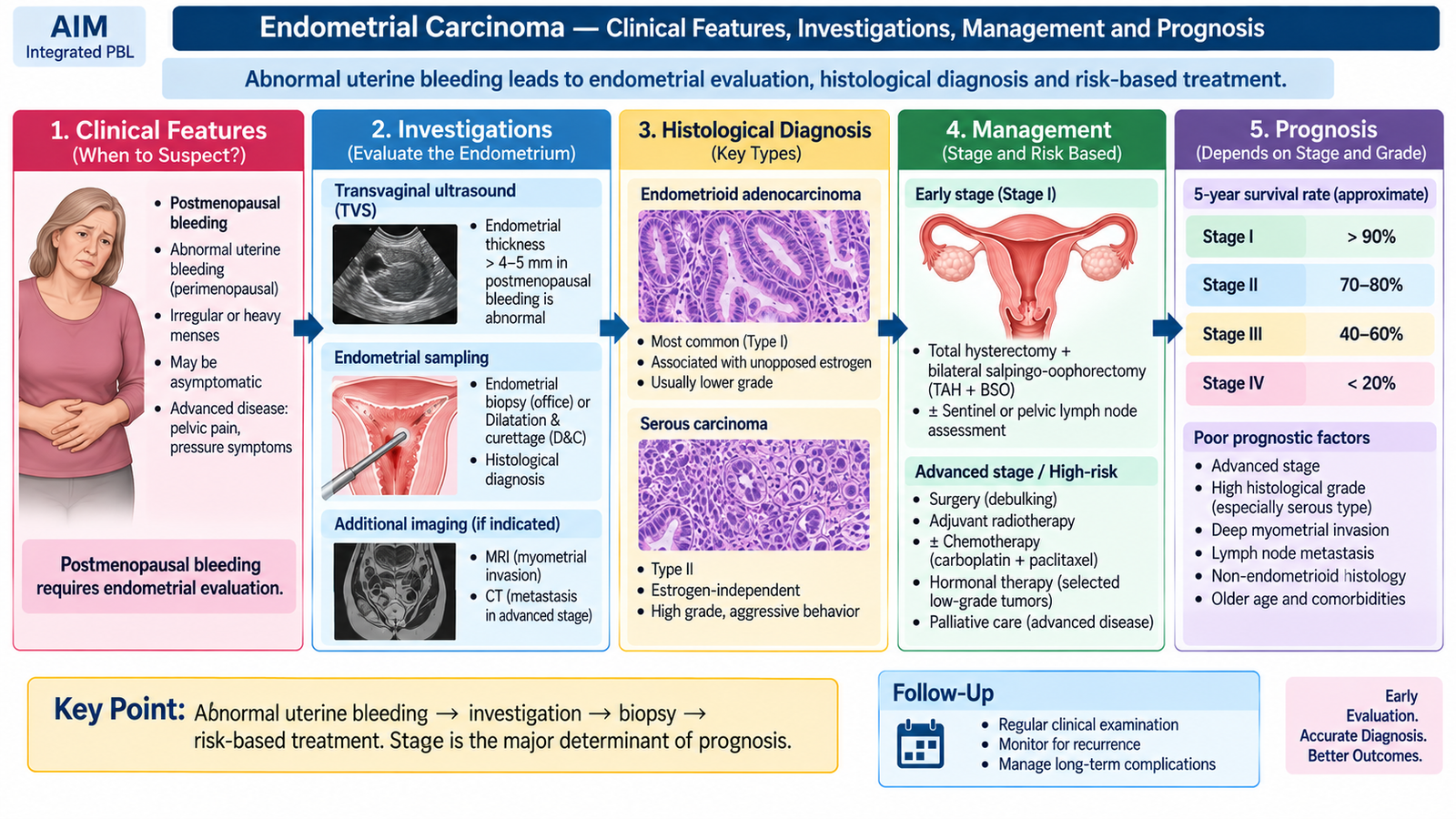
E. Endometrial Hyperplasia and Endometrial Carcinoma — Etiology, Pathogenesis and Morphology
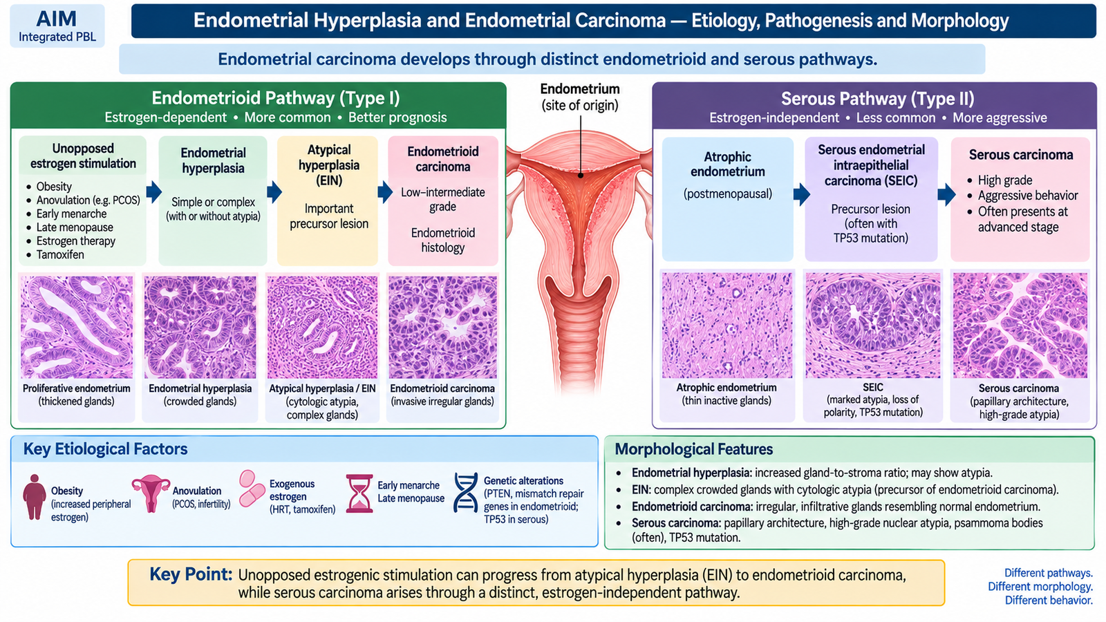
Endometrial carcinoma arises from the lining of the uterine cavity. An important pathway begins with prolonged estrogenic stimulation without adequate progesterone opposition. This promotes repeated endometrial proliferation and can progress through atypical hyperplasia, also termed endometrial intraepithelial neoplasia (EIN), to endometrioid carcinoma. A separate aggressive pathway produces serous carcinoma, usually in an atrophic endometrium and through different molecular abnormalities.
Endometrial hyperplasia
Endometrial hyperplasia is an abnormal increase in endometrial glands relative to stroma. The clinically important distinction is whether cytological atypia is present.
- Hyperplasia without atypia: increased glandular proliferation but without the marked cytological abnormalities associated with a high risk of malignant progression.
- Atypical hyperplasia/EIN: crowded abnormal glands with cytological atypia and a substantially greater relationship with endometrioid carcinoma.
Etiology and risk factors for the estrogen-related pathway
Anything that produces prolonged unopposed estrogen can stimulate endometrial proliferation. Repeated proliferation increases the opportunity for neoplastic clones to arise.
- Obesity, because adipose tissue contributes to peripheral estrogen formation.
- Chronic anovulation, including anovulatory states such as PCOS, because progesterone exposure is reduced.
- Estrogen therapy without adequate progestogenic opposition in a woman with an intact uterus.
- Estrogen-producing ovarian tumors.
- Nulliparity and prolonged lifetime estrogenic exposure.
- Hereditary mismatch-repair abnormalities such as Lynch syndrome increase susceptibility to endometrial carcinoma through a different genetic mechanism.
Pathogenesis
Endometrioid carcinoma frequently shows alterations involving pathways such as PTEN/PI3K signaling and mismatch-repair mechanisms. At undergraduate level, the important point is that the tumor often develops from a recognizable precursor in an estrogen-driven proliferative endometrium.
Serous carcinoma usually develops in older women in an atrophic endometrium and is not primarily driven by estrogenic hyperplasia. TP53 abnormalities are characteristic. It behaves more aggressively and may spread widely even when the uterine lesion is relatively small.
Gross morphology
- Endometrial carcinoma may form a polypoid or exophytic mass projecting into the uterine cavity.
- It may diffusely thicken the endometrium.
- Progressive disease invades the underlying myometrium.
- Deep myometrial invasion increases access to lymphatic channels and therefore worsens prognosis.
Microscopic morphology
Endometrioid carcinoma forms crowded malignant glands resembling endometrial glands but with loss of normal gland-to-stroma organization. Nuclear atypia and solid growth increase with poorer differentiation.
Serous carcinoma typically shows papillary or complex glandular architecture with marked nuclear atypia and high-grade cytological features. Psammoma bodies may be present.
F. Endometrial Carcinoma — Clinical Features, Investigations, Management and Prognosis
Endometrial carcinoma often becomes clinically apparent relatively early because a tumor arising within the uterine cavity disturbs the endometrial surface and causes bleeding. For this reason, abnormal bleeding—particularly after menopause—is an important warning symptom and should not be dismissed.
Clinical features
- Postmenopausal bleeding is the classic presentation.
- Abnormal, heavy or irregular uterine bleeding may occur before menopause.
- Watery or blood-stained vaginal discharge may occur.
- Pelvic pain, pressure symptoms or systemic manifestations are more likely in advanced disease.
A relevant history should include menopausal status, pattern of bleeding, obesity, chronic anovulation, estrogen exposure, family history and hereditary cancer risk where appropriate.
Investigations
Investigation aims to determine whether the endometrium is abnormal and then obtain tissue for definitive diagnosis.
- Transvaginal ultrasonography evaluates the uterus and endometrium and can identify abnormal endometrial thickening or other uterine pathology.
- Endometrial sampling/biopsy provides histological confirmation and is central to diagnosis.
- Hysteroscopic evaluation may help identify and sample a focal intrauterine lesion when required.
- Imaging for staging assesses myometrial invasion, cervical involvement, lymph nodes and extrauterine spread when indicated.
Management principles
Treatment depends on whether the lesion is a precursor or an established carcinoma, and on the extent and pathological risk of the disease.
- Hyperplasia without atypia: progestogenic treatment and appropriate surveillance are commonly used because many lesions can regress when abnormal estrogenic stimulation is opposed.
- Atypical hyperplasia/EIN: hysterectomy is definitive treatment in women who have completed childbearing. Carefully selected patients desiring fertility may be considered for conservative progestogenic management under specialist supervision.
- Endometrial carcinoma: surgery with removal of the uterus and both adnexa forms the main treatment for operable localized disease, with surgical staging as appropriate.
- Adjuvant radiotherapy and/or systemic treatment may be required according to stage and pathological risk factors.
- Hormonal therapy may have a role in selected hormone-responsive advanced or recurrent tumors.
Prognosis
Endometrial carcinoma often has a comparatively favorable prognosis because abnormal bleeding leads to earlier diagnosis. Prognosis worsens with increasing stage, high tumor grade, aggressive histological subtype, deep myometrial invasion, lymphovascular invasion and nodal or distant spread.
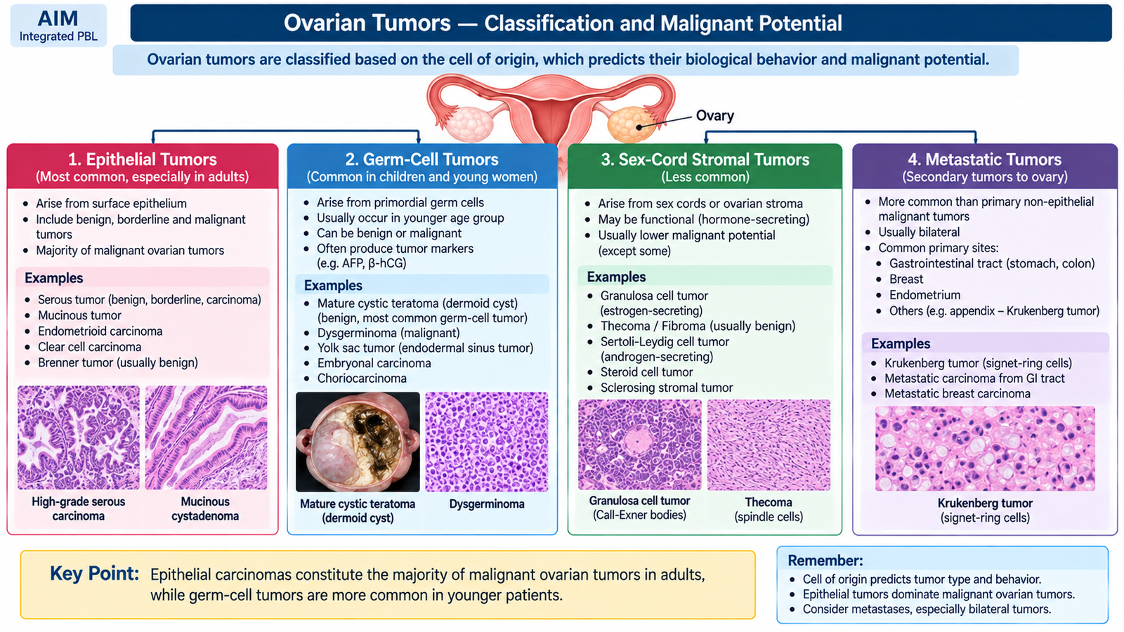
G. Ovarian Tumors — Classification and Malignant Potential
Ovarian tumors arise from several different cell populations, so ovarian neoplasia is more diverse than carcinoma of the cervix or endometrium. Classification is therefore based mainly on the tissue of origin. Knowing the major categories allows the student to predict the usual age group, tumor markers and biological behavior.
| Tumor family | Important benign examples | Important malignant examples |
|---|---|---|
| Surface epithelial tumors | Serous cystadenoma, mucinous cystadenoma, Brenner tumor | Serous carcinoma, mucinous carcinoma, endometrioid carcinoma, clear-cell carcinoma |
| Germ-cell tumors | Mature cystic teratoma | Dysgerminoma, yolk-sac tumor, immature teratoma, non-gestational choriocarcinoma |
| Sex-cord stromal tumors | Fibroma and many thecomas | Granulosa-cell tumor has malignant potential; Sertoli-Leydig tumors may also behave malignantly |
| Metastatic tumors | — | Secondary ovarian deposits, including Krukenberg-type metastases |
Surface epithelial tumors may also have a borderline category. These tumors show epithelial proliferation and atypia greater than a benign tumor but lack destructive stromal invasion. This distinction is particularly important in serous and mucinous tumors.
Important pathological relationships
High-grade serous carcinoma, the major aggressive epithelial ovarian carcinoma, is now understood to arise frequently from precursor epithelial lesions in the distal fallopian tube, particularly serous tubal intraepithelial carcinoma. TP53 abnormalities are characteristic, and hereditary BRCA-associated pathways are important in a proportion of patients.
Endometrioid and clear-cell ovarian carcinomas may arise in association with endometriosis. This illustrates that ovarian carcinomas do not all share a single precursor or molecular pathway.
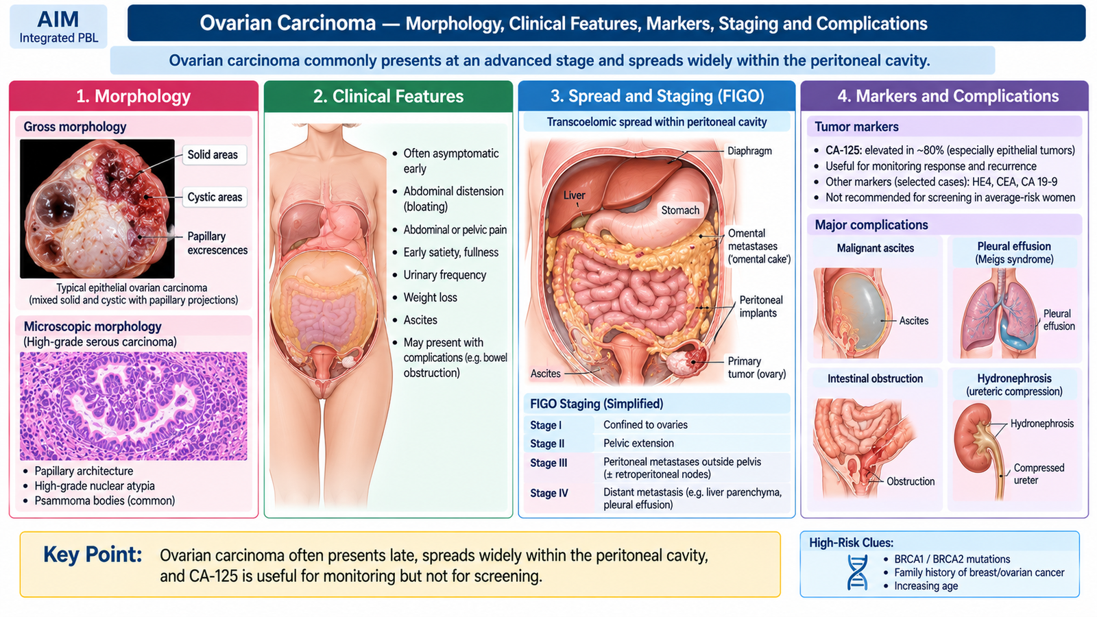
H. Ovarian Carcinoma — Morphology, Clinical Features, Markers, Staging and Complications
Ovarian carcinoma is clinically important because an enlarging tumor can remain relatively silent within the pelvis and abdomen. Malignant epithelial tumors also have ready access to the peritoneal cavity, allowing exfoliated cells to implant widely on peritoneal and omental surfaces. This pattern explains why ascites and diffuse abdominal disease are common in advanced ovarian carcinoma.
Gross morphology
Features suggesting malignancy include a complex solid-cystic mass, irregular surface, papillary excrescences, necrosis, hemorrhage and capsular involvement. Bilaterality is particularly common in serous carcinoma and metastatic tumors.
- Serous carcinoma: commonly cystic and solid with papillary projections; malignant tumors may involve both ovaries.
- Mucinous tumors: often large and multiloculated, containing mucinous material.
- Endometrioid and clear-cell carcinomas: may be associated with endometriotic lesions.
Microscopic morphology
High-grade serous carcinoma typically shows complex papillary, glandular and solid architecture with marked nuclear atypia and frequent mitotic activity. Psammoma bodies may occur. Endometrioid carcinoma forms glands resembling endometrial adenocarcinoma, while clear-cell carcinoma contains cells with clear cytoplasm and characteristic hobnail-type cells in some areas.
Clinical features
Symptoms are often vague because early ovarian enlargement does not necessarily disturb a hollow organ or produce visible bleeding.
- Persistent abdominal or pelvic discomfort.
- Abdominal bloating or increasing abdominal girth.
- Early satiety and reduced appetite due to abdominal mass effect or ascites.
- Urinary frequency or pressure symptoms.
- Adnexal or pelvic mass on examination.
- Ascites in advanced disease.
- Weight loss and constitutional symptoms in advanced malignancy.
Serological markers
Tumor markers support evaluation and follow-up but should not be interpreted as stand-alone proof of malignancy. Their value depends on tumor type and clinical context.
| Marker | Important association | Clinical interpretation |
|---|---|---|
| CA-125 | Especially epithelial ovarian carcinoma | Useful in evaluation and monitoring; may also rise in benign conditions. |
| AFP | Yolk-sac and some other germ-cell tumors | Helps identify and follow appropriate germ-cell tumors. |
| β-hCG | Choriocarcinomatous and selected germ-cell components | Supports characterization of relevant germ-cell malignancy. |
| LDH | Dysgerminoma | Can support diagnosis and disease monitoring. |
| Inhibin | Granulosa-cell tumor | Useful marker for a sex-cord stromal tumor. |
Spread and staging
The characteristic route of epithelial ovarian carcinoma is transcoelomic spread: tumor cells exfoliate into the peritoneal cavity and implant on pelvic and abdominal peritoneal surfaces, particularly the omentum. Lymphatic spread also occurs, while hematogenous spread is more typical of advanced disease.
| Stage | Essential concept |
|---|---|
| I | Disease limited to the ovary/ovaries or fallopian tube/tubes. |
| II | Tumor extends to other structures within the pelvis. |
| III | Peritoneal spread outside the pelvis and/or involvement of retroperitoneal lymph nodes. |
| IV | Distant metastatic disease beyond the usual peritoneal pattern. |
Complications
- Ascites due to widespread peritoneal irritation, tumor implants and impaired fluid drainage.
- Omental and diffuse peritoneal metastases.
- Pleural involvement or malignant pleural effusion in advanced disease.
- Bowel obstruction from extensive peritoneal tumor.
- Urinary obstruction from pelvic disease.
- Venous thromboembolic complications associated with malignancy.
- Malnutrition and cachexia in advanced disease.
I. Ovarian Carcinoma — Diagnosis and Management
Evaluation of a suspected ovarian malignancy combines clinical assessment, imaging and appropriate tumor markers. No single symptom or blood marker can reliably establish the diagnosis. The objective is to determine whether an adnexal mass is suspicious for malignancy, assess the extent of disease and obtain definitive pathological confirmation while planning treatment.
Diagnostic approach
- History: persistent bloating, early satiety, abdominal or pelvic pain, urinary symptoms, weight change and relevant family history.
- Abdominal and pelvic examination: assess for adnexal mass, ascites, abdominal distension and evidence of advanced disease.
- Pelvic ultrasonography: characterizes the adnexal mass and identifies features such as solid components, papillary projections, multiloculation and ascites.
- Serum tumor markers: selected according to age and suspected tumor type.
- Cross-sectional imaging: evaluates disease distribution, peritoneal deposits, lymph nodes and possible distant spread.
- Histopathology: establishes the definitive tumor type and grade.
CA-125 is particularly useful in the assessment and follow-up of many epithelial ovarian cancers, but it is not specific for malignancy. It can rise in several benign gynecological and peritoneal conditions. Therefore, it must be interpreted together with the clinical picture and imaging.
Management principles
The principal treatment of epithelial ovarian carcinoma combines surgery and systemic chemotherapy according to stage, tumor type and patient suitability. The aim of surgery is both accurate staging and removal of as much visible tumor as safely possible.
- Early localized disease: surgical staging and removal of involved reproductive organs as appropriate; fertility-preserving surgery may be considered in carefully selected young patients with suitable early-stage tumors.
- More extensive epithelial ovarian carcinoma: cytoreductive surgery is used to reduce visible tumor burden.
- Systemic chemotherapy: platinum-based chemotherapy, commonly combined with a taxane, is a major treatment principle for epithelial ovarian carcinoma when indicated.
- Neoadjuvant chemotherapy may be considered when optimal primary surgery is not initially feasible or when patient factors make immediate extensive surgery unsuitable.
- Recurrent or advanced disease: further systemic treatment and symptom-directed management are individualized according to tumor biology, previous therapy and general condition.
The prognosis of ovarian carcinoma depends strongly on stage, residual tumor burden after surgery, histological type and response to systemic treatment. Because early ovarian cancer may produce few symptoms, many epithelial malignancies are diagnosed after peritoneal dissemination, which contributes to their less favorable overall prognosis compared with many endometrial cancers.
Integrated Mechanism Flow
Although the initiating pathway differs between organs, the three major malignancies can be understood using the same general progression from carcinogenic drive to clinically detectable invasive disease.
HPV, hormonal drive or tumor-specific genetic pathway
Abnormal survival and proliferation
Present in important cervical and endometrial pathways
Basement membrane and tissue invasion
Local, lymphatic, peritoneal or distant
Diagnosis, staging and appropriate treatment
Important Comparison — Cervical vs Endometrial vs Ovarian Cancer
| Feature | Cervical carcinoma | Endometrial carcinoma | Ovarian carcinoma |
|---|---|---|---|
| Major etiologic concept | Persistent high-risk HPV | Often unopposed estrogen/EIN pathway; serous pathway is distinct | Multiple epithelial and molecular pathways |
| Classic presentation | Postcoital or abnormal vaginal bleeding | Postmenopausal bleeding | Bloating, early satiety, pelvic/abdominal symptoms |
| Key diagnostic principle | Biopsy of suspicious cervical lesion | Endometrial sampling | Imaging + markers + definitive histopathology |
| Characteristic spread | Local pelvic extension and lymphatics | Myometrial invasion, then lymphatic/extrauterine spread | Transcoelomic peritoneal spread |
| Important marker | No serum marker central to routine diagnosis | No serum marker central to routine diagnosis | CA-125 for many epithelial tumors; other markers depend on tumor type |
| Prevention/early detection concept | HPV vaccination + screening | Prompt investigation of abnormal uterine bleeding and control of relevant hormonal risk | Clinical evaluation of suspicious symptoms/masses; serum markers are not stand-alone screening tests |
| Major treatment principle | Stage-based surgery or chemoradiation | Surgery is central for localized disease | Cytoreductive surgery + systemic chemotherapy when indicated |
⭐ AIM High-Yield Review
Gynecologic Malignancies: Cervical, Endometrial and Ovarian Cancer
Watch this video after completing the learning material to reinforce the major gynecologic cancers, their clinical features, diagnosis and treatment principles.