
Topic 6 — Parathyroid Disorders: Hyperparathyroidism and Hypoparathyroidism
Topic Introduction
The parathyroid glands regulate the concentration of calcium in extracellular fluid mainly through parathyroid hormone (PTH). Hyperparathyroidism occurs when PTH secretion is excessive, while hypoparathyroidism results from deficient PTH secretion or, in closely related disorders, failure of tissues to respond normally to PTH. Because PTH acts on bone and kidney and indirectly increases intestinal calcium absorption, abnormalities of this hormone can affect the skeleton, kidneys, gastrointestinal system, nervous system and muscles. In this chapter, you will learn the major types and pathological basis of both disorders, their causes and clinical manifestations, the laboratory pattern used for diagnosis and the main principles of treatment.
A. PTH and Calcium–Phosphate Homeostasis
Understanding parathyroid disease becomes much easier once the normal action of PTH is clear. PTH is secreted by the chief cells of the parathyroid glands. Its major physiological purpose is to prevent the extracellular calcium concentration from falling too low. A decrease in ionized calcium stimulates PTH secretion, while restoration of calcium suppresses further secretion through normal negative feedback.
How PTH raises serum calcium
PTH acts mainly on bone and kidney. Its effects on the intestine occur indirectly through activation of vitamin D.
- Bone: sustained PTH activity promotes osteoclast-mediated bone resorption indirectly through signals produced by osteoblast-lineage cells. Calcium and phosphate are released from mineralized bone.
- Kidney — calcium: PTH increases renal tubular calcium reabsorption, helping conserve calcium.
- Kidney — phosphate: PTH decreases proximal tubular phosphate reabsorption, producing phosphaturia and therefore lowering serum phosphate.
- Vitamin D: PTH stimulates renal formation of active vitamin D, which increases intestinal absorption of calcium.
This relationship explains why excessive PTH generally produces a tendency toward hypercalcemia and reduced serum phosphate, whereas deficient PTH produces hypocalcemia and increased serum phosphate.

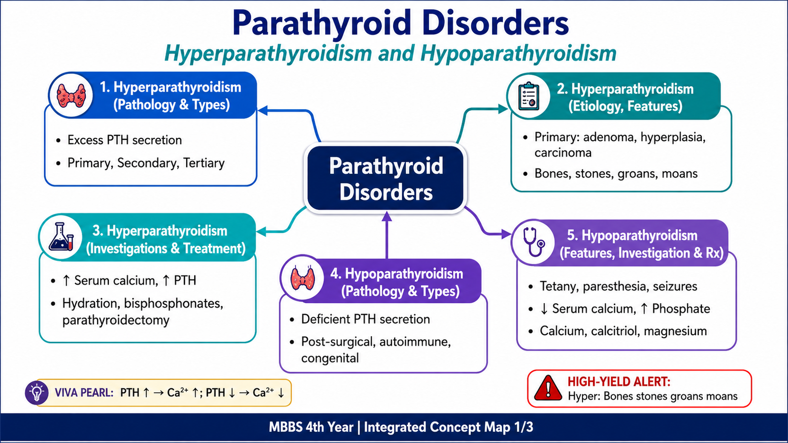
B. Hyperparathyroidism — Pathological Basis and Major Types
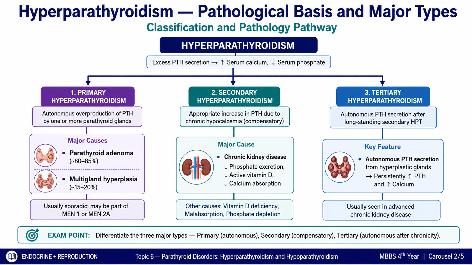
Hyperparathyroidism means excessive secretion of PTH. The disorder is classified according to why the parathyroid glands are producing too much hormone. In primary disease, the abnormality begins within the parathyroid glands themselves. In secondary disease, increased PTH is initially an appropriate compensatory response to a persistent stimulus such as hypocalcemia. In tertiary disease, long-standing stimulation eventually produces autonomous PTH secretion.
1. Primary Hyperparathyroidism
Primary hyperparathyroidism results from an intrinsic abnormality of one or more parathyroid glands. PTH is secreted despite a serum calcium concentration that should normally suppress it.
| Parathyroid lesion | Key pathological concept |
|---|---|
| Parathyroid adenoma | The commonest cause. Usually a solitary benign neoplasm producing PTH independently of normal calcium feedback. |
| Primary parathyroid hyperplasia | Usually involves multiple glands. It may occur sporadically or in inherited endocrine neoplasia syndromes. |
| Parathyroid carcinoma | A rare cause that may produce marked PTH excess and hypercalcemia. Malignancy is established by invasive or metastatic behaviour rather than cellular appearance alone. |
Pathological appearance
A typical adenoma is a well-circumscribed lesion composed mainly of parathyroid chief cells. A rim of compressed normal parathyroid tissue may be present at its edge. In contrast, primary hyperplasia affects more than one gland. Parathyroid carcinoma is important because invasion into surrounding tissue or metastasis supports malignancy; cytological atypia alone is not sufficient.
2. Secondary Hyperparathyroidism
Secondary hyperparathyroidism develops when the parathyroid glands are chronically stimulated by a physiological need to raise serum calcium. The most important setting is chronic kidney disease. Diseased kidneys retain phosphate and form less active vitamin D. Reduced active vitamin D decreases intestinal calcium absorption, while phosphate retention also contributes to lowering ionized calcium. These changes chronically stimulate PTH secretion and cause parathyroid hyperplasia.
Other causes of persistent hypocalcemic stimulation, such as severe vitamin D deficiency or malabsorption, can also produce secondary hyperparathyroidism.
3. Tertiary Hyperparathyroidism
Tertiary hyperparathyroidism develops after long-standing secondary hyperparathyroidism, most often in chronic renal disease. Prolonged stimulation causes enlarged parathyroid tissue to become partly or completely autonomous. The glands then continue secreting large amounts of PTH even when the original stimulus is no longer sufficient to justify it. Unlike typical secondary hyperparathyroidism, tertiary disease is commonly associated with hypercalcemia.
Effect of persistent PTH excess on bone
Continued PTH excess increases bone turnover and resorption. Severe prolonged disease may produce osteitis fibrosa cystica, characterized by increased osteoclastic activity, marrow fibrosis and weakened bone. Local areas of hemorrhage and reactive tissue may form so-called brown tumors. These are not true neoplasms; their brown color reflects hemorrhage and hemosiderin deposition.
C. Hyperparathyroidism — Clinical Features, Investigations and Treatment
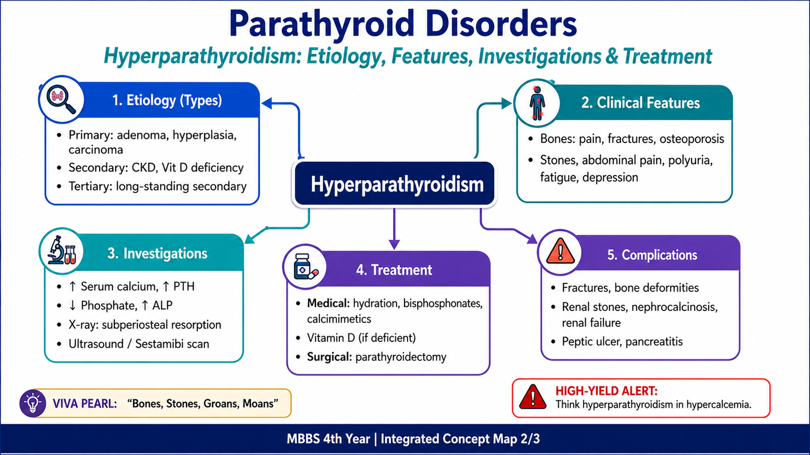
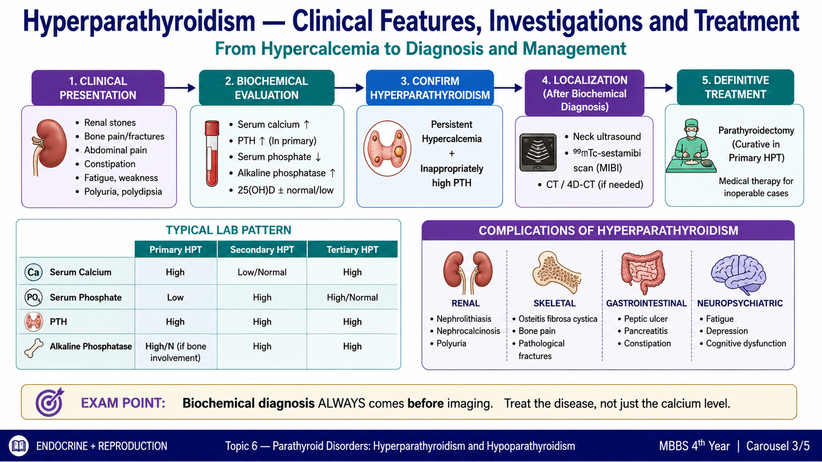
The clinical presentation depends on the type and severity of hyperparathyroidism. Primary hyperparathyroidism is frequently detected when routine laboratory testing shows hypercalcemia, but symptomatic patients may develop manifestations involving the kidneys, skeleton, gastrointestinal tract, muscles and nervous system. Most of these manifestations can be understood as consequences of excess calcium, excess PTH or prolonged bone resorption.
Clinical Features
- Renal: polyuria and polydipsia may occur because hypercalcemia reduces the kidney’s ability to concentrate urine. Increased urinary calcium can contribute to renal calculi and nephrocalcinosis.
- Skeletal: increased bone resorption may cause bone pain, reduced bone strength and fractures. Severe prolonged disease may produce osteitis fibrosa cystica.
- Muscular: proximal muscle weakness and fatigue may occur.
- Gastrointestinal: constipation, anorexia, nausea or abdominal discomfort can accompany hypercalcemia.
- Neuropsychiatric: tiredness, impaired concentration, low mood or confusion may occur, particularly as hypercalcemia becomes more severe.
- Severe hypercalcemia: marked dehydration, altered consciousness and cardiac electrical disturbances may occur and require urgent assessment.
Diagnostic Approach
The central diagnostic principle is to interpret the serum calcium and PTH together. PTH should normally be suppressed when serum calcium is high. Therefore, a high or even inappropriately normal PTH in a patient with confirmed hypercalcemia points toward PTH-dependent hypercalcemia.
Important investigations and what they mean
- Serum calcium: confirms hypercalcemia. Corrected total calcium or ionized calcium can be used when appropriate.
- Serum PTH: establishes whether hypercalcemia is PTH-dependent.
- Serum phosphate: is commonly reduced in primary hyperparathyroidism because PTH promotes renal phosphate excretion.
- Renal function: helps identify chronic kidney disease and assesses renal complications.
- Vitamin D status: may identify associated deficiency and helps interpret secondary elevations of PTH.
- Urinary calcium assessment: may be useful when distinguishing primary hyperparathyroidism from inherited causes of PTH-dependent hypercalcemia such as familial hypocalciuric hypercalcemia.
- Bone assessment: bone mineral density testing helps determine skeletal involvement.
- Renal imaging: may identify calculi or nephrocalcinosis when renal involvement is suspected.
Role of parathyroid imaging
Parathyroid imaging is primarily used for localization before surgery, not for establishing the biochemical diagnosis. Ultrasound and radionuclide localization studies may help identify an abnormal gland after the diagnosis of primary hyperparathyroidism has already been made from clinical and biochemical findings.
Typical biochemical patterns
| Disorder | PTH | Calcium | Phosphate tendency | Main reason |
|---|---|---|---|---|
| Primary HPT | High or inappropriately non-suppressed | High | Often low | Autonomous parathyroid lesion |
| Secondary HPT in CKD | High | Usually low or normal | Often high | Phosphate retention and reduced active vitamin D |
| Tertiary HPT | Markedly high | High | Variable; often influenced by renal disease | Autonomous glands after prolonged secondary stimulation |
Treatment Principles
Treatment depends on the type of hyperparathyroidism, the severity of hypercalcemia and whether there is end-organ damage. Management should correct the underlying mechanism rather than simply lowering PTH without considering its cause.
Primary hyperparathyroidism
- Parathyroidectomy is the definitive treatment for symptomatic primary hyperparathyroidism and for appropriate patients with significant biochemical or end-organ involvement.
- Selected patients with mild disease who do not require or cannot undergo surgery may be managed with clinical and biochemical monitoring.
- Cinacalcet, a calcimimetic, increases the sensitivity of the calcium-sensing receptor to extracellular calcium and can reduce PTH secretion and serum calcium in selected patients.
- Antiresorptive therapy may be used when preservation of bone mineral density is an important objective, although it does not remove the abnormal parathyroid gland.
Severe symptomatic hypercalcemia
Severe hypercalcemia can cause major fluid loss and neurological deterioration. Initial management includes restoration of intravascular volume with intravenous isotonic fluid when appropriate. Additional calcium-lowering therapy may include rapidly acting agents such as calcitonin and longer-acting antiresorptive treatment depending on the clinical situation. Definitive treatment of the underlying hyperparathyroid disorder is then required.
Secondary hyperparathyroidism
Treatment targets the persistent stimulus. In chronic kidney disease this includes management of phosphate balance and abnormalities of vitamin D metabolism. Active vitamin D preparations and calcimimetic therapy may be used in appropriate patients under renal or endocrine care. Severe disease that remains uncontrolled despite medical treatment may require parathyroid surgery.
Tertiary hyperparathyroidism
Because the enlarged glands have become autonomous, simply correcting the original stimulus may not normalize PTH secretion. Parathyroidectomy is therefore an important definitive treatment in significant tertiary disease, while selected patients may also receive medical therapy such as a calcimimetic.
D. Hypoparathyroidism — Pathological Basis and Etiology
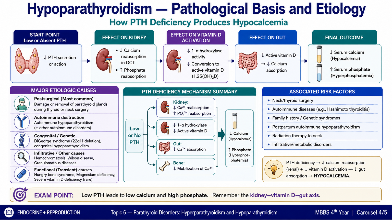
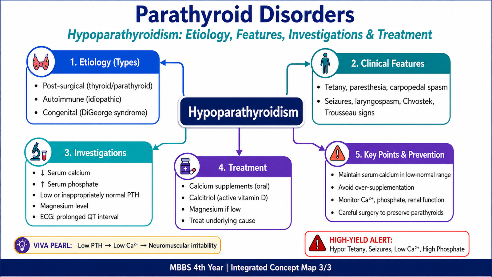
Hypoparathyroidism is a state of inadequate PTH secretion. Because PTH normally maintains serum calcium and promotes renal phosphate excretion, loss of PTH produces the characteristic combination of hypocalcemia and hyperphosphatemia. The pathological basis varies: the glands may be removed or damaged, destroyed by autoimmune disease, absent because of a developmental abnormality or functionally suppressed by a metabolic disturbance.
Major Causes
1. Postsurgical hypoparathyroidism
Damage to, removal of or loss of the blood supply to the parathyroid glands during neck surgery is an important acquired cause. The fall in PTH reduces renal calcium conservation and active vitamin D formation, so serum calcium falls.
2. Autoimmune hypoparathyroidism
Autoimmune destruction may occur as an isolated disorder or as part of an autoimmune endocrine syndrome. Progressive destruction of functional parathyroid tissue reduces PTH production.
3. Congenital or developmental causes
Congenital absence or underdevelopment of parathyroid tissue can produce hypoparathyroidism from childhood. Developmental disorders affecting structures derived from the pharyngeal apparatus, such as DiGeorge syndrome, may include deficient parathyroid tissue and therefore hypocalcemia.
4. Functional suppression due to magnesium disturbance
Severe magnesium deficiency can impair PTH secretion and reduce the response of target tissues to PTH. Hypocalcemia may therefore persist until the magnesium abnormality is corrected.
5. Other destructive causes
Less commonly, parathyroid tissue can be damaged by infiltrative processes or other destructive conditions. The important principle is that loss of functioning parathyroid tissue reduces PTH secretion.
How PTH deficiency causes hypocalcemia
Hypocalcemia increases the excitability of nerves and skeletal muscle. This is why neuromuscular symptoms such as tingling, cramps, carpopedal spasm and tetany are central manifestations of hypoparathyroidism.
E. Hypoparathyroidism — Clinical Features, Investigations and Treatment
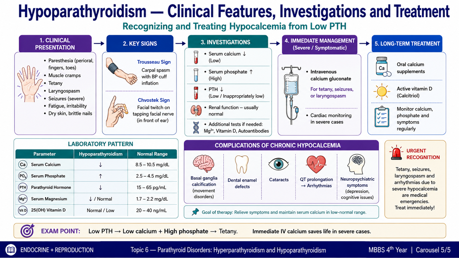
The manifestations of hypoparathyroidism are mainly manifestations of hypocalcemia. Acute falls in calcium are particularly likely to produce neuromuscular symptoms, while chronic disease may produce additional neurological, ocular and ectodermal changes. Recognizing these manifestations and then confirming the calcium–phosphate–PTH pattern is the key diagnostic approach.
Clinical Features
Neuromuscular manifestations
- Perioral numbness and tingling of the fingers or toes.
- Muscle cramps and stiffness.
- Carpopedal spasm.
- Tetany due to increased neuromuscular excitability.
- Seizures in severe hypocalcemia.
- Laryngospasm may occur in severe disease and can threaten the airway.
Clinical signs of latent tetany
Chvostek sign is contraction of facial muscles after tapping over the facial nerve. It may occur in hypocalcemia but is not completely specific.
Trousseau sign is carpal spasm produced by temporarily reducing blood flow to the arm with an inflated blood-pressure cuff. It reflects increased neuromuscular excitability and is a useful clinical clue to hypocalcemia.
Chronic manifestations
Long-standing hypocalcemia and hyperphosphatemia can produce additional abnormalities, including:
- dry or coarse skin and brittle nails;
- abnormal hair growth or alopecia in some patients;
- cataract formation;
- dental abnormalities when disease begins early in life;
- intracranial calcification, particularly involving the basal ganglia, in some patients with long-standing disease.
Cardiac effects
Hypocalcemia can prolong ventricular repolarization and therefore produces a prolonged QT interval on the ECG. Severe electrolyte disturbance may predispose to clinically important cardiac instability.
Investigations
The diagnosis is based on demonstrating hypocalcemia and then deciding whether the PTH response is appropriate. A patient with low calcium should normally increase PTH secretion. Therefore, a low or inappropriately normal PTH in the presence of hypocalcemia strongly supports hypoparathyroidism.
- Serum calcium: reduced.
- PTH: low or inappropriately normal for the degree of hypocalcemia.
- Serum phosphate: typically increased because renal phosphate excretion falls when PTH is deficient.
- Serum magnesium: should be assessed because marked magnesium deficiency can impair PTH secretion and action.
- Renal function: helps evaluate other causes of mineral disturbance and is important during treatment.
- Vitamin D status: may help identify alternative or additional causes of hypocalcemia.
- ECG: may show QT prolongation in significant hypocalcemia.
Treatment
Treatment aims to relieve symptoms of hypocalcemia while maintaining calcium at a safe level and avoiding excessive urinary calcium loss. The urgency of treatment depends on whether hypocalcemia is mild and chronic or severe and symptomatic.
Acute symptomatic hypocalcemia
Severe symptoms such as tetany, seizures, significant cardiac abnormalities or laryngospasm require urgent treatment. Intravenous calcium, commonly given as calcium gluconate, is used with appropriate clinical and ECG monitoring. Associated magnesium deficiency must also be corrected because calcium may remain low if severe magnesium depletion persists.
Chronic hypoparathyroidism
- Oral calcium supplementation provides calcium directly.
- Active vitamin D, such as calcitriol, increases intestinal calcium absorption and compensates for reduced PTH-dependent activation of vitamin D.
- Magnesium deficiency, when present, should be corrected.
- Serum calcium, phosphate and renal function are monitored during therapy.
- Urinary calcium may also require monitoring because patients lacking PTH have reduced renal calcium conservation, and excessive treatment can therefore promote hypercalciuria and renal complications.
Specialist therapies, including PTH replacement in selected difficult cases, may be considered when conventional treatment does not provide satisfactory control. These are not substitutes for careful biochemical monitoring.
Integrated Mechanism Flow
Important Comparison — Hyperparathyroidism vs Hypoparathyroidism
| Feature | Hyperparathyroidism | Hypoparathyroidism |
|---|---|---|
| Basic abnormality | Excess PTH secretion | Deficient PTH secretion |
| Serum calcium | Usually high in primary and tertiary disease | Low |
| Serum phosphate | Often low in primary disease; may be high in CKD-associated secondary disease | High |
| Typical causes | Adenoma, multigland hyperplasia, CKD-related secondary stimulation, autonomous tertiary disease | Postsurgical damage, autoimmune destruction, congenital deficiency, severe magnesium disturbance |
| Major manifestations | Renal stones, polyuria, bone disease, weakness, constipation, neurocognitive symptoms | Paresthesia, cramps, tetany, carpopedal spasm, seizures |
| Characteristic examination clues | Findings largely reflect hypercalcemia and end-organ disease | Trousseau sign and Chvostek sign |
| ECG tendency | Hypercalcemia can shorten QT | Hypocalcemia prolongs QT |
| Main treatment principle | Control hypercalcemia and treat the source of excess PTH | Replace calcium, support calcium absorption and correct associated magnesium deficiency |