
Diabetes Mellitus: Pathogenesis, Diagnosis, Chronic Management and Population Prevention
An integrated 4th Year chapter linking the pathological basis of diabetes with clinical recognition, diagnostic reasoning, glucose-lowering pharmacology and population-level prevention.
1. Topic Introduction
Diabetes mellitus is a group of metabolic disorders characterized by persistent hyperglycaemia caused by inadequate insulin secretion, impaired insulin action, or both. Although raised blood glucose is the central laboratory abnormality, diabetes affects much more than glucose metabolism. Progressive metabolic and vascular injury can damage the kidneys, retina, peripheral nerves and large arteries. Understanding diabetes therefore requires linking its classification and pathogenesis with clinical presentation, diagnosis, complications, treatment and prevention. In this chapter you will learn how insulin resistance and β-cell failure produce type 2 diabetes, how major forms of diabetes are recognized, how commonly used glucose-lowering drugs work, how insulin preparations differ, and how screening and preventive care can reduce the burden of disease.
A. Diabetes Mellitus: Core Concept, Classification and Diagnostic Criteria
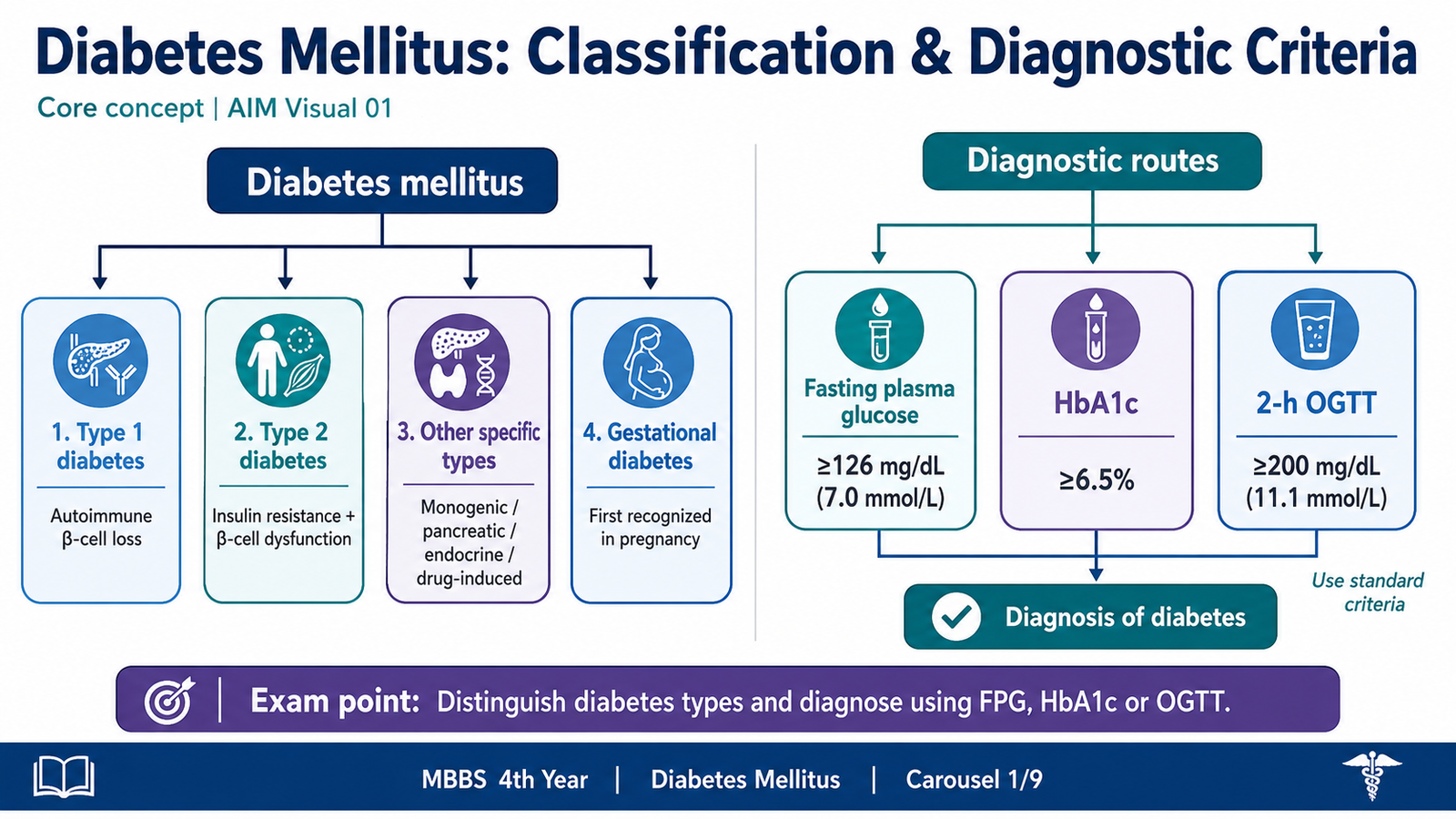
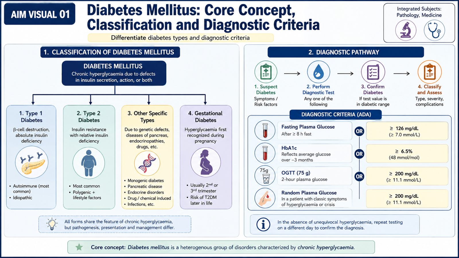
Diabetes mellitus is not a single disease. It is a group of disorders that share chronic hyperglycaemia but differ in their underlying cause. The most useful classification therefore depends on the mechanism responsible for inadequate insulin action. Correct classification matters because it influences treatment, the tendency to develop ketoacidosis and the expected course of disease.
Classification
- Type 1 diabetes mellitus: destruction of pancreatic β-cells produces severe or absolute insulin deficiency. Most cases are immune-mediated.
- Type 2 diabetes mellitus: insulin resistance is combined with progressive β-cell dysfunction. It is the most common form in adults, although it can occur at younger ages.
- Other specific types: diabetes may occur because of defined genetic abnormalities, pancreatic disease, endocrine disorders or drugs that disturb glucose regulation.
- Gestational diabetes mellitus: hyperglycaemia first recognized during pregnancy that does not represent clearly established pre-existing diabetes.
Type 1 and type 2 diabetes should not be distinguished by age alone. Type 1 diabetes can present in adults, while type 2 diabetes increasingly occurs in younger people. The mechanism, clinical pattern and laboratory evidence are more useful than age by itself.
Diagnostic Criteria
Diabetes is diagnosed by demonstrating hyperglycaemia using a validated plasma glucose measurement or HbA1c. HbA1c reflects average glycaemic exposure over the preceding several weeks because glucose becomes non-enzymatically attached to haemoglobin during the lifespan of circulating red cells.
| Diagnostic Test | Value Consistent with Diabetes | Interpretation |
|---|---|---|
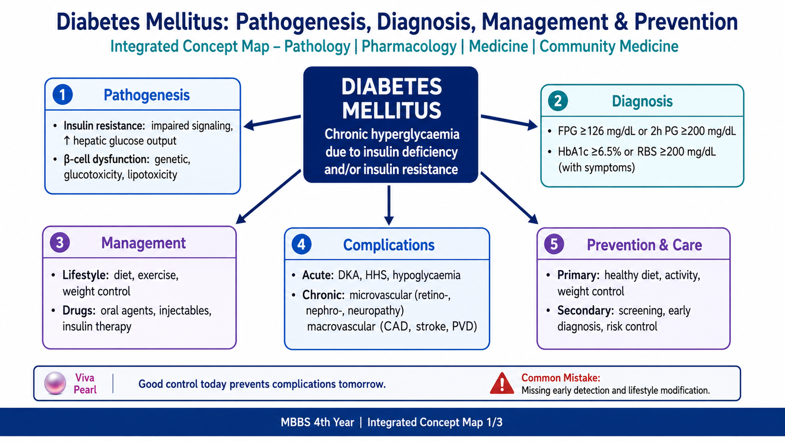
| Fasting plasma glucose | ≥126 mg/dL (7.0 mmol/L) | Measured after an overnight fast. |
| 2-hour plasma glucose during a 75-g oral glucose tolerance test | ≥200 mg/dL (11.1 mmol/L) | Demonstrates impaired handling of a defined glucose load. |
| HbA1c | ≥6.5% | Requires an appropriately standardized laboratory method. |
| Random plasma glucose | ≥200 mg/dL (11.1 mmol/L) | Diagnostic when accompanied by classic hyperglycaemic symptoms or hyperglycaemic crisis. |
In an asymptomatic patient without unequivocal hyperglycaemia, an abnormal result is generally confirmed by repeat testing. HbA1c can become unreliable when red-cell survival or haemoglobin composition is substantially altered. In such situations, plasma glucose-based testing is more appropriate.
B. Pathogenesis: Insulin Resistance and β-Cell Dysfunction
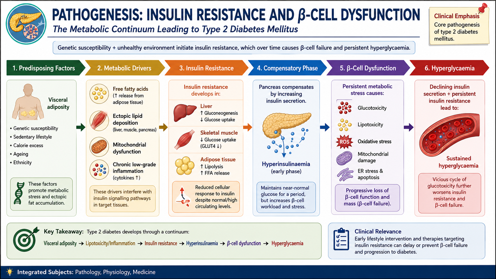
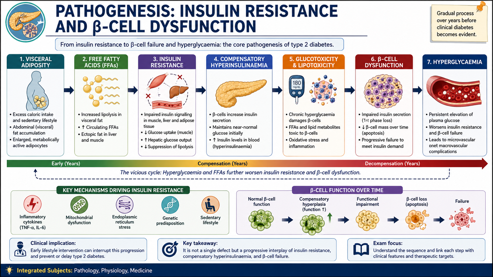
Normal insulin maintains glucose homeostasis by promoting glucose uptake in skeletal muscle and adipose tissue, reducing hepatic glucose production and supporting anabolic metabolism. Diabetes develops when the amount of effective insulin becomes insufficient for the body’s metabolic requirements. In type 1 diabetes the dominant problem is β-cell destruction. In type 2 diabetes the two central abnormalities are insulin resistance and progressive β-cell dysfunction.
Insulin Resistance
Insulin resistance means that normal concentrations of insulin produce a weaker-than-normal biological response. Skeletal muscle therefore removes less glucose from the blood, while the liver fails to suppress glucose production adequately. Adipose tissue also becomes resistant to insulin’s anti-lipolytic action and releases increased free fatty acids.
At first, pancreatic β-cells compensate by secreting more insulin. This may keep glucose near normal despite marked insulin resistance. Diabetes appears when the β-cells can no longer produce enough insulin to overcome that resistance.
β-Cell Dysfunction in Type 2 Diabetes
β-cell failure develops gradually. Genetic susceptibility influences β-cell reserve, while persistent metabolic stress further impairs β-cell function. Chronic hyperglycaemia produces glucotoxicity, and excess fatty acids produce lipotoxicity. Both reduce efficient insulin synthesis and secretion. Islet amyloid derived from islet amyloid polypeptide may also accumulate in type 2 diabetes and is associated with β-cell loss.
An early abnormality is often loss of the normal rapid insulin response to a meal. Postprandial glucose therefore rises first. As β-cell failure progresses, fasting glucose also increases because insulin can no longer restrain hepatic glucose production adequately.
β-Cell Destruction in Type 1 Diabetes
Most type 1 diabetes results from autoimmune destruction of pancreatic β-cells in a genetically susceptible person. T-lymphocyte-mediated injury progressively reduces β-cell mass. Circulating autoantibodies may act as markers of this autoimmune process, although they are not themselves the main cause of β-cell destruction. When β-cell reserve becomes critically low, absolute insulin deficiency produces symptomatic hyperglycaemia and a marked tendency to ketosis.
C. Clinical Presentation, Insulin-Resistance Syndromes and Diagnostic Work-Up
The clinical presentation of diabetes depends on the degree and rate of development of hyperglycaemia. Rapid loss of insulin action tends to produce obvious symptoms, while slowly developing type 2 diabetes may remain unnoticed for years. A good clinical assessment therefore identifies both the symptoms of hyperglycaemia and clues to the mechanism causing it.
Clinical Features
- Polyuria: when filtered glucose exceeds renal reabsorptive capacity, glucose remains in the tubular fluid and causes osmotic diuresis.
- Polydipsia: urinary water loss produces dehydration and stimulates thirst.
- Weight loss: particularly in marked insulin deficiency, the body cannot use glucose effectively and increases breakdown of fat and protein.
- Fatigue and weakness: result from disturbed fuel utilization and dehydration.
- Blurred vision: acute changes in extracellular glucose alter fluid movement within ocular tissues and can temporarily change refraction.
- Recurrent infections: chronic hyperglycaemia impairs several host-defence mechanisms and creates favourable conditions for some infections.
- Slow wound healing: vascular dysfunction, neuropathy and impaired immune responses contribute.
Type 1 diabetes often has a relatively rapid symptomatic onset and may first present with diabetic ketoacidosis. Type 2 diabetes usually evolves more slowly and may first be detected during routine testing, evaluation of another illness or investigation of a complication.
Insulin-Resistance Syndromes
Insulin resistance exists on a spectrum. The commonest clinical pattern is associated with visceral obesity and the metabolic syndrome, in which hyperglycaemia occurs together with abnormalities such as hypertension and atherogenic dyslipidaemia. Some patients show acanthosis nigricans, a dark, thickened, velvety appearance of skin commonly seen around the neck or flexures. High insulin concentrations can stimulate growth pathways in the skin and contribute to this appearance.
Women with insulin resistance may also have features associated with polycystic ovary syndrome. Much less commonly, severe insulin resistance may occur with lipodystrophy or uncommon defects affecting insulin action. Atypical severe disease, especially in a young patient without the usual obesity-related pattern, warrants specialist assessment.
Management of insulin resistance focuses on the mechanism: reduce excess adiposity when present, increase physical activity, improve dietary quality and treat associated cardiovascular risk factors. Glucose-lowering therapy is selected according to the patient’s diabetes type and clinical situation. Severe or unusual insulin-resistance syndromes require treatment of the underlying disorder as well as control of hyperglycaemia.
Diagnostic Work-Up
The diagnostic work-up should answer three questions: Does the patient have diabetes? What type is most likely? Has diabetes already produced complications?
- Confirm hyperglycaemia using fasting plasma glucose, HbA1c or an oral glucose tolerance test as appropriate.
- Assess the clinical pattern: onset, weight change, ketosis, family history, medication exposure, pregnancy and evidence of insulin resistance.
- Look for metabolic decompensation if the patient is acutely unwell. Blood or urine ketones, electrolytes, renal function and acid-base assessment are important when ketoacidosis is suspected.
- Clarify the diabetes type when uncertain. Pancreatic autoantibodies can support autoimmune type 1 diabetes, while C-peptide provides information about endogenous insulin secretion. These tests are not required routinely in every patient.
- Assess associated risk and complications. Blood pressure, lipid status, renal function, urinary albumin assessment, foot examination and retinal assessment help identify disease that may already be clinically silent.
D. Acute and Chronic Complications of Diabetes
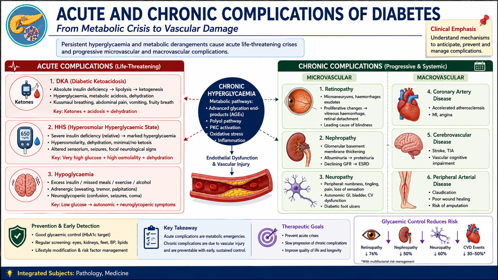
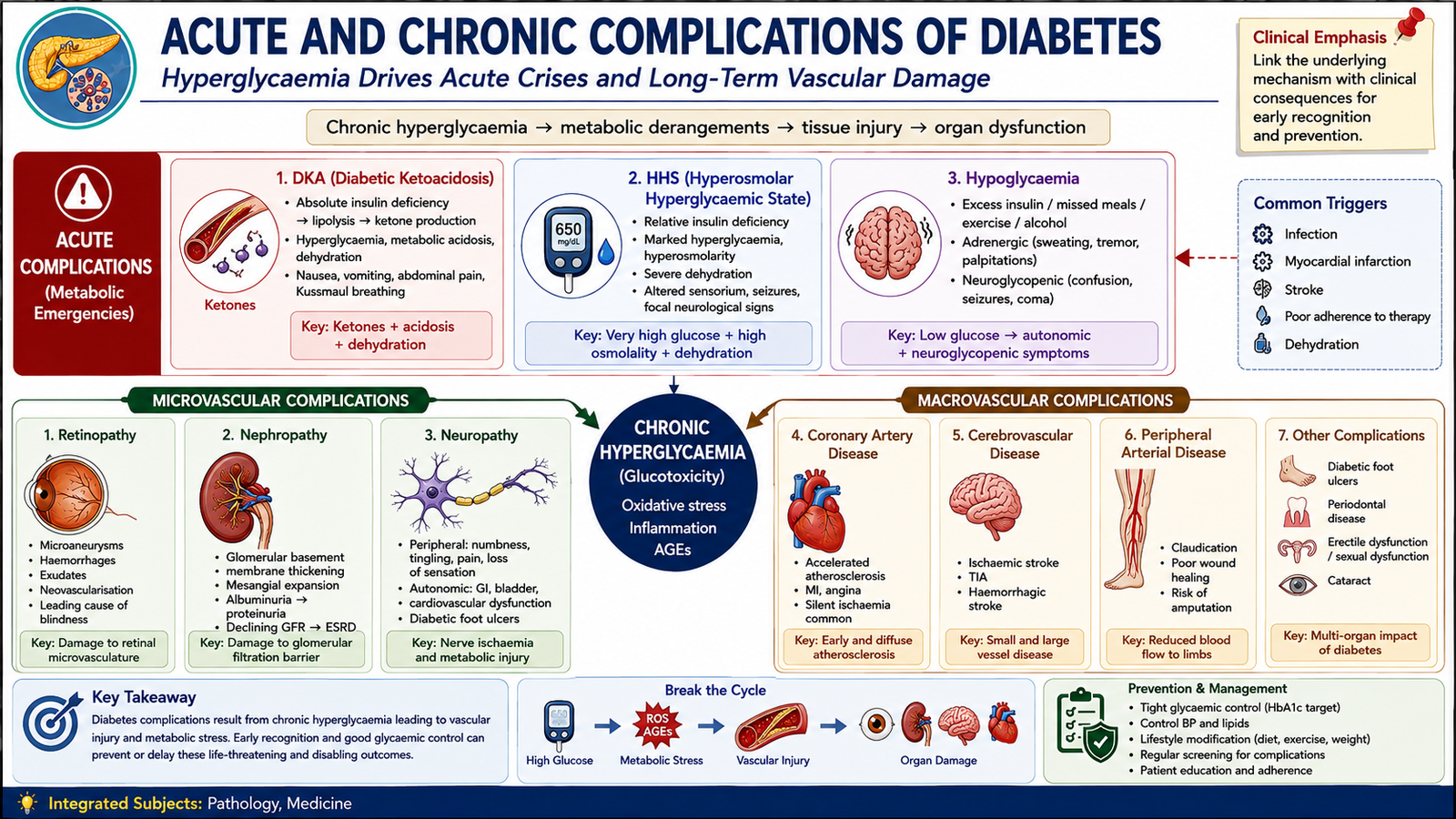
Diabetic complications arise through two broad mechanisms. Acute complications result from sudden severe disturbances of glucose and fuel metabolism. Chronic complications develop after prolonged metabolic and vascular injury. Understanding the mechanism explains why diabetes can affect organs that initially appear unrelated to pancreatic function.
Diabetic Ketoacidosis
Diabetic ketoacidosis, or DKA, occurs when insulin activity becomes severely deficient. Without adequate insulin, adipose tissue releases free fatty acids. The liver converts these fatty acids into ketone bodies. Accumulation of acidic ketones causes metabolic acidosis, while hyperglycaemia causes osmotic diuresis and severe water and electrolyte loss.
Severe hyperglycaemia → glycosuria → osmotic diuresis → dehydration and electrolyte depletion
Patients may develop polyuria, thirst, vomiting, abdominal discomfort, dehydration, deep rapid breathing, altered consciousness and the characteristic metabolic consequences of acidosis. DKA occurs most often in type 1 diabetes but can occur in other forms under severe metabolic stress. Management requires fluid replacement, insulin, careful electrolyte monitoring—especially potassium—and treatment of the precipitating illness.
Hyperosmolar Hyperglycaemic State
Hyperosmolar hyperglycaemic state, or HHS, usually develops in type 2 diabetes. There is enough insulin activity to limit major ketone formation but not enough to control glucose effectively. Extreme hyperglycaemia produces prolonged osmotic diuresis, profound dehydration and increased plasma osmolality. Neurological manifestations such as confusion, lethargy or reduced consciousness may become prominent. Treatment is based on careful fluid replacement, insulin, correction of electrolyte abnormalities and treatment of the precipitating cause.
Hypoglycaemia
Hypoglycaemia is usually a complication of diabetes treatment rather than the untreated disease itself. It is particularly associated with insulin and drugs that directly stimulate insulin secretion. Adrenergic symptoms include sweating, tremor and palpitations, while inadequate glucose supply to the brain causes confusion, behavioural change, seizures or loss of consciousness. An awake patient who can swallow requires prompt carbohydrate treatment; severe hypoglycaemia may require parenteral glucose or glucagon.
Pathological Basis of Chronic Complications
Persistent hyperglycaemia damages tissues through interacting biochemical pathways. Advanced glycation end-products form when glucose attaches non-enzymatically to proteins and lipids. These products alter extracellular matrix proteins and activate inflammatory and endothelial pathways. Hyperglycaemia also promotes oxidative stress, abnormal protein kinase C signalling and excessive intracellular glucose metabolism through pathways such as the polyol pathway. The combined result is endothelial dysfunction, basement-membrane abnormalities and impaired tissue perfusion.
Small vessels and capillaries develop basement-membrane thickening and functional injury.
- Retinopathy: capillary injury causes microaneurysms, leakage and retinal ischaemia; severe ischaemia stimulates pathological neovascularization.
- Nephropathy: glomerular basement-membrane thickening and mesangial expansion progress to glomerulosclerosis. Nodular glomerulosclerosis is a characteristic advanced lesion.
- Neuropathy: metabolic injury and damage to the small vessels supplying nerves contribute to sensory, motor and autonomic dysfunction.
Diabetes accelerates atherosclerosis through endothelial dysfunction and interaction with hypertension, dyslipidaemia and other cardiovascular risk factors.
- Coronary artery disease and myocardial infarction
- Cerebrovascular disease and stroke
- Peripheral arterial disease
Clinical Significance
Chronic complications may remain silent until substantial injury has occurred. This is why diabetes care includes active surveillance rather than waiting for symptoms. Neuropathy combined with peripheral arterial disease is particularly important in the diabetic foot: loss of protective sensation allows unnoticed trauma, while poor perfusion and impaired healing increase the risk of ulceration, infection and tissue loss.
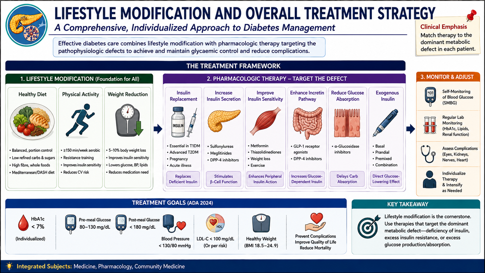
E. Lifestyle Modification and Overall Treatment Strategy
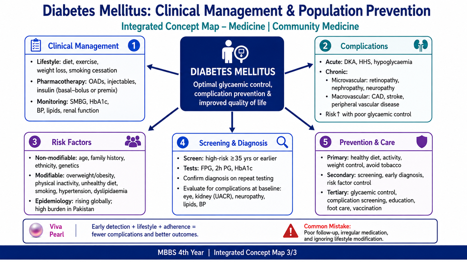
Diabetes treatment aims to correct abnormal glucose metabolism while reducing long-term vascular and functional complications. Management is therefore broader than prescribing a glucose-lowering drug. The treatment plan depends on diabetes type, degree of hyperglycaemia, risk of hypoglycaemia, body weight, associated disease, renal and hepatic function and the patient’s ability to use the treatment safely.
Lifestyle Modification
Lifestyle intervention is central to type 2 diabetes and insulin resistance. It also remains important in people treated with medicines or insulin because medication does not remove the effects of excess energy intake, physical inactivity or cardiovascular risk factors.
- Healthy eating: emphasize portion control, nutritionally balanced meals, vegetables, fibre-rich foods and reduction of excessive refined carbohydrates and energy-dense foods.
- Physical activity: improves skeletal-muscle glucose uptake and insulin sensitivity and supports cardiovascular health.
- Weight management: reducing excess body weight lowers insulin resistance and may substantially improve glucose control.
- Tobacco avoidance: is important because diabetes already increases vascular risk.
- Education and self-management: patients should understand symptoms of hyperglycaemia and hypoglycaemia, medication use and the importance of continued follow-up.
Pharmacological Approaches
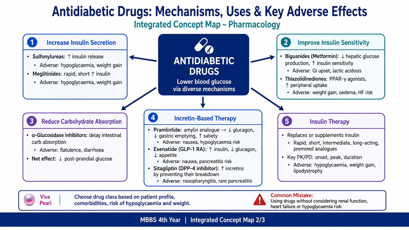
Drug therapy can be classified according to the physiological defect it targets.
- Replace deficient insulin: insulin preparations and insulin analogues.
- Stimulate pancreatic insulin release: sulfonylureas and meglitinides.
- Improve insulin sensitivity or reduce hepatic glucose production: metformin and thiazolidinediones.
- Slow carbohydrate digestion and absorption: α-glucosidase inhibitors.
- Enhance incretin pathways: GLP-1 receptor agonists such as exenatide and DPP-4 inhibitors such as sitagliptin.
- Increase urinary glucose excretion: sodium-glucose cotransporter-2 inhibitors.
- Modify postprandial physiology: the amylin analogue pramlintide.
Type 1 diabetes requires insulin replacement because endogenous insulin secretion is severely deficient. In type 2 diabetes, lifestyle measures and non-insulin drugs may provide adequate control for a period, but progressive β-cell failure means that some patients eventually require insulin. Insulin is also appropriate when marked hyperglycaemia, catabolic symptoms or acute metabolic decompensation requires rapid and reliable glucose control.
F. Insulin and Pancreatic-Hormone Pharmacology
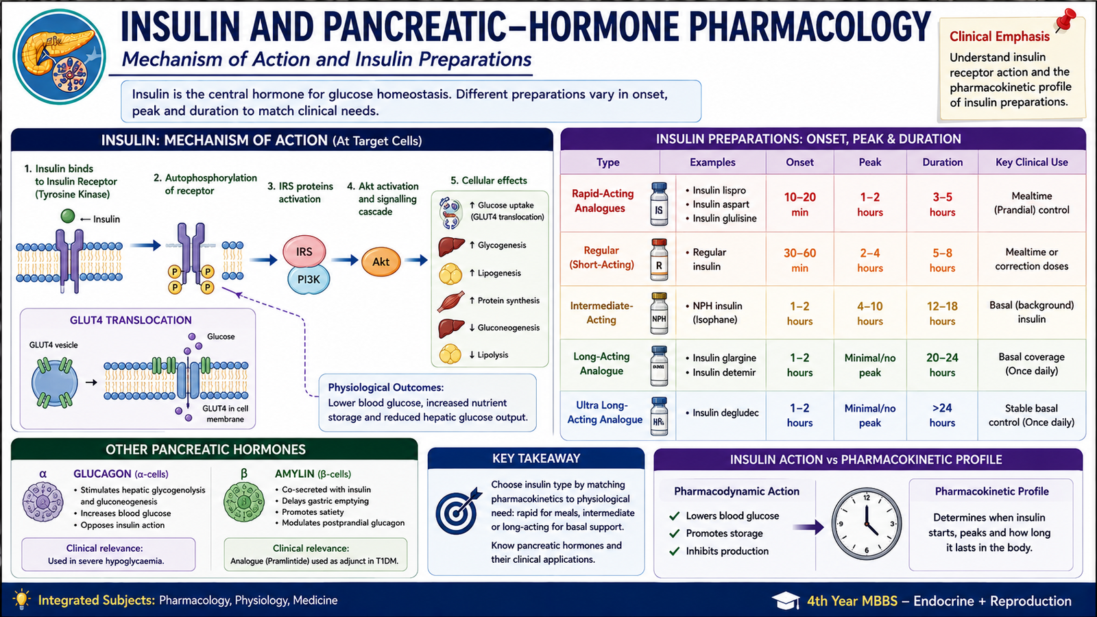
Insulin is a peptide hormone and cannot be given as an ordinary oral tablet because gastrointestinal enzymes would digest it. For chronic treatment it is generally delivered into subcutaneous tissue, from which it is absorbed into the circulation. The different insulin preparations act on the same insulin receptor; their major differences are pharmacokinetic—particularly how quickly they enter the blood and how long their effect lasts.
Pharmacodynamics of Insulin
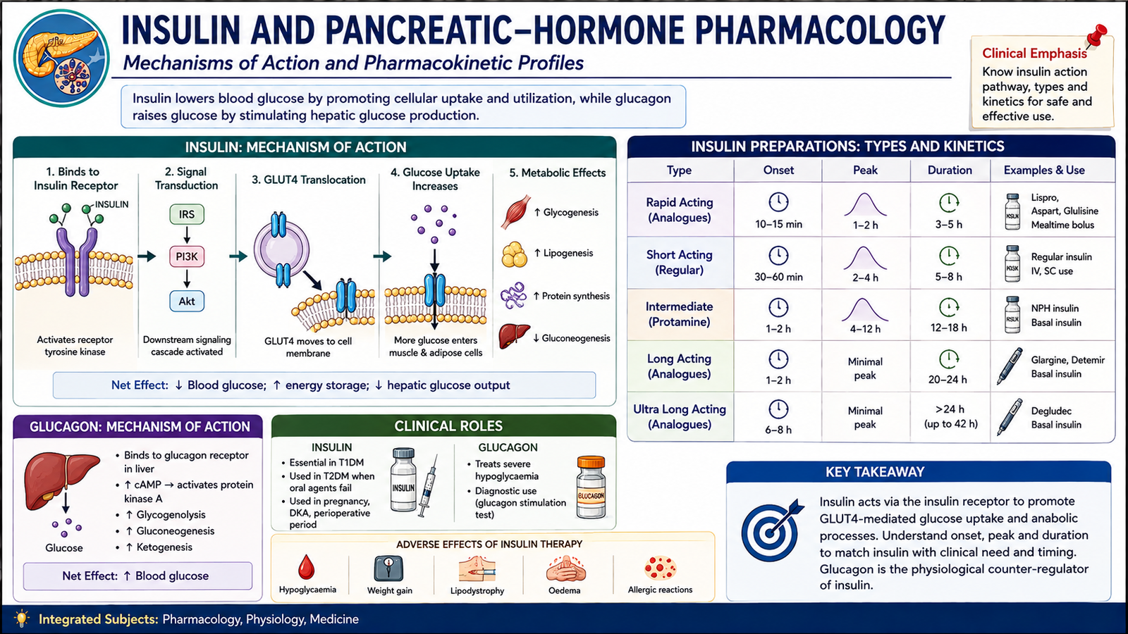
Insulin binds to a transmembrane receptor with intrinsic tyrosine-kinase activity. Receptor activation produces intracellular signalling that increases glucose transport into skeletal muscle and adipose tissue, promotes storage of nutrients and suppresses hepatic glucose production.
Insulin also → reduces hepatic glucose production, lipolysis and ketogenesis.
Major Insulin Preparations
| Group | Examples | Main Pharmacokinetic Principle | Clinical Role |
|---|---|---|---|
| Rapid acting | Lispro, aspart, glulisine | Rapid subcutaneous absorption | Mealtime glucose control |
| Short acting | Regular insulin | Slower subcutaneous absorption than rapid analogues; can also be given intravenously when required | Mealtime or acute-care use |
| Intermediate acting | NPH insulin | Protamine delays absorption | Basal insulin coverage |
| Long acting | Glargine, detemir, degludec | Molecular modifications prolong absorption or persistence | Sustained basal insulin effect |
Rapid-acting analogues are designed to reduce molecular self-association and therefore enter the circulation quickly after subcutaneous injection. Long-acting analogues use modifications that delay absorption or prolong persistence, producing a flatter basal effect. Insulin is metabolized mainly by the liver and kidneys, so reduced renal clearance may increase the risk of hypoglycaemia.
Adverse Effects of Insulin
- Hypoglycaemia: the most important adverse effect; excessive insulin activity lowers plasma glucose below the requirement of the brain and other tissues.
- Weight gain: improved glucose utilization and reduced urinary calorie loss favour energy storage.
- Lipohypertrophy at injection sites: repeated injections into the same area can stimulate local fat growth and make insulin absorption less predictable.
- Hypokalaemia: insulin promotes movement of potassium into cells and can lower extracellular potassium, especially during intensive treatment.
Glucagon as a Counter-Regulatory Hormone
Glucagon increases hepatic glucose output and opposes insulin during hypoglycaemia. Pharmacological glucagon can therefore be used in severe hypoglycaemia when a patient cannot safely take oral carbohydrate and appropriate parenteral rescue is required.
G. Oral Glucose-Lowering Drugs
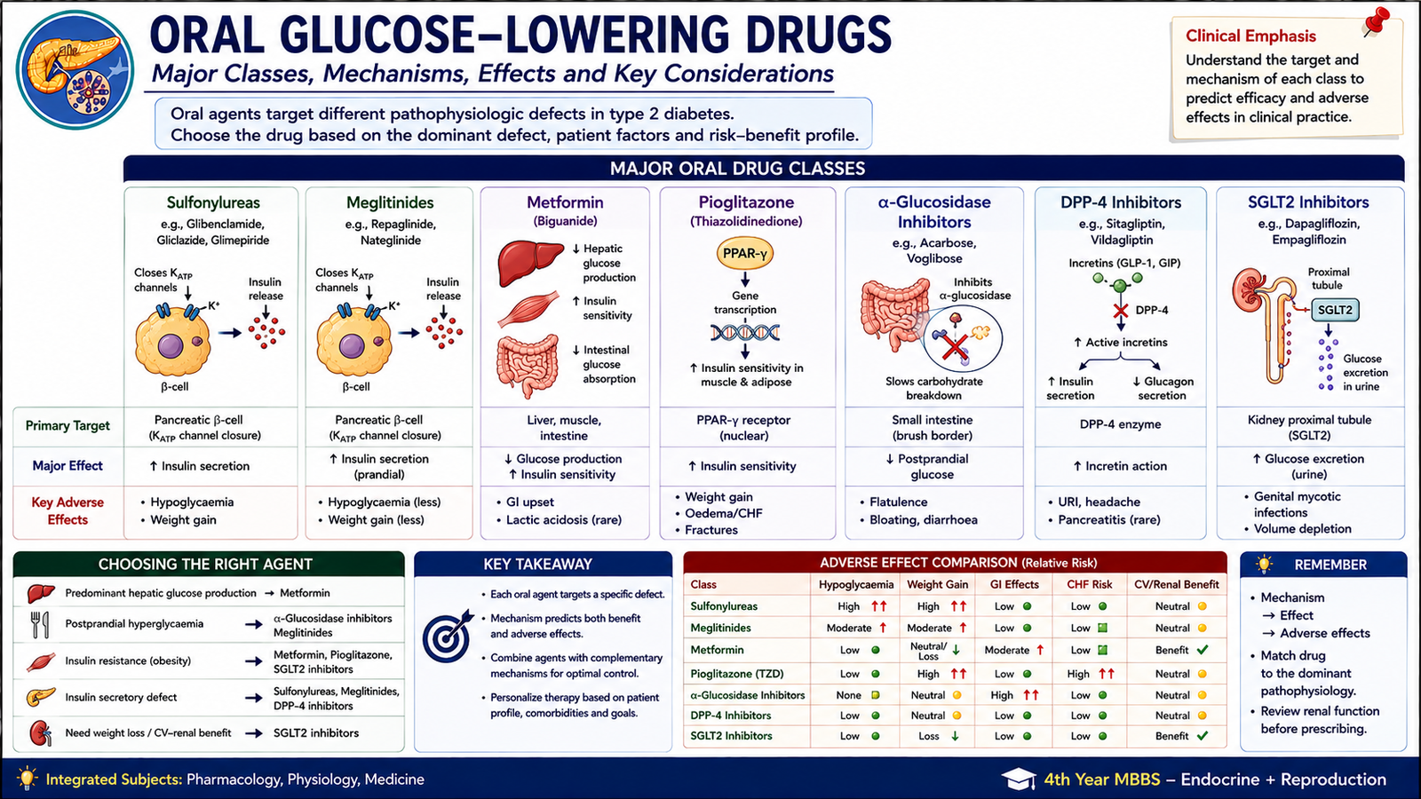
Oral glucose-lowering medicines act at different points in glucose regulation. Some increase insulin secretion, some improve insulin sensitivity, and others reduce the rate at which glucose enters the circulation. Their mechanisms explain both their therapeutic effects and their characteristic adverse effects.
Sulfonylureas
Sulfonylureas require functioning pancreatic β-cells. They bind to the sulfonylurea receptor component of ATP-sensitive potassium channels in the β-cell membrane.
Common examples include gliclazide, glimepiride and glibenclamide. They are useful in type 2 diabetes when endogenous β-cell function remains. Because insulin release is stimulated independently of immediate glucose need, hypoglycaemia is the major adverse effect. They can also cause weight gain. Prolonged hypoglycaemia is particularly concerning when drug clearance is reduced.
Meglitinides
Repaglinide and nateglinide also close β-cell ATP-sensitive potassium channels and stimulate insulin release. Their action is relatively rapid and short, so they are particularly suited to controlling postprandial glucose when taken in relation to meals. Hypoglycaemia and weight gain can occur, although their shorter action can make prolonged hypoglycaemia less likely than with longer-acting insulin secretagogues.
Biguanides: Metformin
Metformin is the major biguanide used in diabetes. It lowers glucose primarily by reducing hepatic glucose production and also improves insulin sensitivity in peripheral tissues. Activation of cellular energy-sensing pathways, including AMP-activated protein kinase, contributes to its metabolic effects.
Because metformin does not directly force pancreatic insulin release, it has a low risk of hypoglycaemia when used alone and usually does not promote weight gain. Its major adverse effects are gastrointestinal symptoms such as nausea, abdominal discomfort and diarrhoea. Long-term use may be associated with vitamin B12 deficiency. Lactic acidosis is rare but serious, so significant conditions that markedly impair drug elimination or produce severe tissue hypoxia require particular caution.
Thiazolidinediones
Thiazolidinediones, represented by pioglitazone, activate the nuclear receptor PPAR-γ. This alters transcription of genes involved in glucose and lipid metabolism and increases insulin sensitivity, especially in adipose tissue. Because gene transcription must change, the therapeutic effect develops more slowly than that of insulin secretagogues.
Important adverse effects include weight gain, fluid retention and oedema. Fluid retention can worsen heart failure, making this an important clinical caution. Increased fracture risk is another recognized concern.
α-Glucosidase Inhibitors
Acarbose and related drugs inhibit intestinal brush-border α-glucosidase enzymes that normally break complex carbohydrates into absorbable monosaccharides. Carbohydrate digestion is therefore delayed, reducing the rapid rise in glucose after a meal.
The unabsorbed carbohydrate continues into the distal intestine, where bacterial fermentation produces flatulence, abdominal discomfort and diarrhoea. These drugs do not usually cause hypoglycaemia when used alone because they do not increase insulin secretion.
| Class | Main Target | Main Effect | Important Adverse Effect |
|---|---|---|---|
| Sulfonylureas | β-cell KATP channel | Increase insulin secretion | Hypoglycaemia, weight gain |
| Meglitinides | β-cell KATP channel | Short mealtime insulin release | Hypoglycaemia |
| Metformin | Hepatic metabolism and insulin sensitivity | Reduces hepatic glucose production | GI effects; rare lactic acidosis |
| Thiazolidinediones | PPAR-γ | Increase insulin sensitivity | Oedema, weight gain |
| α-Glucosidase inhibitors | Intestinal carbohydrate digestion | Reduce postprandial glucose rise | Flatulence, diarrhoea |
H. Incretin-Based Therapy, Pramlintide and Euglycaemic Drugs
After a meal, intestinal hormones amplify glucose-dependent insulin release. This is called the incretin effect. Two important incretin hormones are GLP-1 and GIP. The pharmacological value of this system is that insulin secretion rises mainly when glucose is elevated, so therapies acting through the incretin pathway have relatively little tendency to produce hypoglycaemia when used without insulin or insulin secretagogues.
Exenatide
Exenatide is a GLP-1 receptor agonist. It mimics important actions of endogenous GLP-1.
It is used in type 2 diabetes and can improve postprandial and overall glycaemic control. Reduced appetite and delayed gastric emptying can support weight reduction. Gastrointestinal adverse effects, particularly nausea, are common early in treatment.
Sitagliptin
Sitagliptin inhibits dipeptidyl peptidase-4 (DPP-4), the enzyme that normally degrades endogenous incretin hormones.
Sitagliptin is an oral treatment for type 2 diabetes. It is generally weight neutral and has a low risk of hypoglycaemia when used without a drug that independently increases insulin activity.
Pramlintide
Amylin is normally secreted together with insulin by pancreatic β-cells. Pramlintide is an amylin analogue given by subcutaneous injection. It slows gastric emptying, suppresses inappropriate postprandial glucagon release and increases satiety. These actions reduce the rapid rise in blood glucose after meals.
Pramlintide can be used as an adjunct to mealtime insulin in selected patients with type 1 or type 2 diabetes. Nausea is common, and because it is used together with insulin, clinically important hypoglycaemia can occur unless insulin therapy and meal intake are managed appropriately.
What Are Euglycaemic Drugs?
The term euglycaemic drugs is commonly used in undergraduate pharmacology for glucose-lowering agents that do not directly force insulin release and therefore have a low tendency to produce hypoglycaemia when used alone. They lower abnormally high glucose while usually allowing normal counter-regulatory mechanisms to protect against an excessive fall.
Important examples include:
- Metformin
- Thiazolidinediones
- α-Glucosidase inhibitors
- DPP-4 inhibitors such as sitagliptin
- GLP-1 receptor agonists such as exenatide
- SGLT2 inhibitors
By contrast, insulin, sulfonylureas and meglitinides can directly produce hypoglycaemia because they increase insulin activity even when blood glucose is falling.

I. Epidemiology, Risk Factors, Screening and Prevention
Diabetes is a major non-communicable disease worldwide, and its prevalence has increased markedly over recent decades. Type 2 diabetes accounts for most of this burden. Pakistan also carries a particularly high and increasing burden of diabetes, including a substantial number of people whose disease has not yet been diagnosed. Exact prevalence estimates vary with the population studied, diagnostic method and year of assessment, so the epidemiological principle is more important than memorizing a changing isolated figure.
Epidemiological Determinants
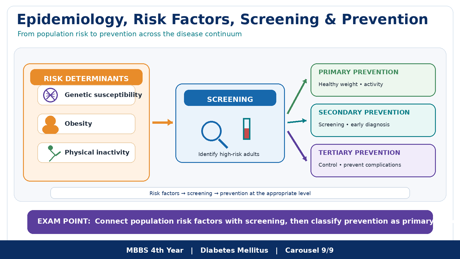
The population burden of type 2 diabetes reflects interaction between genetic susceptibility and environmental exposure. Urbanization, reduced physical activity, increasing obesity, calorie-dense diets and population ageing increase the opportunity for genetically susceptible individuals to develop insulin resistance and β-cell failure. Diabetes therefore represents both an individual clinical problem and a population-health problem.
Risk Factors
- Increasing age
- Family history and genetic susceptibility
- Previous gestational diabetes
- Population or ethnic susceptibility
- Overweight and central obesity
- Physical inactivity
- Unhealthy dietary pattern and excessive energy intake
- Tobacco use
- Associated hypertension and dyslipidaemia
Risk factors often cluster. Central obesity, hypertension, dyslipidaemia and insulin resistance may therefore occur in the same person and multiply cardiovascular risk rather than acting as completely separate problems.
Screening
Screening aims to detect diabetes in an apparently healthy person before symptoms or complications make the diagnosis obvious. This is important because type 2 diabetes may remain asymptomatic for a prolonged period.
Common screening methods include:
- Fasting plasma glucose
- HbA1c
- Oral glucose tolerance testing when clinically appropriate
- Risk assessment to identify people in whom biochemical screening is especially important
Urine glucose is not an adequate diagnostic screening test because renal glucose handling varies between individuals. Screening is most useful when people with abnormal results can receive confirmatory assessment, counselling, treatment and continuing follow-up.
Levels of Prevention
| Level | Aim | Application to Diabetes |
|---|---|---|
| Primary prevention | Prevent or delay development of disease | Healthy diet, physical activity, prevention or reduction of obesity, tobacco avoidance and risk-factor education. |
| Secondary prevention | Detect disease early and prevent progression | Screening, early diagnosis, appropriate glucose management and surveillance for retinal, renal, neural and vascular complications. |
| Tertiary prevention | Limit disability from established complications | Treatment of diabetic foot disease, renal and eye complications, cardiovascular disease, rehabilitation and measures that preserve function and independence. |
Effective diabetes care therefore spans the entire disease course: prevention in high-risk populations, early detection of asymptomatic disease, continued clinical management and active prevention of disability in patients who already have complications.
Integrated Mechanism Flow
Important Comparison — Type 1 versus Type 2 Diabetes Mellitus
| Feature | Type 1 Diabetes | Type 2 Diabetes |
|---|---|---|
| Dominant mechanism | β-cell destruction → absolute insulin deficiency | Insulin resistance + progressive β-cell dysfunction |
| Typical onset | Often relatively rapid | Usually gradual |
| Insulin resistance | Not the principal primary defect | Major early abnormality |
| Endogenous insulin | Markedly deficient | May initially be high, then falls as β-cells fail |
| Autoimmunity | Common in immune-mediated disease | Not the usual mechanism |
| Ketosis tendency | High if insulin is absent | Lower in usual stable disease, but not impossible |
| Essential treatment principle | Insulin replacement is required | Lifestyle + individualized non-insulin drugs and/or insulin |