
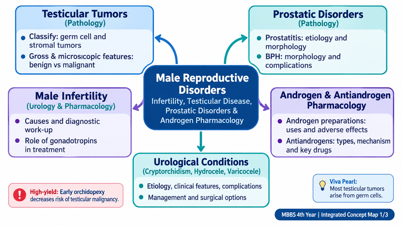
This chapter follows the KMU learning outcomes in a logical sequence. First understand how the reproductive disorders, pathological changes and drug mechanisms connect; then use the final AIM High-Yield Review for rapid revision.
Topic 14 — Male Reproductive Disorders: Infertility, Testicular Disease, Prostatic Disorders and Androgen Pharmacology
An integrated approach to male infertility, scrotal and testicular disorders, prostatic disease, testicular tumors and the pharmacology of androgens, gonadotropins and antiandrogen therapy.
Topic Introduction
Male reproductive disorders range from problems of testicular descent and sperm production to scrotal swellings, testicular tumors and diseases of the prostate. Understanding them requires a clear connection between structure, function, pathology and treatment. In this chapter, you will learn how male infertility is investigated, why cryptorchidism and varicocele can affect fertility, how common testicular tumors are classified and recognized, and how prostatitis, benign prostatic hyperplasia and prostate carcinoma differ. The final part explains androgen pharmacology, gonadotropin therapy, antiandrogens and the pharmacological approaches studied for male contraception. The aim is to understand the reasoning behind the findings rather than memorize isolated facts.
A. Male Infertility: Causes and Diagnostic Work-up
Male infertility means inability of the male partner to contribute successfully to conception. Infertility is traditionally evaluated when a couple has been unable to achieve pregnancy despite regular unprotected intercourse for about one year. Because fertility depends on both partners, assessment should not focus on the man in isolation. In the male, normal fertility requires an intact hypothalamic–pituitary–testicular axis, normally functioning testes, patent reproductive ducts, adequate sexual function and successful deposition of semen.
How normal spermatogenesis is controlled
The hypothalamus releases gonadotropin-releasing hormone, which stimulates pituitary secretion of LH and FSH. LH acts mainly on Leydig cells and stimulates testosterone production. FSH acts on Sertoli cells and supports spermatogenesis. High intratesticular testosterone is also essential for normal sperm production.
Hypothalamic GnRH → pituitary LH + FSH → Leydig cell testosterone + Sertoli cell support → spermatogenesis.
Causes of male infertility
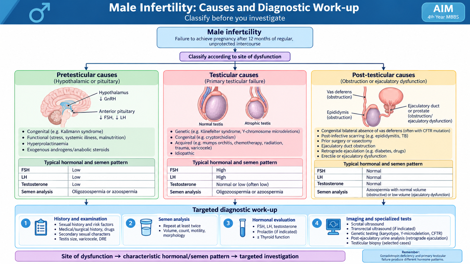
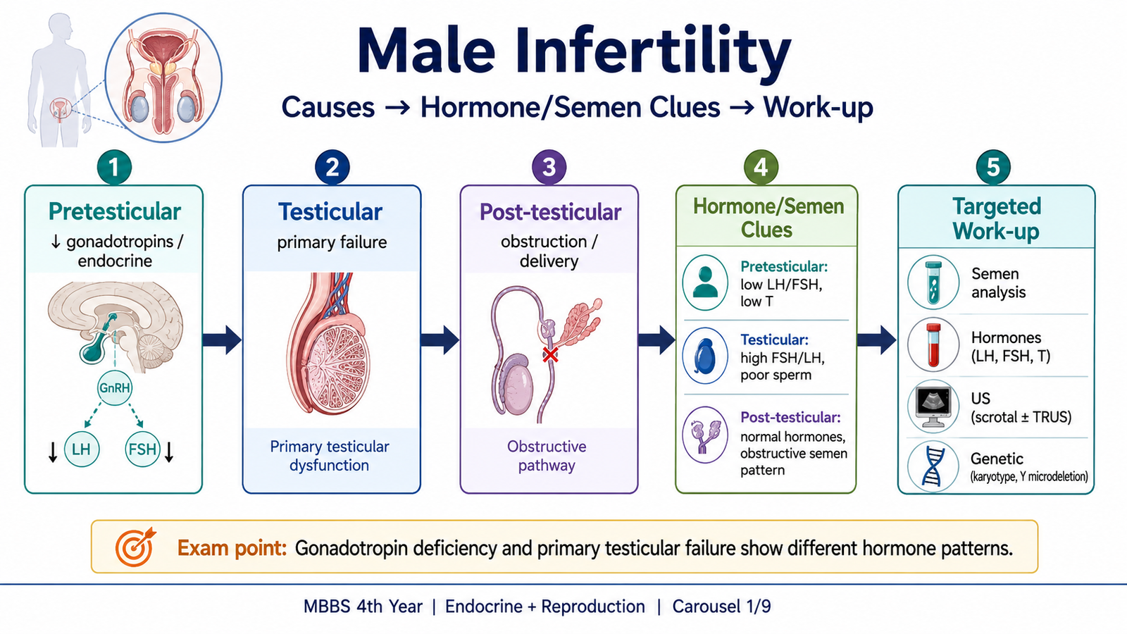
A useful way to understand the causes is to ask whether the problem occurs before the testis, within the testis or after sperm have been produced.
- Pretesticular causes: hypothalamic or pituitary disorders causing deficient gonadotropin stimulation, endocrine disturbances and some systemic illnesses.
- Testicular causes: primary testicular failure, cryptorchidism, varicocele, previous severe testicular inflammation, testicular trauma, genetic disorders, cytotoxic treatment and other causes that damage spermatogenesis.
- Post-testicular causes: obstruction of the epididymis, vas deferens or ejaculatory ducts, congenital absence of reproductive ducts, and ejaculatory disorders.
- Sexual or functional causes: erectile dysfunction, failure of ejaculation or retrograde ejaculation.
- Drug and exposure-related causes: exogenous androgens or anabolic steroids can suppress LH and FSH and thereby suppress spermatogenesis.
An important examination concept is that infertility does not necessarily mean androgen deficiency. A man may have apparently normal virilization and sexual function but still have severely impaired spermatogenesis.
Clinical assessment
The diagnostic work-up begins with a careful history. The aim is to identify congenital, endocrine, testicular, obstructive, sexual or treatment-related factors before ordering specialized tests.
- Duration of infertility and whether any previous conception has occurred.
- Frequency of intercourse and sexual or ejaculatory problems.
- History of undescended testis, testicular surgery, trauma or infection.
- Previous chemotherapy, radiotherapy or drugs that may interfere with reproductive function.
- Use of testosterone or anabolic steroids.
- Features suggesting pituitary or other endocrine disease.
Physical examination assesses secondary sexual characteristics, testicular size and consistency, epididymis and vas deferens, and the presence of a varicocele or other scrotal abnormality. Reduced testicular volume may indicate impaired seminiferous tubular function, while absent vasa deferentia suggest an obstructive congenital disorder.
Semen analysis
Semen analysis is the basic laboratory investigation of male infertility. It evaluates the ejaculate rather than diagnosing a specific disease by itself. Important features include semen volume, sperm concentration, motility and morphology. Because semen parameters vary biologically, an abnormal result is usually confirmed rather than interpreted from a single isolated specimen.
Important descriptive abnormalities include:
- Azoospermia: no sperm detected in the ejaculate.
- Oligozoospermia: reduced sperm concentration.
- Asthenozoospermia: reduced sperm motility.
- Teratozoospermia: increased proportion of morphologically abnormal sperm.
Hormonal and further investigations
Hormonal testing is interpreted according to the clinical picture. FSH, LH and testosterone help distinguish central gonadotropin deficiency from primary testicular failure.
- Low testosterone with low or inappropriately normal LH/FSH suggests hypothalamic or pituitary dysfunction.
- Impaired testicular function with raised gonadotropins suggests primary testicular failure because the normal negative feedback is lost.
- Prolactin is investigated when the history suggests a relevant pituitary or gonadal-axis disturbance.
- Ultrasonography may identify varicocele, testicular pathology or other scrotal abnormalities when indicated.
- Genetic evaluation may be required in selected men with severe sperm-production failure or congenital reproductive-tract abnormalities.
- Further tests for obstruction or retrograde ejaculation are selected according to the clinical findings.
Management principle
Treatment is directed at the identified cause. A surgically correctable obstruction or clinically important varicocele requires urological assessment. Gonadotropin treatment is useful when infertility results from deficient pituitary stimulation, but it cannot restore sperm production when the seminiferous tissue itself has irreversibly failed. Exogenous testosterone should not be used as a treatment for a man actively seeking fertility because it suppresses pituitary gonadotropins and may further reduce sperm production.
B. Cryptorchidism
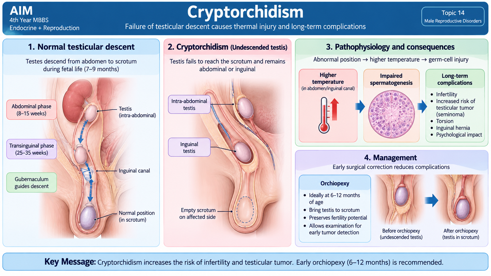
Cryptorchidism is failure of one or both testes to descend completely into the scrotum. A testis may remain in the abdomen, inguinal canal or another point along its normal route of descent. This is important because the scrotum provides a temperature lower than core body temperature, which is necessary for optimal spermatogenesis.
Etiology and core mechanism
Normal testicular descent depends on several interacting factors, including normal fetal development, hormonal signaling and anatomical guidance. Disturbance of these processes may result in an undescended testis. Prematurity and developmental abnormalities may be associated with failure of complete descent.
Clinical features
The main finding is an absent testis from the corresponding side of the scrotum. The testis may be palpable in the inguinal region or may be non-palpable if it lies within the abdomen. Examination should therefore determine whether the testis is palpable and whether the opposite testis is normal.
Complications
- Subfertility or infertility: prolonged abnormal temperature damages germ cells and impairs spermatogenesis.
- Increased risk of testicular germ-cell tumor: the association remains clinically important even after the testis is brought into the scrotum.
- Testicular torsion: abnormal fixation and position may predispose to twisting.
- Associated inguinal hernia: persistence of the processus vaginalis may accompany cryptorchidism.
- Trauma: an ectopic or inguinal testis may be more exposed to injury.
Management
Persistent cryptorchidism requires surgical assessment. Orchiopexy brings the testis into the scrotum and fixes it there. Performing correction early in childhood reduces prolonged thermal injury and also makes later examination of the testis easier. In a post-pubertal patient with a severely atrophic intra-abdominal testis, removal may be considered after specialist assessment because fertility potential may be poor and malignancy risk remains relevant.
Orchiopexy does not completely abolish the increased malignancy risk. Long-term awareness and clinical examination therefore remain important.
C. Hydrocele and Varicocele
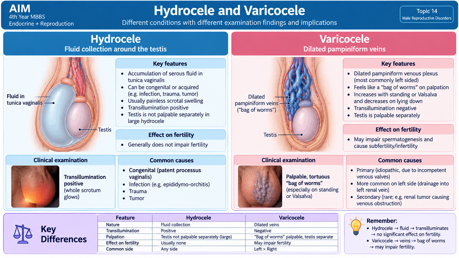
Hydrocele and varicocele are common causes of scrotal swelling, but they arise from very different mechanisms. A hydrocele is a collection of serous fluid around the testis, while a varicocele is abnormal dilatation and tortuosity of the veins of the pampiniform plexus. Distinguishing them clinically is important because varicocele may affect fertility whereas hydrocele mainly causes swelling and discomfort unless it is secondary to another disease.
Hydrocele
Fluid normally exists in a small amount between the layers of the tunica vaginalis. Hydrocele develops when fluid accumulates excessively because of abnormal communication, increased production or reduced absorption.
Causes
- Congenital hydrocele: related to persistence of the processus vaginalis.
- Primary adult hydrocele: no obvious underlying testicular disease is identified.
- Secondary hydrocele: may occur with testicular or epididymal inflammation, trauma or an underlying testicular lesion.
Clinical features
A hydrocele usually presents as a painless or mildly uncomfortable scrotal swelling. The swelling is typically cystic, and the testis may be difficult to palpate separately when a large amount of fluid surrounds it. Clear hydrocele fluid usually permits transillumination, producing a characteristic glow when a light source is placed behind the swelling.
Transillumination supports the diagnosis but does not replace proper examination. A secondary cause must be considered when the onset or examination is atypical.
Management
Small uncomplicated hydroceles may be observed when they cause no important symptoms. Persistent, large or symptomatic hydroceles are treated surgically. Hydrocelectomy deals with the hydrocele sac by techniques based on excision, eversion or plication. The operative details are less important at this level than the principle that persistent symptomatic fluid collection is treated definitively and that any underlying testicular disorder must not be missed.
Varicocele
A varicocele is caused by venous dilatation within the pampiniform plexus. It occurs more commonly on the left side because of differences in venous drainage and pressure. Failure of effective venous drainage allows blood to pool around the testis.
Clinical features
- Dragging scrotal discomfort or heaviness.
- A soft, compressible mass classically described as a “bag of worms.”
- The swelling becomes more obvious when standing or during Valsalva.
- The affected testis may become smaller in longstanding disease.
- Some patients present during investigation of infertility.
Why can varicocele cause infertility?
The pampiniform plexus normally helps cool arterial blood entering the testis. Venous dilatation interferes with this heat-exchange function and may increase testicular temperature. Venous stasis and altered local environment can then impair spermatogenesis.
Management
Asymptomatic varicoceles without testicular or fertility consequences may be observed. Intervention may be considered when there is persistent pain, impaired testicular growth or fertility-related evidence of testicular dysfunction. Treatment interrupts abnormal venous reflux by surgical ligation or radiological embolization. A newly appearing unusual right-sided or non-reducing varicocele deserves assessment for a secondary cause rather than being assumed to be an ordinary primary varicocele.
D. Testicular Tumors: Classification, Morphology, Staging and Prognosis
Most clinically important testicular neoplasms arise from germ cells. Testicular tumors are particularly important because they often affect younger men and many malignant germ-cell tumors are highly curable when appropriately treated. A painless enlarging testicular mass should therefore be taken seriously and evaluated promptly.
Classification
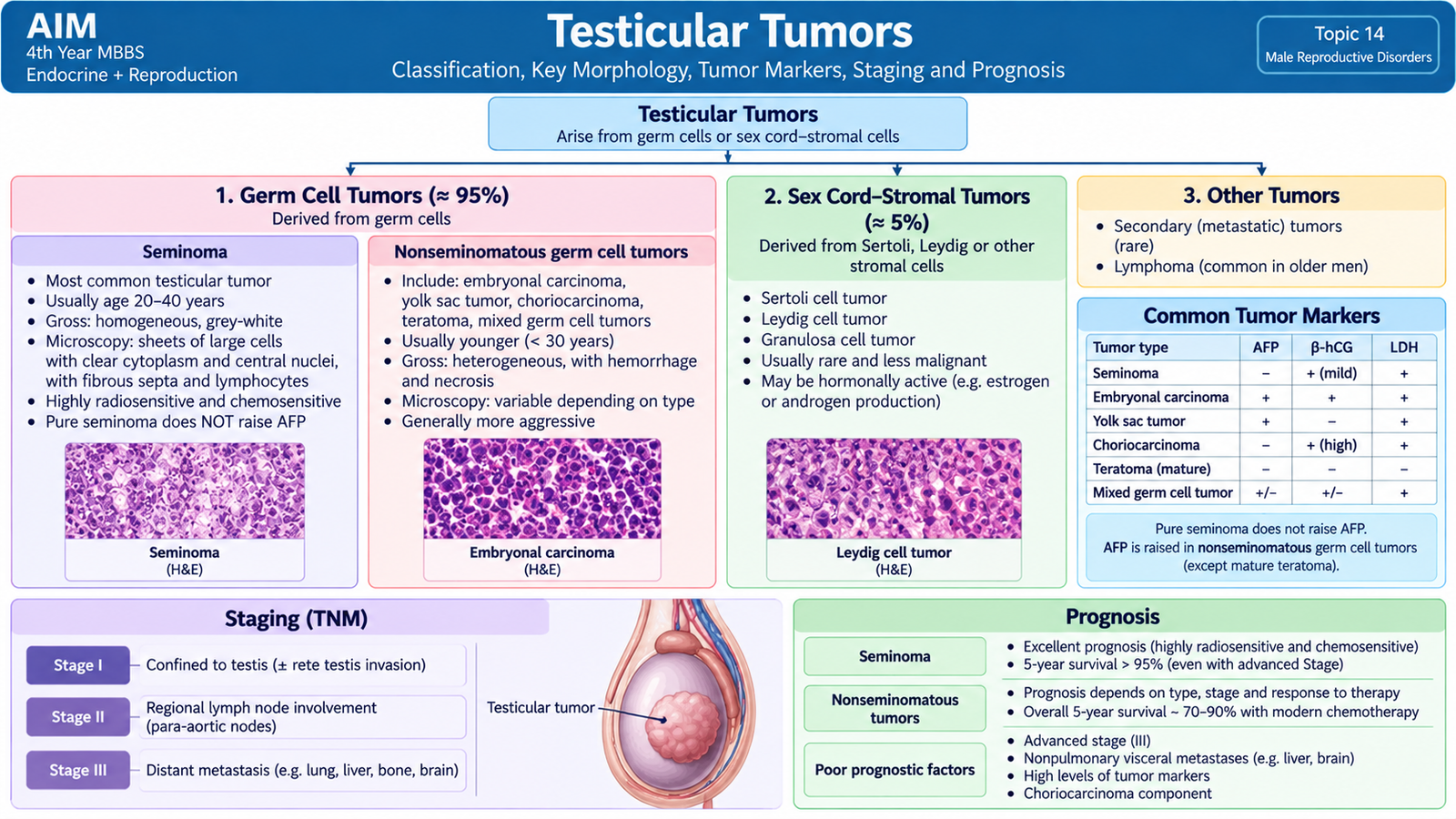
Testicular tumors can be organized into two major groups according to the cell of origin.
| Major Group | Important Types | Key Point |
|---|---|---|
| Germ-cell tumors | Seminoma, embryonal carcinoma, yolk-sac tumor, choriocarcinoma, teratoma and mixed germ-cell tumors | Most important malignant testicular neoplasms |
| Sex cord–stromal tumors | Leydig-cell tumor and Sertoli-cell tumor | Often hormonally active and frequently benign |
Among adult germ-cell tumors, seminoma is considered separately from the nonseminomatous germ-cell tumors because their biological behavior and therapeutic approach differ.
Seminoma
Seminoma is composed of malignant germ cells that retain a relatively uniform appearance. It commonly forms a well-defined testicular mass and tends to spread first through lymphatic channels.
Gross morphology
- Usually a homogeneous, pale grey-white mass.
- Often lobulated and bulging on cut surface.
- Extensive hemorrhage and necrosis are usually less prominent than in many nonseminomatous tumors.
Microscopic morphology
The tumor is made of relatively uniform large cells arranged in sheets or lobules. The cells typically have clear or pale cytoplasm, distinct cell borders and centrally placed nuclei. Fibrous septa often divide the tumor into lobules and may contain lymphocytes.
Pure seminoma does not produce alpha-fetoprotein. A minority contain syncytiotrophoblastic cells that can produce β-hCG.
Embryonal carcinoma
Embryonal carcinoma is a more aggressive nonseminomatous germ-cell tumor. It often invades beyond the testicular parenchyma and may occur as a component of a mixed germ-cell tumor.
Morphology
Grossly, it is typically a poorly circumscribed heterogeneous mass with areas of hemorrhage and necrosis. Microscopically, primitive anaplastic epithelial-like cells form solid, glandular or papillary patterns. The cells show greater pleomorphism and less uniformity than seminoma.
Yolk-sac tumor
Yolk-sac tumor shows differentiation resembling extraembryonic yolk-sac structures and characteristically produces alpha-fetoprotein.
Microscopically, different patterns may occur, but the classic examination finding is the Schiller-Duval body, in which tumor cells surround a central blood vessel within a space resembling a primitive glomerulus.
Choriocarcinoma
Choriocarcinoma is an aggressive germ-cell tumor with trophoblastic differentiation. It is often small in the testis despite widespread metastatic disease because it spreads early through the bloodstream.
Grossly, hemorrhage and necrosis are prominent. Microscopically, both cytotrophoblasts and syncytiotrophoblasts are present. Syncytiotrophoblasts produce β-hCG, which may be markedly elevated.
Teratoma and mixed germ-cell tumors
Teratoma contains tissues derived from more than one embryonic germ layer. In post-pubertal males, a testicular teratoma is regarded as having malignant potential even when the tissues appear mature microscopically. Many adult testicular germ-cell tumors contain more than one histological component and are therefore classified as mixed germ-cell tumors.
Leydig-cell and Sertoli-cell tumors
Leydig-cell tumors arise from androgen-producing interstitial cells. They are usually well-circumscribed, yellow-brown lesions and are frequently benign. They may produce androgens or, through altered steroid metabolism, estrogenic manifestations. Microscopically, the cells resemble normal Leydig cells, with eosinophilic granular cytoplasm; characteristic crystalline structures called Reinke crystals may be seen.
Sertoli-cell tumors arise from supporting cells of the seminiferous tubules. They are less common and are usually benign. Microscopically they may form cord-like or tubular structures.
Clinical presentation and diagnosis
The typical presentation is a painless testicular mass or enlargement. Some patients describe heaviness or discomfort. A solid intratesticular lesion should be considered potentially malignant until proven otherwise.
Ultrasonography establishes whether a mass is intratesticular and helps characterize it. Serum tumor markers such as AFP, β-hCG and LDH contribute to diagnosis, staging, prognosis and follow-up. Histological diagnosis is obtained after appropriate urological surgical management rather than routine trans-scrotal biopsy, because the normal lymphatic drainage pattern should not be disrupted.
Pattern of spread
Testicular lymphatics drain toward retroperitoneal para-aortic lymph nodes. Therefore, malignant germ-cell tumors commonly spread first to retroperitoneal nodes rather than superficial inguinal nodes. Choriocarcinoma is an important exception in behavior because early hematogenous dissemination is particularly characteristic.
Staging
Formal staging uses the TNM system together with relevant serum tumor-marker information. For undergraduate understanding, the clinical stages can be remembered in a simpler form:
- Stage I: disease limited to the testis and closely related local structures.
- Stage II: spread to regional retroperitoneal lymph nodes.
- Stage III: distant metastatic disease or spread beyond the regional nodal field.
Prognosis
Stage, tumor type and serum-marker burden are major prognostic factors. Seminoma generally has an excellent prognosis and is highly sensitive to treatment. Nonseminomatous germ-cell tumors tend to behave more aggressively, but many remain highly curable with modern combined treatment. Earlier-stage disease has a better prognosis than widely metastatic disease.
E. Prostatitis: Etiology and Morphology
Prostatitis refers to inflammatory disorders involving the prostate. Some forms are caused by bacterial infection, whereas others produce chronic pelvic symptoms without demonstrable bacterial infection. The pathological appearance depends on whether inflammation is acute or chronic.
Major clinical-pathological forms
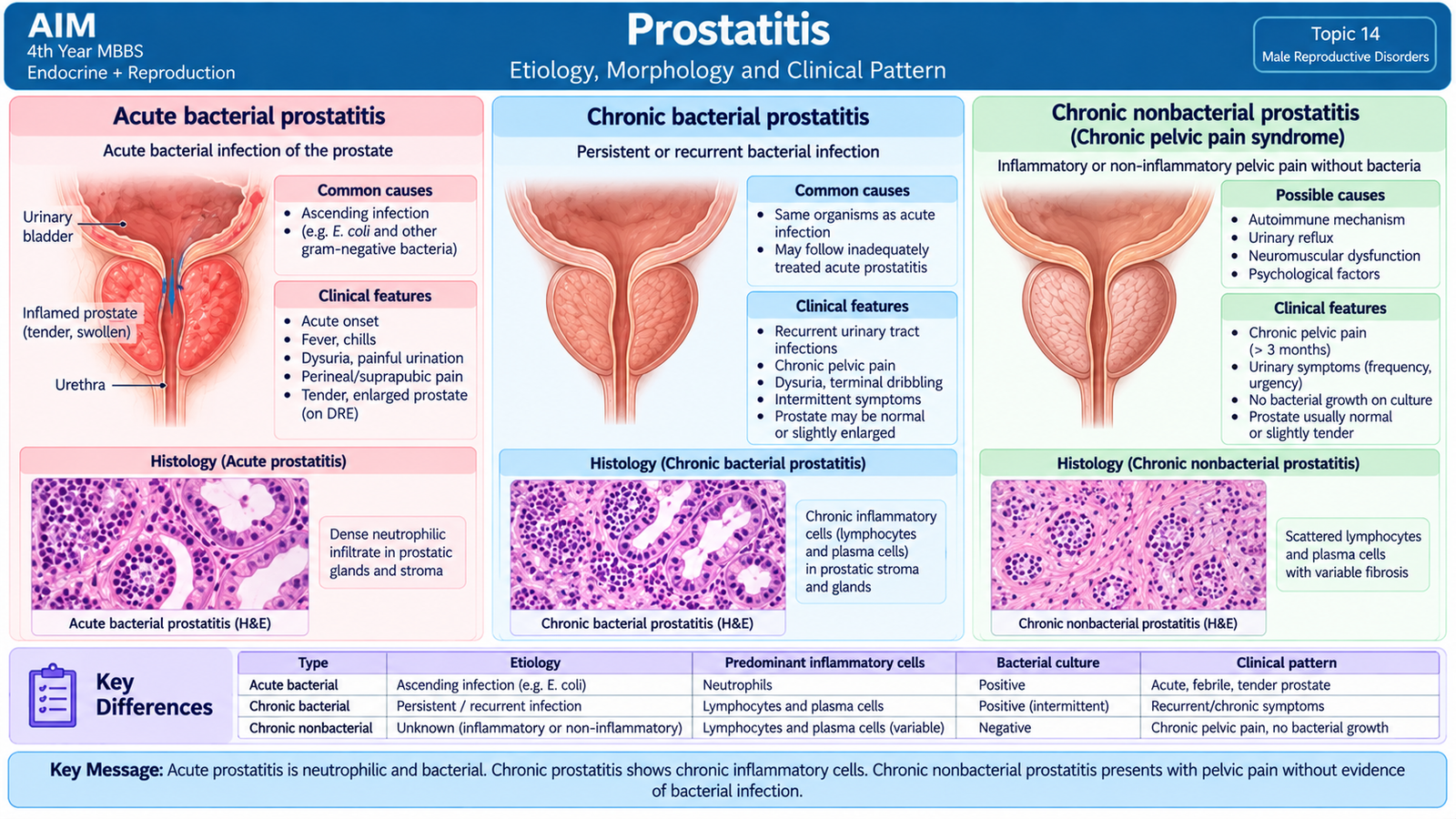
- Acute bacterial prostatitis
- Chronic bacterial prostatitis
- Chronic prostatitis/chronic pelvic pain syndrome
- Asymptomatic inflammatory prostatitis
Acute bacterial prostatitis
Acute prostatitis usually results from bacterial infection reaching the prostate from the urinary tract or through prostatic ducts. Enteric gram-negative organisms are common causes.
Acute infection triggers a strong neutrophilic inflammatory response. Neutrophils enter prostatic glands and stroma, producing edema and, in more severe disease, small abscesses.
Patients may have fever, urinary symptoms, pelvic discomfort and a tender swollen prostate. Vigorous prostatic massage is avoided in acute bacterial prostatitis because manipulation of an acutely infected gland may worsen bacteremia.
Chronic bacterial prostatitis
Chronic bacterial prostatitis is a persistent or recurrent bacterial infection of the prostate. Patients may have recurrent urinary infection with pelvic discomfort, although symptoms can be less dramatic than in acute disease.
Microscopically, chronic inflammatory cells, particularly lymphocytes and plasma cells, infiltrate the gland and stroma. Chronic inflammation may produce fibrosis and structural distortion.
Chronic pelvic pain syndrome
This group produces persistent pelvic, perineal or genitourinary discomfort but does not behave like a conventional recurrent bacterial infection. Inflammatory cells may be present in some cases and absent in others. Its importance is that chronic pelvic symptoms do not automatically prove bacterial prostatitis.
Morphological distinction
| Form | Dominant Morphology | Clinical Pattern |
|---|---|---|
| Acute bacterial | Neutrophils, edema, possible microabscesses | Acute systemic and urinary symptoms |
| Chronic bacterial | Lymphocytes, plasma cells and possible fibrosis | Recurrent infection or persistent symptoms |
F. Benign Prostatic Hyperplasia
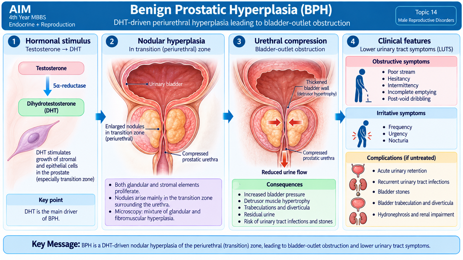
Benign prostatic hyperplasia (BPH) is a non-neoplastic increase in the number of epithelial and stromal cells of the prostate. It develops mainly in the transition and periurethral regions. Although the process is benign, the location is clinically important because expanding nodules can compress the prostatic urethra and obstruct urinary flow.
Etiology and hormonal basis
BPH is strongly related to aging and continued androgen action. Within prostatic stromal cells, testosterone is converted by 5-alpha reductase to dihydrotestosterone (DHT). DHT binds the androgen receptor with high affinity and promotes signals that support stromal and glandular growth.
Gross morphology
The prostate becomes enlarged and contains multiple well-circumscribed nodules, particularly around the urethra. Some nodules are predominantly glandular and appear softer and yellow-pink, whereas stromal nodules are firmer and paler. The nodules may compress the urethral lumen into a narrow slit.
Microscopic morphology
Microscopy shows hyperplasia of both glands and fibromuscular stroma. The glands vary in size and often show papillary infoldings. Importantly, the process remains benign and maintains the basic glandular organization rather than showing the destructive invasion characteristic of carcinoma.
Lower urinary tract symptoms
Symptoms occur because BPH causes both a static component and a dynamic component of obstruction. The static component comes from the enlarged tissue physically narrowing the urethra. The dynamic component results from alpha-adrenergic smooth-muscle tone in the prostate and bladder neck.
Voiding symptoms
- Difficulty initiating urination.
- Weak urinary stream.
- Intermittent flow.
- Straining.
- Sensation of incomplete emptying.
Storage symptoms
- Frequency.
- Urgency.
- Nocturia.
Examination and investigations
Digital rectal examination typically reveals a smoothly enlarged, firm or rubbery prostate without the irregular hard nodules that raise concern for carcinoma.
Urinalysis helps identify infection or hematuria. Renal function is assessed when obstruction or renal involvement is suspected. PSA may be used when assessment for prostate cancer is clinically relevant, but PSA is not specific for carcinoma and may also rise in benign prostatic disease or inflammation. Ultrasound and measurement of residual urine can help assess the consequences of obstruction.
Complications
- Acute urinary retention from severe obstruction.
- Recurrent urinary infection because residual urine promotes bacterial growth.
- Bladder-wall hypertrophy and trabeculation because the detrusor works against increased resistance.
- Bladder diverticula from prolonged high intravesical pressure.
- Bladder stones promoted by urinary stasis.
- Hydroureter and hydronephrosis in severe longstanding obstruction.
- Renal dysfunction if upper urinary tract obstruction becomes significant.
Management
Management depends on symptom severity, complications and the degree of obstruction. Mild uncomplicated disease may be observed with follow-up. Drug treatment targets either smooth-muscle tone or DHT-dependent prostate growth.
Alpha-1 blockers relax prostate and bladder-neck smooth muscle → relatively rapid improvement in urinary flow.
5-alpha-reductase inhibitors reduce DHT → gradual reduction in androgen-dependent prostate enlargement.
Finasteride is especially relevant because it inhibits 5-alpha reductase and therefore reduces conversion of testosterone to DHT. Surgical or endoscopic intervention is considered when symptoms remain troublesome despite appropriate medical treatment or when important complications such as recurrent retention, significant obstruction, stones or renal consequences develop. Procedures such as transurethral removal or other prostate-debulking techniques relieve the mechanical obstruction; detailed operative technique is beyond the required undergraduate scope.
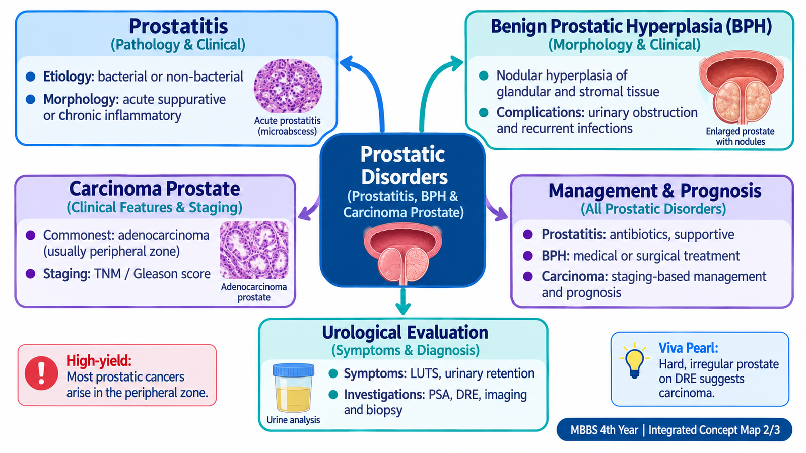
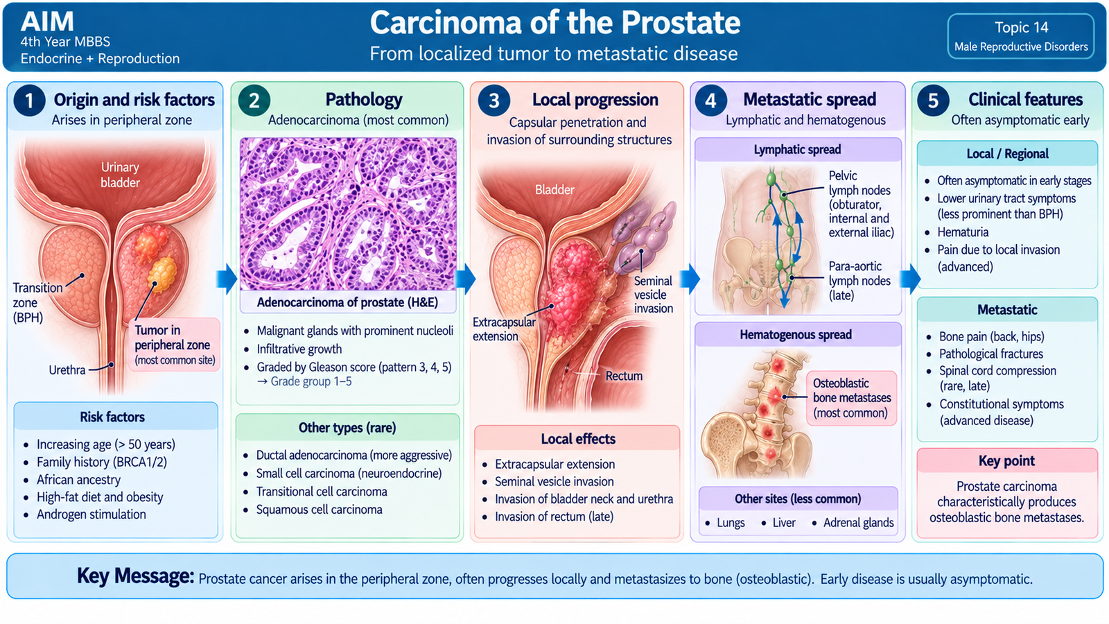
G. Carcinoma of the Prostate
Carcinoma of the prostate is a malignant epithelial tumor of the prostate, most commonly an adenocarcinoma. Unlike BPH, which develops mainly in the transition/periurethral region, most conventional prostatic adenocarcinomas originate in the peripheral zone. This explains why an early cancer may be palpable on digital rectal examination while producing little urinary obstruction.
Etiology and important risk factors
Prostate carcinoma is strongly associated with increasing age and androgen-dependent biology. Familial and inherited susceptibility is important in some patients. The development and progression of conventional prostate adenocarcinoma depend substantially on androgen-receptor signaling, which is why suppression of androgen action is a major treatment strategy in advanced disease.
Types
The common form is acinar adenocarcinoma. Less common pathological types include ductal adenocarcinoma and neuroendocrine or small-cell carcinoma. These uncommon variants differ biologically from the usual androgen-dependent acinar tumor, but the essential undergraduate emphasis is recognition of conventional adenocarcinoma.
Pathological and clinical recognition
Prostatic adenocarcinoma tends to form small, crowded malignant glands that infiltrate the prostatic stroma. In contrast with normal glands, the malignant glands lack the normal basal-cell layer. Histological assessment also evaluates the architectural pattern of tumor growth.
Because many tumors arise peripherally, early disease may be asymptomatic. Later presentations can include lower urinary tract symptoms, hematuria or complications of local spread. Metastatic disease has a particular tendency to involve the axial skeleton.
Bone metastasis
Prostate carcinoma commonly spreads to bone, especially the vertebral column, pelvis and other parts of the axial skeleton. These metastases are characteristically associated with an osteoblastic or sclerotic reaction. Therefore, persistent back or bone pain in a patient with advanced prostate carcinoma is an important clinical clue.
Examination
Digital rectal examination may reveal a hard, irregular or nodular area, particularly when the peripheral part of the gland is involved. This contrasts with the smooth enlargement more typical of uncomplicated BPH.
Investigations
Prostate-specific antigen (PSA) is produced by prostatic epithelium. It is useful in assessment and follow-up but is not specific for cancer. BPH, inflammation and manipulation may also influence PSA levels. PSA therefore requires interpretation in the clinical context rather than being treated as a stand-alone diagnosis.
Multiparametric MRI helps define suspicious lesions, local extent and biopsy targets. Histological confirmation is obtained by prostate biopsy when indicated. Once carcinoma is diagnosed, the biopsy also provides information about tumor grade.
Gleason grading
The Gleason system evaluates the architectural pattern of prostatic adenocarcinoma. Tumors that retain more organized gland formation behave less aggressively than tumors that have lost glandular differentiation. Modern reporting groups these patterns into prognostic grade groups, but the basic principle is simple:
Increasing architectural disorder → higher grade → more aggressive biological behavior.
Staging
Prostate carcinoma is staged using the TNM system. At undergraduate level, the important idea is to determine whether the cancer is confined to the prostate, has extended locally, involves lymph nodes or has produced distant metastases.
- T category: extent of the primary prostatic tumor and local extension.
- N category: regional lymph-node involvement.
- M category: distant metastasis, especially bone involvement.
Clinical decisions are therefore often described in practical terms as localized disease, locally advanced disease or metastatic disease.
Complications
- Local urinary obstruction.
- Hematuria or local tissue invasion.
- Ureteric obstruction and renal complications in advanced local disease.
- Lymph-node metastasis.
- Bone metastasis causing pain, skeletal complications and neurological problems if the spine is involved.
Management principles
Management depends on stage, grade, expected tumor behavior, patient factors and overall fitness. Selected low-risk localized tumors may be observed under structured active surveillance. Clinically significant localized tumors can be treated with curative-intent surgery or radiotherapy. Locally advanced or metastatic androgen-dependent tumors are treated using strategies that suppress androgen production or block androgen-receptor signaling.
Androgen deprivation may be achieved by suppressing pituitary gonadotropin drive with GnRH-based therapy or by surgical removal of testicular androgen production. Androgen-receptor antagonists and other agents that interfere with androgen signaling may be added according to the clinical situation. Systemic anticancer therapy is considered for more advanced disease under specialist care.
Prognosis
Prognosis depends mainly on tumor stage, histological grade and overall disease burden. A localized low-grade tumor has a substantially better outlook than high-grade metastatic disease. Bone or visceral metastasis indicates advanced-stage disease.
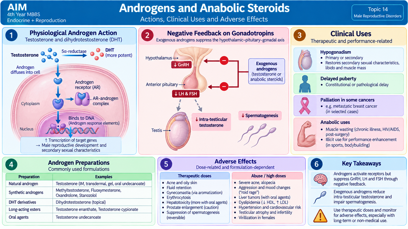
H. Androgens and Anabolic Steroids
Androgens are hormones that produce and maintain male reproductive development, secondary sexual characteristics and anabolic effects. Testosterone is the principal natural androgen. Pharmacological androgen preparations are used when clinically important androgen deficiency is present. Anabolic steroids are related compounds designed to retain anabolic actions, although complete separation of anabolic and androgenic effects is not possible.
Androgen preparations
Natural testosterone has a relatively short duration when given without pharmaceutical modification and undergoes substantial first-pass metabolism if swallowed in ordinary form. Different preparations therefore modify the route or molecular structure to improve delivery.
- Testosterone preparations: injectable testosterone esters, transdermal testosterone preparations and selected oral formulations.
- Longer-acting ester preparations: testosterone enanthate, cypionate and undecanoate are examples.
- Oral androgen derivatives: methyltestosterone is a classic example of a 17-alpha-alkylated androgen.
- Anabolic steroids: examples include nandrolone, oxandrolone and stanozolol.
Mechanism of action
Testosterone diffuses into target cells and binds the intracellular androgen receptor. The activated receptor interacts with nuclear DNA and changes gene transcription, producing androgen-dependent proteins and cellular responses.
In some tissues, testosterone undergoes further metabolism:
- 5-alpha reductase converts testosterone to DHT, a more potent androgen in tissues such as the prostate and external genital structures.
- Aromatase converts some testosterone to estradiol, which contributes to effects such as bone maturation and some feedback actions.
Pharmacological effects
- Development and maintenance of male secondary sexual characteristics.
- Support of male accessory reproductive organs.
- Increase in protein synthesis and lean body mass.
- Effects on bone growth and maturation.
- Stimulation of erythropoiesis.
- Negative feedback on hypothalamic and pituitary GnRH, LH and FSH secretion.
Why exogenous testosterone can cause infertility
This is an important pharmacodynamic concept. Administration of testosterone raises circulating androgen activity, which suppresses hypothalamic GnRH and pituitary LH and FSH through negative feedback. Intratesticular testosterone then falls because Leydig-cell stimulation is reduced. The result can be marked suppression of spermatogenesis despite adequate or high circulating androgen levels.
Pharmacokinetic principles
Unmodified testosterone has poor usefulness as a conventional oral drug because of extensive hepatic first-pass metabolism. Esterification allows slow release from intramuscular depot preparations. Transdermal formulations provide absorption through the skin and can produce more sustained exposure. Seventeen-alpha-alkylated oral anabolic-androgenic steroids resist hepatic breakdown but carry greater risk of liver toxicity.
Clinical uses
The major therapeutic role is androgen replacement in documented male androgen deficiency when restoration of androgen action is appropriate. Testosterone may also be used in selected boys with delayed pubertal development when specialist evaluation shows an appropriate indication. Exogenous testosterone is not a fertility treatment for a man trying to produce sperm.
Adverse effects
Many adverse effects follow directly from excessive androgen action or suppression of the normal hypothalamic–pituitary–testicular axis.
- Suppression of spermatogenesis and infertility.
- Testicular atrophy during prolonged high-dose exposure.
- Acne and oily skin.
- Fluid retention.
- Increase in red-cell mass.
- Gynecomastia may occur because some androgen is aromatized to estrogen.
- Acceleration of androgen-dependent prostatic disease.
- Premature epiphyseal closure if excessive exposure occurs before completion of growth.
- Hepatic injury is especially associated with some orally active 17-alpha-alkylated anabolic steroids.
Anabolic-steroid misuse
Non-medical anabolic-steroid use may produce substantial muscular effects, but the endocrine system responds to high androgen exposure by suppressing endogenous LH and FSH. Therefore, testicular atrophy and reduced sperm production can occur even in a person who appears strongly virilized.
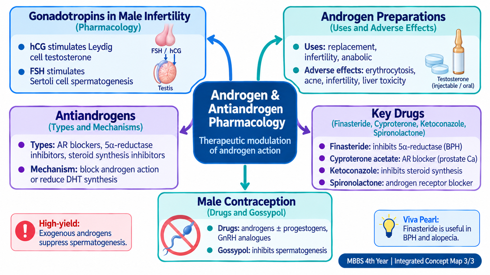
I. Gonadotropins, Antiandrogens and Male Contraception
This group of drugs acts at different points along the hypothalamic–pituitary–testicular and androgen pathways. Gonadotropins can stimulate testosterone production and spermatogenesis when pituitary stimulation is deficient. Antiandrogens reduce androgen production, conversion or receptor activity. Conversely, experimental hormonal male-contraceptive approaches deliberately suppress gonadotropins in order to reduce sperm production.
Gonadotropins in male infertility
Gonadotropin treatment is most logical in a man whose testes are potentially functional but are not receiving adequate LH and FSH stimulation, as in hypogonadotropic hypogonadism.
Human chorionic gonadotropin
hCG has LH-like activity. It stimulates LH receptors on Leydig cells and thereby increases testicular testosterone production.
FSH preparations
FSH preparations act primarily on Sertoli cells and promote the environment required for spermatogenesis. Human menopausal gonadotropin provides gonadotropin activity, while recombinant FSH preparations can provide more direct FSH stimulation.
In appropriate gonadotropin-deficient infertility, hCG is used to restore Leydig-cell testosterone stimulation and FSH activity is added when direct support of spermatogenesis is required.
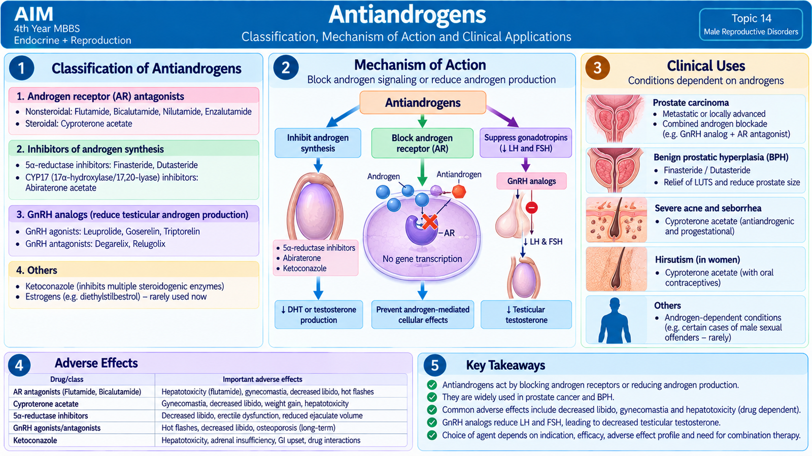
Classification of antiandrogen approaches
Antiandrogen therapy can reduce androgen effects by blocking the androgen receptor, preventing formation of the more potent androgen DHT, reducing steroid synthesis or suppressing testicular androgen production.
| Mechanism | Examples | Main Effect |
|---|---|---|
| Androgen-receptor antagonism | Bicalutamide, flutamide, nilutamide; cyproterone acetate | Blocks androgen signaling in target tissues |
| 5-alpha-reductase inhibition | Finasteride, dutasteride | Reduces testosterone conversion to DHT |
| Steroid-synthesis inhibition | Ketoconazole | Reduces androgen synthesis |
| Suppression of pituitary LH | GnRH agonist or antagonist approaches | Reduces testicular testosterone production |
Nonsteroidal androgen-receptor antagonists
Drugs such as flutamide, bicalutamide and nilutamide compete with androgens at the androgen receptor. The receptor cannot produce its normal transcriptional response, so androgen-dependent tissues receive less androgenic stimulation. Their major clinical relevance is androgen blockade in prostate carcinoma.
Because blocking androgen action can disturb the androgen-estrogen balance, adverse effects may include gynecomastia, reduced libido and sexual dysfunction. Hepatic toxicity is an important concern with some members of the class, particularly flutamide.
Finasteride
Finasteride inhibits 5-alpha reductase and reduces conversion of testosterone to DHT. Because DHT is an important growth signal within the prostate, reducing DHT gradually decreases the androgen-dependent component of prostatic enlargement.
Its important clinical uses include BPH and androgen-dependent hair loss. Adverse effects may include reduced libido, erectile dysfunction, reduced ejaculatory volume and occasionally gynecomastia. Finasteride also lowers PSA, so its effect must be considered when PSA is interpreted clinically.
Cyproterone acetate
Cyproterone acetate is a steroidal antiandrogen. It directly antagonizes the androgen receptor and also has progestational activity that suppresses pituitary gonadotropin secretion. It therefore reduces androgen effects both at the receptor and by decreasing testicular androgen stimulation.
It has been used in androgen-dependent clinical conditions, including suppression of androgen action in selected patients with prostate carcinoma and other states in which excessive androgen effect needs to be reduced. Adverse effects reflect loss of androgen action and its hormonal activity and may include reduced libido, sexual dysfunction, gynecomastia and suppression of spermatogenesis. Hepatic adverse effects are also clinically important.
Spironolactone as an androgen-receptor blocker
Spironolactone is best known as a mineralocorticoid-receptor antagonist, but it also has antiandrogen actions. It can competitively interfere with androgen receptors and reduce androgen synthesis to some extent. This explains antiandrogenic adverse effects in men, including gynecomastia and sexual dysfunction, and also explains its therapeutic use in selected hyperandrogenic states.
Ketoconazole as an inhibitor of steroid synthesis
Ketoconazole is an azole antifungal drug, but at sufficiently high exposure it also inhibits cytochrome P450 enzymes involved in steroid synthesis. This reduces production of testicular and adrenal androgens.
This property has historically allowed ketoconazole to be used when rapid suppression of androgen synthesis was required, particularly in androgen-dependent prostate cancer. Its usefulness is limited by significant adverse effects, including hepatic toxicity, endocrine disturbance and important drug interactions. Its role here should therefore be understood mainly as a pharmacological example of steroid-synthesis inhibition.
GnRH-based androgen suppression
Continuous administration of a GnRH agonist initially stimulates LH and testosterone release but subsequently causes pituitary GnRH-receptor down-regulation and marked suppression of LH secretion. This produces a fall in testicular testosterone after the initial stimulatory phase.
GnRH antagonists block pituitary GnRH receptors directly and therefore suppress LH and testosterone without the same initial stimulation. Both approaches are relevant to androgen deprivation in prostate carcinoma.
Drugs investigated for male contraception
The pharmacological principle of hormonal male contraception is almost the opposite of gonadotropin treatment for infertility. Instead of stimulating LH and FSH, the aim is to suppress them enough to markedly reduce sperm production while maintaining adequate peripheral androgen activity.
Agents and approaches that have been studied include:
- Testosterone-based regimens.
- Androgen plus progestin combinations.
- Gonadotropin-suppressing hormonal approaches.
- Non-hormonal agents such as gossypol.
These pharmacological approaches should be understood as concepts investigated for male contraception rather than being assumed to represent routine universally used contraceptive drug regimens.
Gossypol
Gossypol is a naturally occurring polyphenolic compound obtained from the cotton plant. It was investigated as an oral non-hormonal male contraceptive because it can impair sperm production and sperm function.
Its clinical development was limited by important toxicity and reversibility concerns. Reported problems include hypokalemia and persistent or potentially irreversible suppression of spermatogenesis in some men. For this reason, gossypol is mainly important in the curriculum as a historical and pharmacological example rather than as a standard routine male contraceptive.
Important Comparison — BPH versus Carcinoma Prostate
BPH and prostate carcinoma can both occur in older men and may both be associated with urinary symptoms or an elevated PSA. Their anatomical origin, pathological behavior and examination findings are different, making this a high-value examination comparison.
| Feature | BPH | Prostatic Carcinoma |
|---|---|---|
| Nature | Benign nodular hyperplasia | Malignant epithelial tumor, usually adenocarcinoma |
| Usual zone | Transition/periurethral region | Peripheral zone |
| Early urinary obstruction | Common because urethra is compressed | May be absent in early peripheral tumors |
| DRE | Smooth, symmetrically enlarged, firm/rubbery | Hard, irregular or nodular lesion |
| Metastasis | Does not metastasize | May spread to lymph nodes and bone |
| PSA | May be increased | May be increased; interpreted with clinical context |
| Key hormonal link | DHT promotes nodular growth | Androgen-receptor signaling supports many conventional tumors |
⭐ AIM High-Yield Review
Topic 14 — Male Reproductive Disorders
Use these selected videos after reading the AIM learning material to reinforce male infertility, testicular tumors, prostatic disorders and androgen pharmacology.
1. Male Infertility — Causes & Evaluation
Strong Medicine — clinical approach to reproductive physiology, causes and evaluation of male infertility.
2. Testicular Tumors — Seminoma & Nonseminoma
Pathology-focused review of seminoma, embryonal carcinoma, yolk-sac tumor, choriocarcinoma, teratoma and mixed tumors.
3. Benign Prostatic Hyperplasia
Medical-student overview of BPH pathophysiology, lower urinary tract symptoms, diagnosis and treatment.
4. Prostate Carcinoma — Pathology
Pathology teaching video reinforcing the microscopic appearance and pathological basis of prostatic adenocarcinoma.
5. Antiandrogens — Pharmacology
Classification, mechanisms, clinical uses and adverse effects of antiandrogen drugs relevant to this topic.