
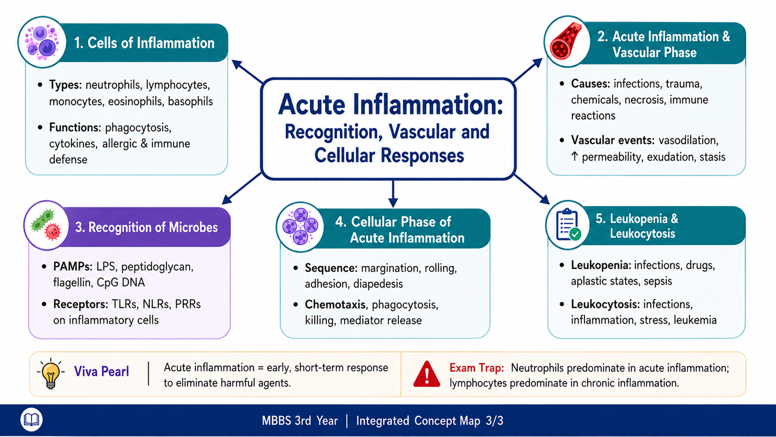
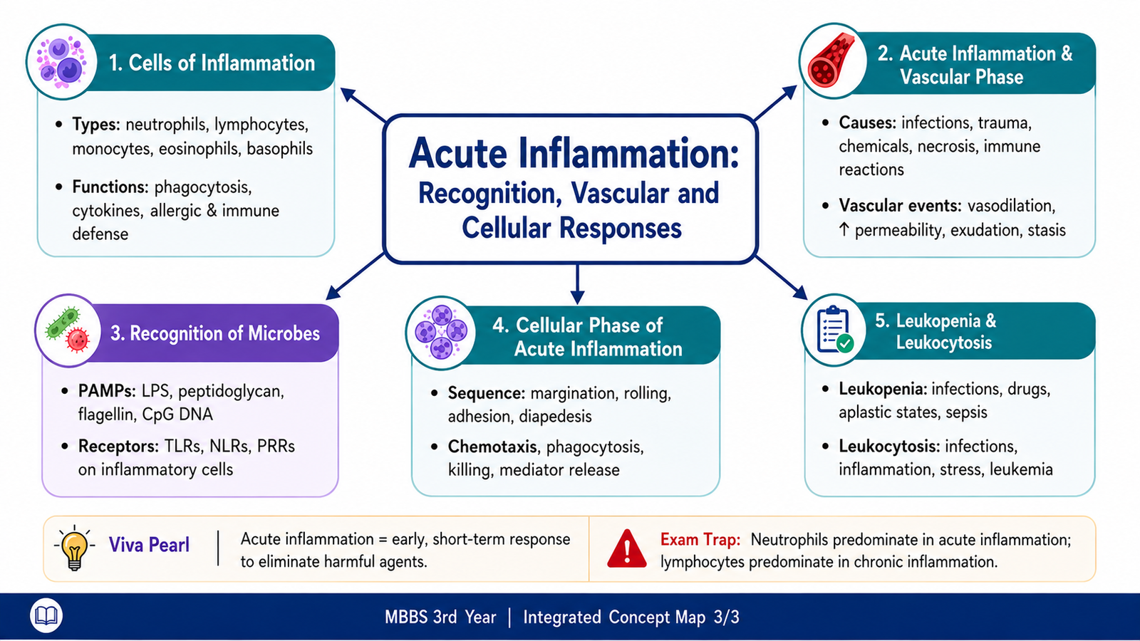
This chapter follows the supplied KMU learning outcomes in a logical sequence. First understand how inflammation is recognized and initiated, then follow the vascular and cellular events before revising the high-yield points at the end.
Acute Inflammation: Recognition, Vascular and Cellular Responses
Understand how inflammatory cells recognize harmful stimuli, how blood vessels react, and how leukocytes are recruited to the site of acute inflammation.
Topic Introduction
Acute inflammation is the rapid protective response of vascularized tissues to infection, tissue injury and other harmful stimuli. Its purpose is to bring plasma proteins and leukocytes, especially neutrophils, to the affected area so that the cause of injury can be removed and damaged tissue can begin to recover. The response starts when inflammatory cells recognize microbial or tissue-derived signals. This recognition triggers changes in blood vessels, followed by movement of leukocytes from the circulation into tissues. In this chapter, you will learn the major inflammatory cells and their functions, the causes and vascular events of acute inflammation, the receptors that recognize microbes, and the complete sequence of the cellular phase.
A. Cells of Inflammation and Their Functions
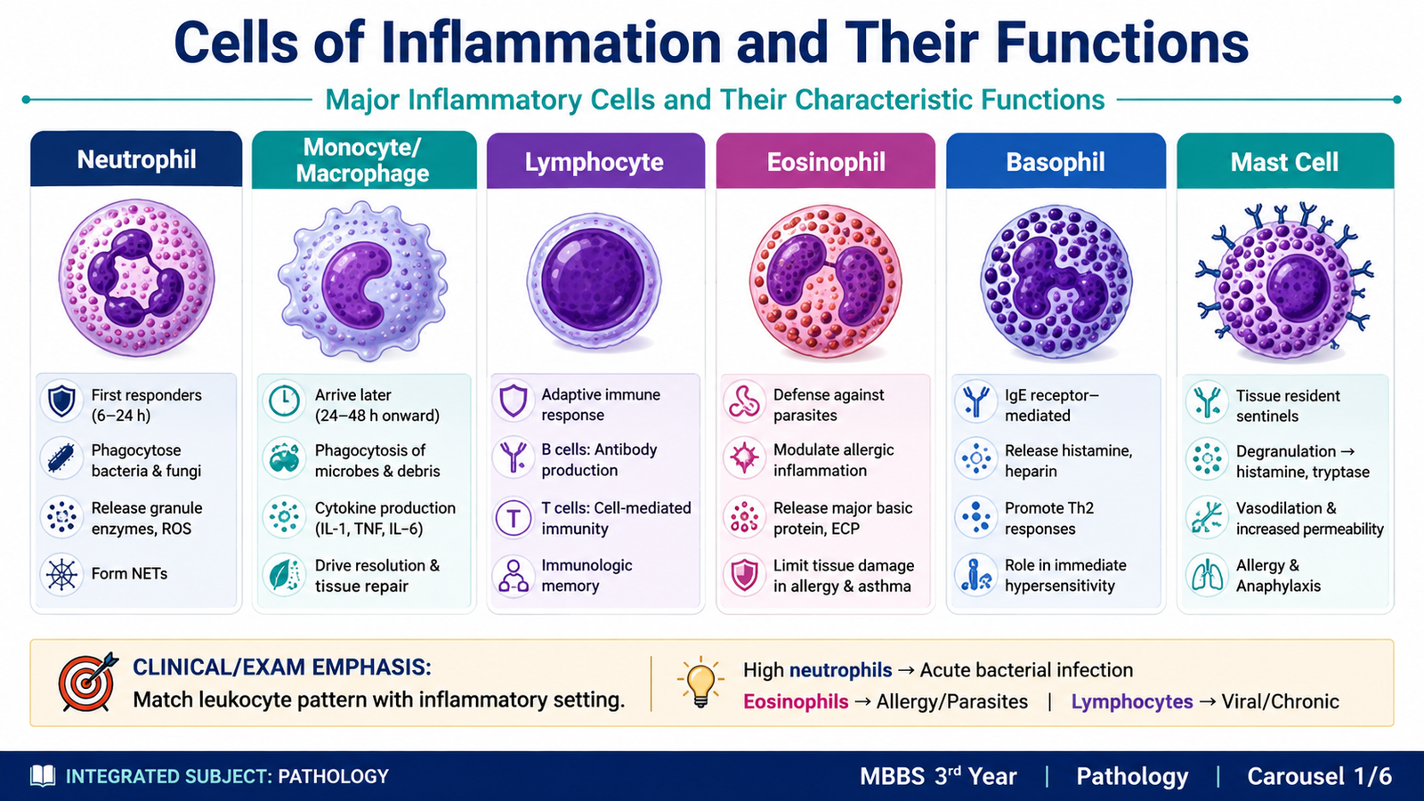
Inflammation depends on several circulating and tissue-resident cells. Each cell has a characteristic role, and the dominant cell population may change according to the type and duration of the inflammatory stimulus. In acute inflammation, neutrophils are usually the most important early leukocytes, but monocytes, macrophages, lymphocytes, eosinophils, basophils and mast cells also contribute in particular situations.
Neutrophils
Neutrophils are rapidly recruited from blood to sites of bacterial infection and tissue injury. They engulf microorganisms and dead material by phagocytosis and kill microbes using lysosomal enzymes, reactive oxygen species and other antimicrobial mechanisms. Because they respond quickly and survive for a relatively short time in tissues, they are especially prominent during the early phase of many acute inflammatory reactions.
Monocytes and Macrophages
Monocytes circulate in blood and migrate into tissues, where they differentiate into macrophages. Macrophages phagocytose microbes and dead cells, produce inflammatory mediators and help coordinate later stages of inflammation. They also participate in tissue repair after the damaging stimulus has been controlled.
Lymphocytes
Lymphocytes are central cells of adaptive immunity. T lymphocytes regulate immune responses and may activate macrophages, while B lymphocytes can differentiate into plasma cells that produce antibodies. Although lymphocytes are more characteristic of chronic immune responses, they can participate when acute inflammation is driven by certain infections or immune mechanisms.
Eosinophils
Eosinophils are particularly important in inflammatory responses associated with parasitic infections and allergic disorders. Their granules contain proteins and enzymes that can damage parasites but can also contribute to tissue injury during allergic inflammation.
Basophils and Mast Cells
Basophils circulate in small numbers in blood, whereas mast cells are mainly found in tissues. Both contain granules rich in mediators such as histamine. Mast-cell activation can rapidly increase vascular permeability and is particularly important in immediate hypersensitivity reactions.
| Cell | Major Role | Common Association |
|---|---|---|
| Neutrophil | Rapid phagocytosis and microbial killing | Acute bacterial inflammation |
| Monocyte/macrophage | Phagocytosis, mediator production, repair | Later inflammatory response |
| Lymphocyte | Adaptive immune regulation | Immune and selected infectious responses |
| Eosinophil | Granule-mediated inflammatory response | Parasites and allergy |
| Basophil/mast cell | Release of histamine and other mediators | Immediate hypersensitivity |
Leukocytosis and Leukopenia
Leukocytosis means an increase in the number of circulating white blood cells, whereas leukopenia means a reduction. The pattern can involve a particular leukocyte type and may provide a clue to the underlying process.
- Neutrophilia: commonly associated with acute bacterial inflammation, tissue necrosis and some stress responses.
- Neutropenia: may result from reduced marrow production, severe overwhelming infection, marrow suppression or increased peripheral destruction.
- Lymphocytosis: often accompanies selected viral infections and some immune responses.
- Lymphopenia: may occur with impaired lymphocyte production, destruction or immunosuppressive states.
- Monocytosis: may be seen in prolonged infections and inflammatory conditions.
- Monocytopenia: can occur with severe marrow suppression or reduced production.
- Eosinophilia: classically associated with allergic disorders and many parasitic infections.
- Eosinopenia: may occur during physiological stress or states associated with increased corticosteroid activity.
- Basophilia: may occur in hypersensitivity states and selected hematological disorders.
- Basopenia: is less commonly emphasized clinically and may occur when circulating basophil numbers fall during stress or corticosteroid effects.
B. Acute Inflammation: Definition, Purpose and Causes
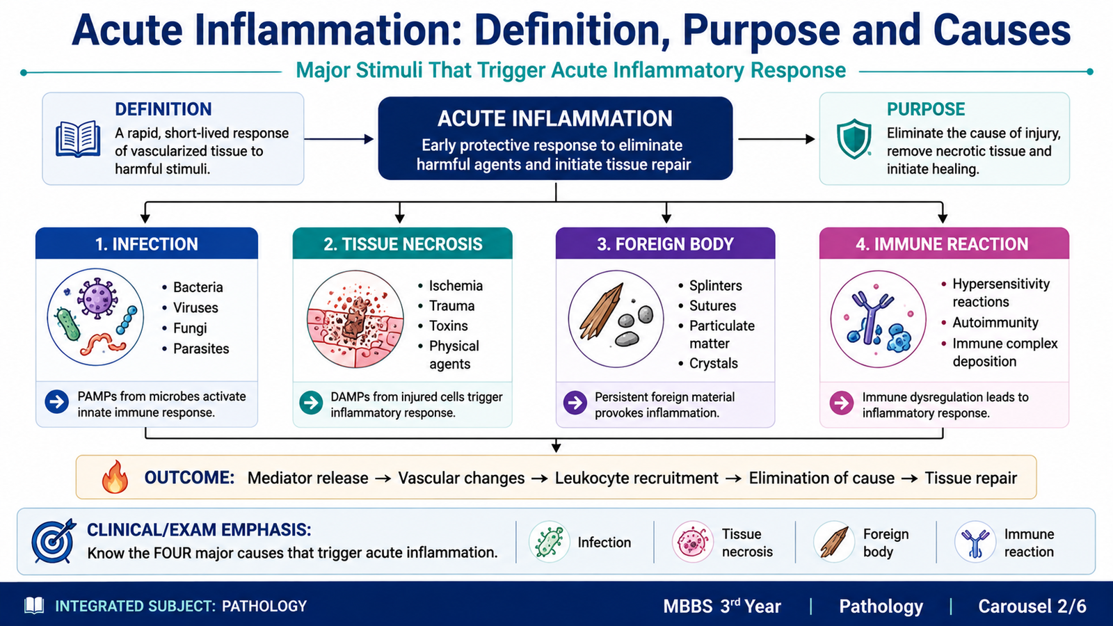
Acute inflammation is a rapid response of vascularized tissues to infection, tissue injury or other harmful stimuli. It usually develops over minutes to hours and is characterized mainly by movement of fluid and plasma proteins into tissues together with recruitment of leukocytes, particularly neutrophils.
The response is protective. Its major purpose is to deliver defensive cells and proteins to the site of injury, destroy or contain the harmful agent, remove damaged tissue and prepare the area for repair. However, the same inflammatory response can also damage normal tissue when it is excessive or prolonged.
Major Causes
- Infections: bacteria, viruses, fungi and parasites can initiate inflammation through microbial products and tissue injury.
- Tissue necrosis: ischemia, trauma and physical or chemical injury release intracellular substances that stimulate inflammation.
- Foreign bodies: splinters, sutures, crystals and other foreign material may directly damage tissue or stimulate inflammatory cells.
- Immune reactions: inappropriate or excessive immune responses against environmental substances or self-tissues can produce inflammation.
The important concept is that different causes can activate similar inflammatory pathways. Once harmful material is recognized, inflammatory cells release mediators that alter local blood vessels and recruit additional leukocytes.
C. Recognition of Microbes and Initiation of Inflammation
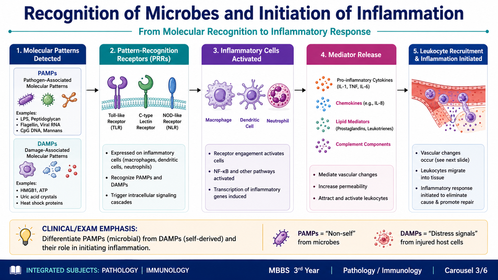
Inflammatory cells must first recognize that a harmful stimulus is present. Cells of innate immunity, including macrophages and other tissue cells, carry receptors that detect characteristic molecular patterns. These receptors allow the body to identify microbes rapidly without requiring previous exposure.
Molecular Patterns
Microorganisms contain structures that are shared by groups of microbes but are not normally present in human cells. These microbial structures are called pathogen-associated molecular patterns (PAMPs). Examples include microbial cell-wall components and microbial nucleic acids.
Damaged or necrotic host cells can also release molecules that signal tissue injury. These are called damage-associated molecular patterns (DAMPs). Together, PAMPs and DAMPs alert inflammatory cells to infection or tissue damage.
Pattern-Recognition Receptors
Inflammatory cells detect PAMPs and DAMPs through pattern-recognition receptors (PRRs). Important receptor families include Toll-like receptors and cytoplasmic receptors that recognize microbial or damage-related molecules in different cellular locations.
This arrangement is useful because microbes may be present outside cells, within intracellular vesicles or in the cytoplasm. Receptors positioned at these sites allow inflammatory cells to detect the location of the threat and activate an appropriate response.
How Recognition Initiates Inflammation
Binding of a microbial or damage-associated pattern to its receptor activates the inflammatory cell. The activated cell then produces mediators that promote vascular changes and leukocyte recruitment. Therefore, recognition is the link between the presence of a harmful stimulus and the visible inflammatory reaction.
PAMP or DAMP → pattern-recognition receptor → inflammatory-cell activation → mediator release → vascular response and leukocyte recruitment
D. Vascular Events of Acute Inflammation
The vascular phase begins very early after tissue injury. Its purpose is to increase local blood delivery and then allow fluid and plasma proteins to leave the circulation and enter the affected tissue. These changes explain important visible features of inflammation such as redness, warmth and swelling.
1. Vasodilation
One of the earliest important changes is dilation of small blood vessels, particularly arterioles. Increased vessel diameter raises local blood flow, a change known as hyperemia. This increased blood flow contributes to the redness and warmth of an inflamed area.
2. Increased Vascular Permeability
The endothelial barrier becomes more permeable, especially in post-capillary venules. Protein-rich fluid then moves from the circulation into the extravascular tissue. This inflammatory fluid is called an exudate.
Loss of intravascular fluid increases the concentration of red cells within small vessels and raises blood viscosity. At the same time, accumulation of fluid in tissues produces inflammatory edema and therefore contributes to swelling.
Exudate and Transudate
| Feature | Exudate | Transudate |
|---|---|---|
| Main mechanism | Increased vascular permeability | Altered hydrostatic or osmotic forces |
| Protein content | Relatively protein-rich | Relatively protein-poor |
| Inflammatory significance | Typical of inflammation | Usually not caused by inflammation |
3. Slowing of Blood Flow and Stasis
As fluid leaves the vessels, the blood becomes more concentrated and moves more slowly. This slowing is called stasis. Stasis brings circulating leukocytes closer to the endothelial surface, preparing them for the cellular phase of inflammation.

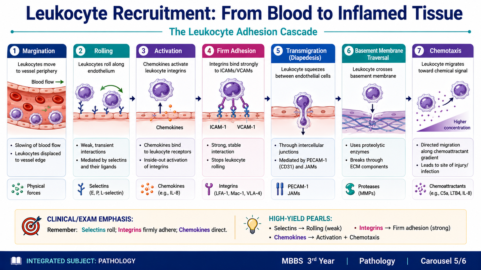
E. Leukocyte Recruitment: From Blood to Inflamed Tissue
After vascular changes have occurred, leukocytes must leave the circulation and reach the exact site where microbes or damaged tissue are present. This process occurs in an organized sequence. In acute inflammation, neutrophils usually dominate the early response because they are rapidly recruited from blood.
Margination
During normal rapid blood flow, red cells occupy much of the central portion of the vessel. When blood flow slows during inflammation, leukocytes move toward the endothelial surface. This peripheral positioning of leukocytes is called margination.
Rolling
Marginal leukocytes make repeated weak attachments to endothelial cells. They detach and attach again, producing a rolling movement along the vessel wall. These weak interactions are mainly mediated by molecules called selectins.
Firm Adhesion
Inflammatory signals activate leukocytes and increase the binding strength of leukocyte integrins. These integrins attach firmly to corresponding endothelial adhesion molecules. The leukocyte then stops rolling and becomes firmly attached to the endothelial surface.
Transmigration
After adhesion, leukocytes pass between endothelial cells, mainly in post-capillary venules. This movement across the vessel wall is called transmigration or diapedesis. Leukocytes then cross the basement membrane and enter the extravascular tissue.
Chemotaxis
Once outside the vessel, leukocytes move toward the site of injury by following a chemical concentration gradient. This directed movement is called chemotaxis. Chemotactic substances may come from microbes or be generated within the host inflammatory response.
F. Leukocyte Activation, Phagocytosis and Microbial Killing
Recruitment brings leukocytes to the site of inflammation, but the cells must then recognize and remove the offending agent. Neutrophils and macrophages become activated after encountering microbes, damaged tissue or inflammatory mediators. Their major functions at the site are recognition, engulfment and destruction of harmful material.
Recognition and Attachment
Microbes can be recognized directly, but phagocytosis becomes much more efficient when particles are coated by host proteins known as opsonins. Opsonins act as molecular tags that allow phagocytes to attach strongly to the target.
Engulfment
The phagocyte extends its cell membrane around the attached particle. The particle is enclosed within an intracellular vesicle called a phagosome. The phagosome then fuses with lysosomes, forming a phagolysosomal compartment in which killing and degradation occur.
Killing and Degradation
Microorganisms are killed by reactive oxygen species, lysosomal enzymes and other antimicrobial substances. The degraded material is then removed. These mechanisms are protective, but leakage of leukocyte enzymes or reactive products into surrounding tissue can also injure normal host cells.
Recognition and attachment → engulfment → phagosome formation → fusion with lysosomes → microbial killing and degradation
This completes the cellular response: leukocytes are recruited from blood, directed toward the inflammatory stimulus, activated and then used to remove microbes and damaged material.

Integrated Mechanism Flow
↓
2. PAMPs or DAMPs are recognized by pattern-recognition receptors
↓
3. Inflammatory cells become activated and release mediators
↓
4. Vasodilation and increased vascular permeability develop
↓
5. Fluid leaves vessels, blood flow slows and leukocytes move toward endothelium
↓
6. Leukocytes roll, adhere, transmigrate and follow chemotactic signals
↓
7. Phagocytes engulf and destroy microbes or damaged material
⭐ AIM High-Yield Review
- Acute inflammation is a rapid protective response of vascularized tissue to harmful stimuli.
- Important causes include infection, tissue necrosis, foreign bodies and immune reactions.
- Neutrophils are the major early leukocytes in many acute inflammatory reactions.
- Macrophages remove microbes and dead tissue and also participate in later repair.
- PAMPs are microbial molecular patterns; DAMPs arise from damaged host cells.
- PAMPs and DAMPs are recognized by pattern-recognition receptors.
- Vasodilation increases local blood flow and contributes to redness and warmth.
- Increased vascular permeability produces a protein-rich exudate and inflammatory edema.
- Fluid loss from vessels causes hemoconcentration and stasis, promoting leukocyte margination.
- Leukocyte recruitment follows: margination → rolling → adhesion → transmigration → chemotaxis.
- ⭐ Selectins are particularly important in rolling.
- ⭐ Integrins are particularly important in firm leukocyte adhesion.
- Phagocytosis involves recognition and attachment, engulfment and intracellular killing.
- Opsonization makes microbial recognition and phagocytosis more efficient.
- Eosinophilia classically suggests allergic inflammation or parasitic infection, whereas neutrophilia commonly accompanies acute bacterial inflammation.
Acute Inflammation — Vascular and Cellular Events
Watch this video after completing the learning material to reinforce vascular changes, leukocyte recruitment and the cellular response in acute inflammation.