
This chapter follows the KMU learning outcomes in a logical sequence. First understand how chronic inflammation develops and which cells and mediators drive it; then revise the morphology, granulomatous pattern and systemic effects using the high-yield section at the end.
Topic 3 — Chronic and Granulomatous Inflammation and Systemic Inflammatory Response
Theme: Infection and Inflammation
Understand how persistent inflammation differs from the acute response, how macrophages and lymphocytes maintain tissue injury and repair, why granulomas form, and how inflammatory mediators produce systemic effects such as fever and leukocytosis.
Topic Introduction
Inflammation is a protective response designed to eliminate harmful agents and begin tissue repair. When the injurious stimulus cannot be removed quickly, the inflammatory response may continue for weeks, months or even years and becomes chronic inflammation. Chronic inflammation is characterized by infiltration with mononuclear cells, continuing tissue destruction and simultaneous attempts at healing. A specialized form called granulomatous inflammation develops when macrophages accumulate in organized collections called granulomas. Inflammation can also affect the whole body. Cytokines released at the inflammatory site may produce fever, leukocytosis and increased acute-phase proteins. This chapter connects these processes so that their causes, cells, mediators, morphology and systemic consequences can be understood as one continuous inflammatory response.
A. Chronic Inflammation: Definition and Difference from Acute Inflammation
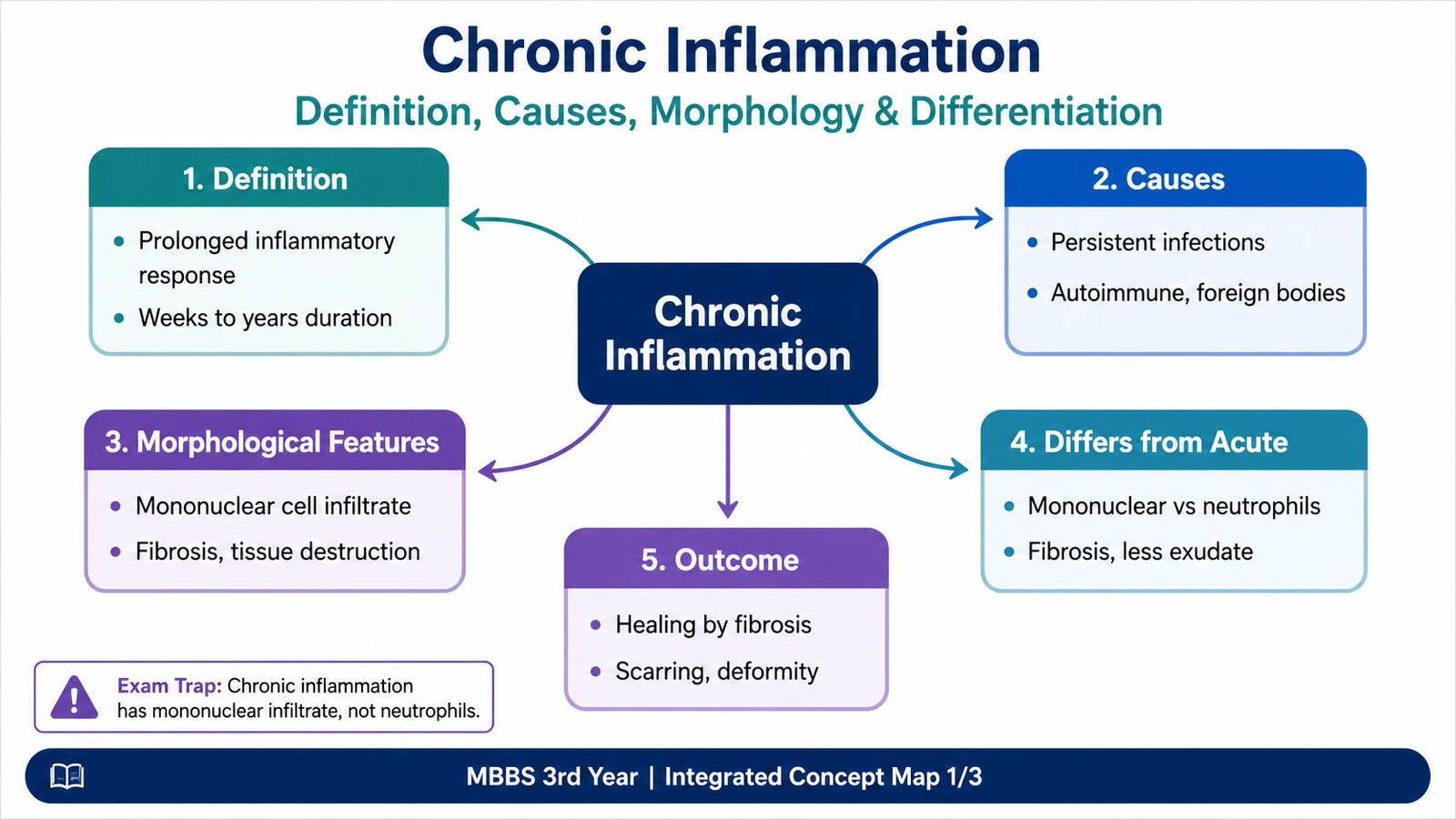
Chronic inflammation is a prolonged inflammatory response in which active inflammation, tissue injury and attempts at repair occur at the same time. It develops when the injurious stimulus persists or when the inflammatory response cannot completely eliminate it.
Unlike acute inflammation, which usually develops rapidly and is dominated by vascular changes and neutrophils, chronic inflammation evolves over a longer period and is dominated by macrophages, lymphocytes and plasma cells. Fibrosis and new blood-vessel formation may become prominent because tissue repair is occurring while damage continues.
| Feature | Acute Inflammation | Chronic Inflammation |
|---|---|---|
| Onset | Rapid | Usually slower or follows persistent injury |
| Duration | Short | Prolonged |
| Predominant cells | Neutrophils | Macrophages, lymphocytes and plasma cells |
| Main tissue pattern | Vascular leakage, edema and leukocyte recruitment | Persistent inflammation, tissue destruction and repair |
| Repair | Usually begins after removal of the stimulus | Often occurs simultaneously with continuing injury |
The distinction is useful but not absolute. Some inflammatory conditions begin as acute inflammation and later become chronic, while others produce chronic inflammation from the beginning.

B. Causes and Development of Chronic Inflammation
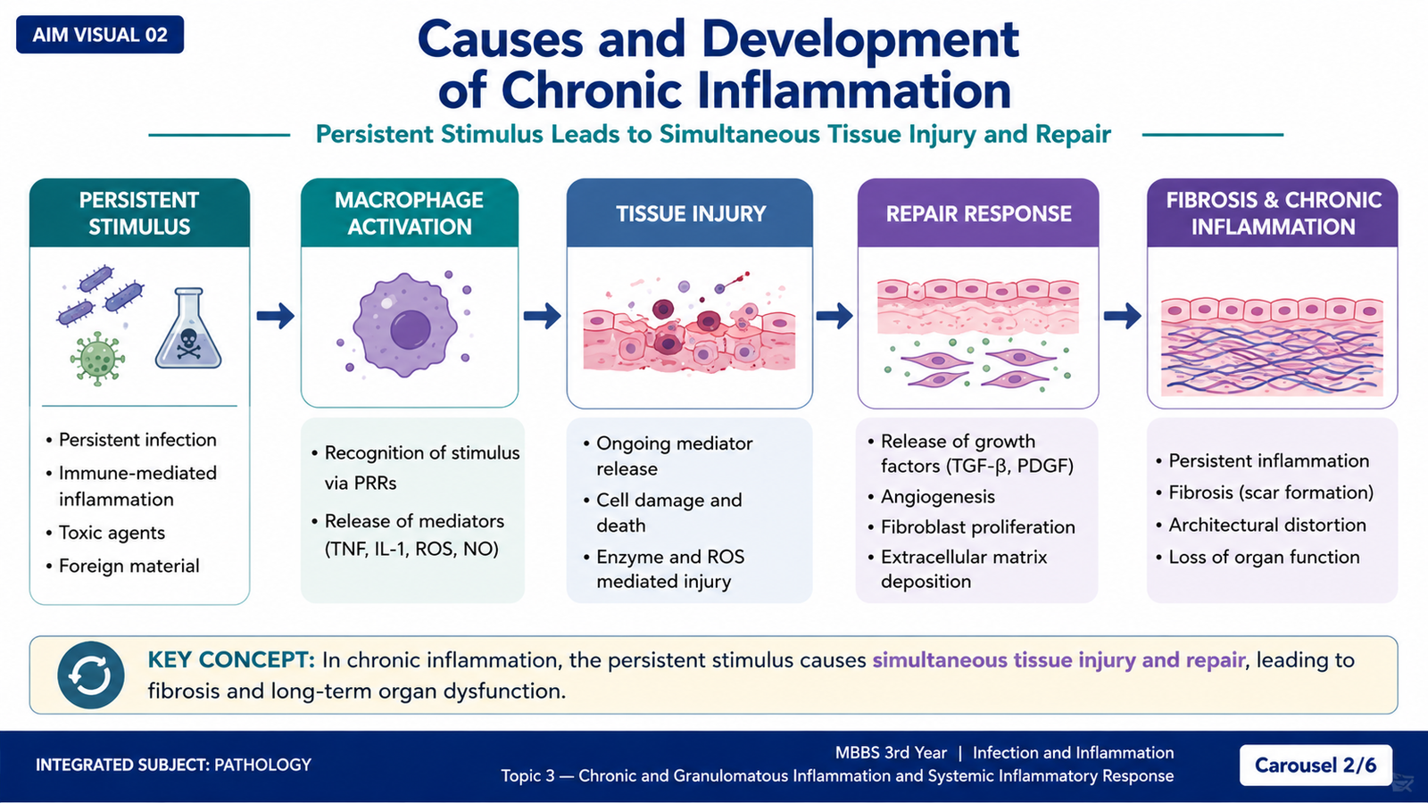
Chronic inflammation develops when the inflammatory stimulus remains present or when repeated injury keeps activating inflammatory cells. Because the stimulus persists, macrophages and lymphocytes remain activated and continuously release mediators. These mediators may destroy tissue but also stimulate repair, especially angiogenesis and fibrosis.
Major Causes
- Persistent infections: Some microorganisms are difficult to eradicate and therefore maintain prolonged immune activation. Certain persistent infections may produce granulomatous inflammation.
- Immune-mediated inflammatory diseases: Persistent or inappropriate immune reactions may repeatedly activate lymphocytes and macrophages.
- Prolonged exposure to potentially toxic agents: Substances that are difficult to remove may continue to stimulate inflammatory responses.
Why Tissue Injury and Repair Occur Together
Activated inflammatory cells release substances that can injure surrounding cells and extracellular matrix. At the same time, macrophages release growth factors that stimulate fibroblasts and blood-vessel formation. Therefore, a chronically inflamed tissue may show active cell injury in one area and fibrosis or healing in another.
Persistent stimulus → continued activation of macrophages and lymphocytes → mediator release → tissue injury → attempts at repair → fibrosis and structural alteration when the process continues.
C. Cells and Mediators of Chronic Inflammation

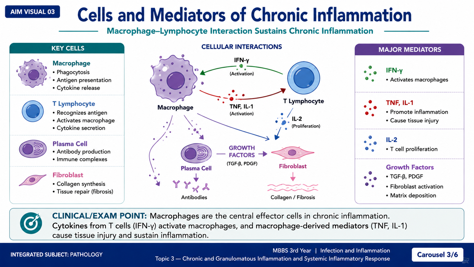
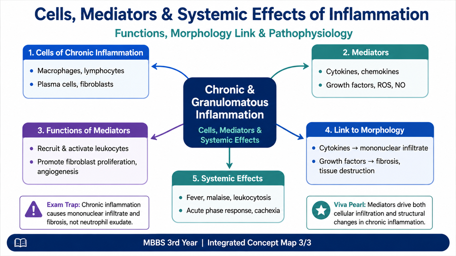
The cellular pattern of chronic inflammation reflects prolonged interaction between the innate and adaptive immune systems. Macrophages are the central effector cells, while lymphocytes regulate and sustain macrophage activation. Plasma cells, eosinophils and mast cells may contribute depending on the cause of inflammation.
1. Macrophages
Macrophages develop from circulating monocytes that enter tissues and differentiate. They are important because they can remove microbes and damaged material, release inflammatory mediators, cause tissue injury and promote repair.
Macrophages may be activated through different functional pathways.
Classical Macrophage Activation
Classically activated macrophages are stimulated mainly by microbial products and signals from activated lymphocytes such as interferon-gamma (IFN-γ). Their main role is antimicrobial and inflammatory.
They generate substances including reactive oxygen species, nitric oxide and inflammatory cytokines. These products help destroy microbes but may also damage surrounding tissue.
Alternative Macrophage Activation
Alternatively activated macrophages are more strongly associated with tissue repair. They release growth factors that stimulate fibroblasts, collagen formation and tissue remodeling.
The balance between these macrophage functions helps explain why chronic inflammation contains both ongoing tissue destruction and fibrosis.
2. Lymphocytes
Lymphocytes and macrophages communicate with each other in chronic inflammation. Activated T lymphocytes release cytokines that recruit and activate macrophages. Activated macrophages, in turn, present antigens and release cytokines that stimulate lymphocytes. This creates a cycle that can maintain inflammation for a long time.
3. Plasma Cells
Plasma cells arise from activated B lymphocytes and produce antibodies. They are commonly present when persistent antigenic stimulation causes a prolonged humoral immune response.
4. Eosinophils
Eosinophils may become prominent when the inflammatory process is associated with IgE-mediated immune responses or certain parasitic infections. Their granules contain proteins that can damage parasites but may also injure host tissues.
5. Mast Cells
Mast cells can participate in both acute and chronic inflammatory responses. When activated, they release mediators that influence vascular responses and leukocyte recruitment and may contribute to persistent inflammation.
Important Mediators
The mediators of chronic inflammation are produced mainly by activated macrophages and lymphocytes. Their effects determine whether inflammation predominates, whether tissue injury progresses, or whether repair and fibrosis become more prominent.
| Mediator / Group | Important Function | Result in Chronic Inflammation |
|---|---|---|
| TNF and IL-1 | Promote leukocyte recruitment and systemic inflammatory responses | Maintain inflammation and contribute to systemic effects |
| Chemokines | Recruit leukocytes | Maintain mononuclear-cell infiltration |
D. Morphological Features of Chronic Inflammation
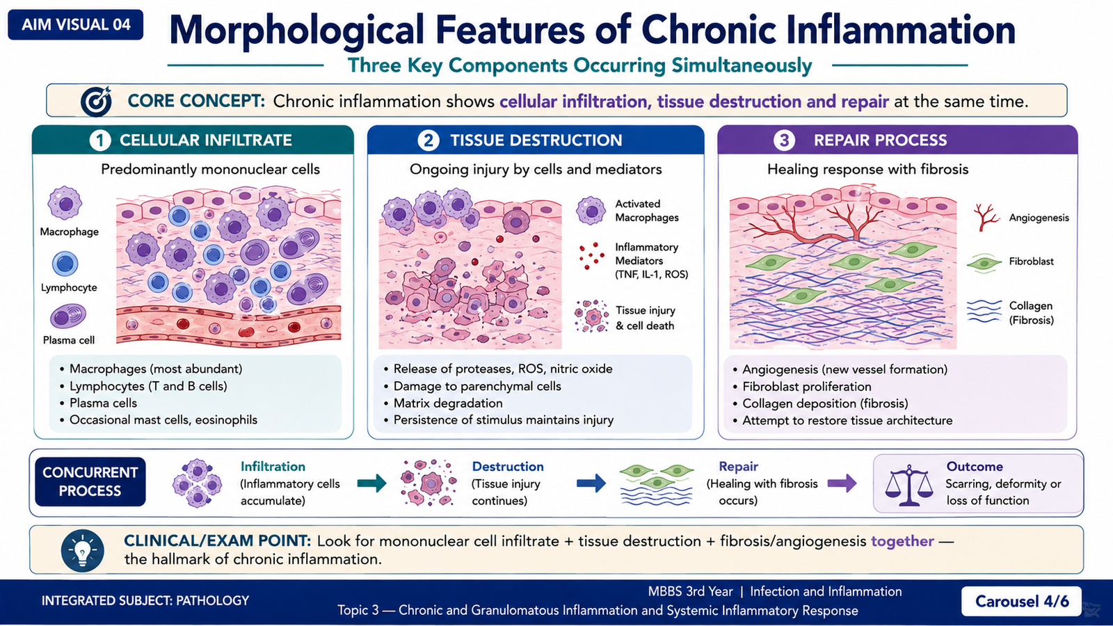
The morphology of chronic inflammation reflects three processes occurring together: mononuclear-cell infiltration, tissue destruction and healing by connective-tissue replacement. Recognizing these three features is important because they distinguish chronic inflammation from a purely acute inflammatory reaction.
1. Mononuclear-Cell Infiltration
The inflammatory infiltrate typically contains macrophages, lymphocytes and plasma cells. These cells predominate because the inflammatory process is persistent and involves continuing activation of both innate and adaptive immune responses.
2. Tissue Destruction
The persistent injurious agent may directly damage tissue, while activated inflammatory cells add further injury through enzymes, reactive oxygen species and other mediators. As a result, normal tissue architecture may progressively deteriorate.
3. Attempts at Healing
At the same time, growth factors released mainly by macrophages stimulate formation of new blood vessels and activation of fibroblasts. Fibroblasts deposit extracellular matrix, especially collagen. If the stimulus persists, this repair response may lead to fibrosis.
Persistent inflammation → mononuclear infiltrate → continuing tissue injury → fibroblast activation and angiogenesis → collagen deposition → fibrosis and altered tissue architecture.
Thus, the microscopic picture of chronic inflammation is not simply a collection of chronic inflammatory cells. The surrounding tissue often shows evidence of both destruction and repair, which reflects the mediator activity occurring within the lesion.
E. Granulomatous Inflammation
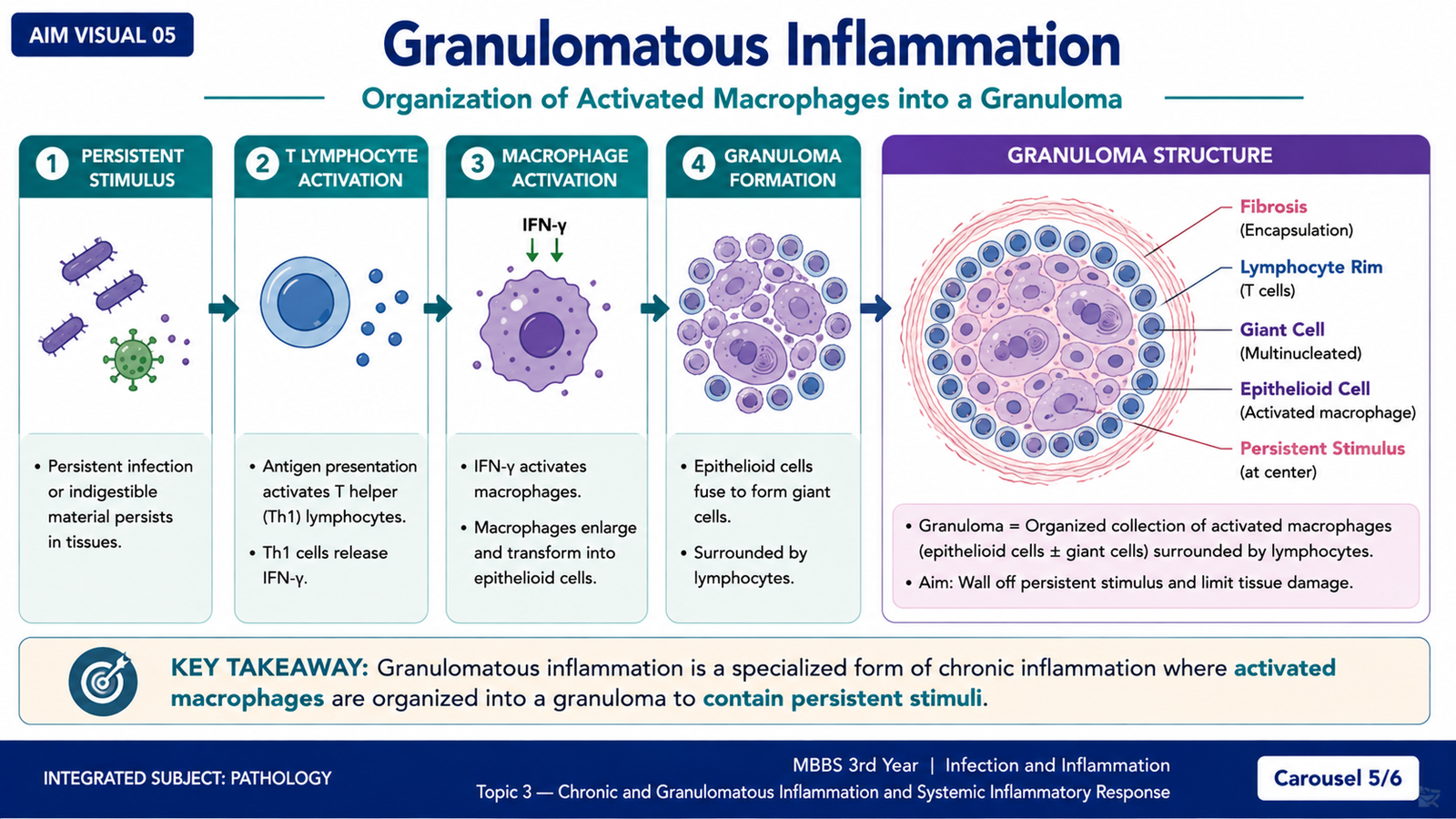
Granulomatous inflammation is a distinctive pattern of chronic inflammation characterized by collections of activated macrophages, usually surrounded by lymphocytes. The activated macrophages acquire an epithelial-like appearance and are therefore called epithelioid cells. Granulomas form when the immune system attempts to contain an agent that is difficult to eliminate.
Why a Granuloma Forms
If macrophages cannot easily destroy or remove a persistent agent, prolonged T-cell-mediated macrophage activation may occur. Activated T lymphocytes release cytokines, particularly IFN-γ, which maintains macrophage activation. The macrophages then gather into organized collections rather than remaining diffusely distributed throughout the tissue.
Morphological Features
A typical granuloma contains a central collection of epithelioid macrophages. Some activated macrophages fuse to form multinucleated giant cells. A surrounding rim of lymphocytes is often present. With persistence, fibroblasts and connective tissue may develop around the lesion.
- Epithelioid cells: activated macrophages with abundant cytoplasm and an epithelial-like appearance.
- Multinucleated giant cells: large cells produced by fusion of activated macrophages.
- Lymphocytes: commonly surround the macrophage collection and help maintain macrophage activation.
- Fibrosis: may develop around older or persistent granulomas.
- Central necrosis: may be present in some granulomatous reactions, but it is not an essential feature of every granuloma.
Immune and Foreign-Body Granulomas
Granulomas can arise through somewhat different mechanisms. In an immune granuloma, a persistent antigen produces a T-cell-mediated response with continuing macrophage activation. In a foreign-body granuloma, macrophages accumulate around material that is too large or resistant to be removed by ordinary phagocytosis.
| Feature | Immune Granuloma | Foreign-Body Granuloma |
|---|---|---|
| Main stimulus | Persistent antigen capable of stimulating an immune response | Indigestible or difficult-to-phagocytose foreign material |
| T-cell role | Prominent | Less central to formation |
| Macrophage response | Cytokine-driven activation | Accumulation around foreign material |
F. Systemic Effects of Inflammation
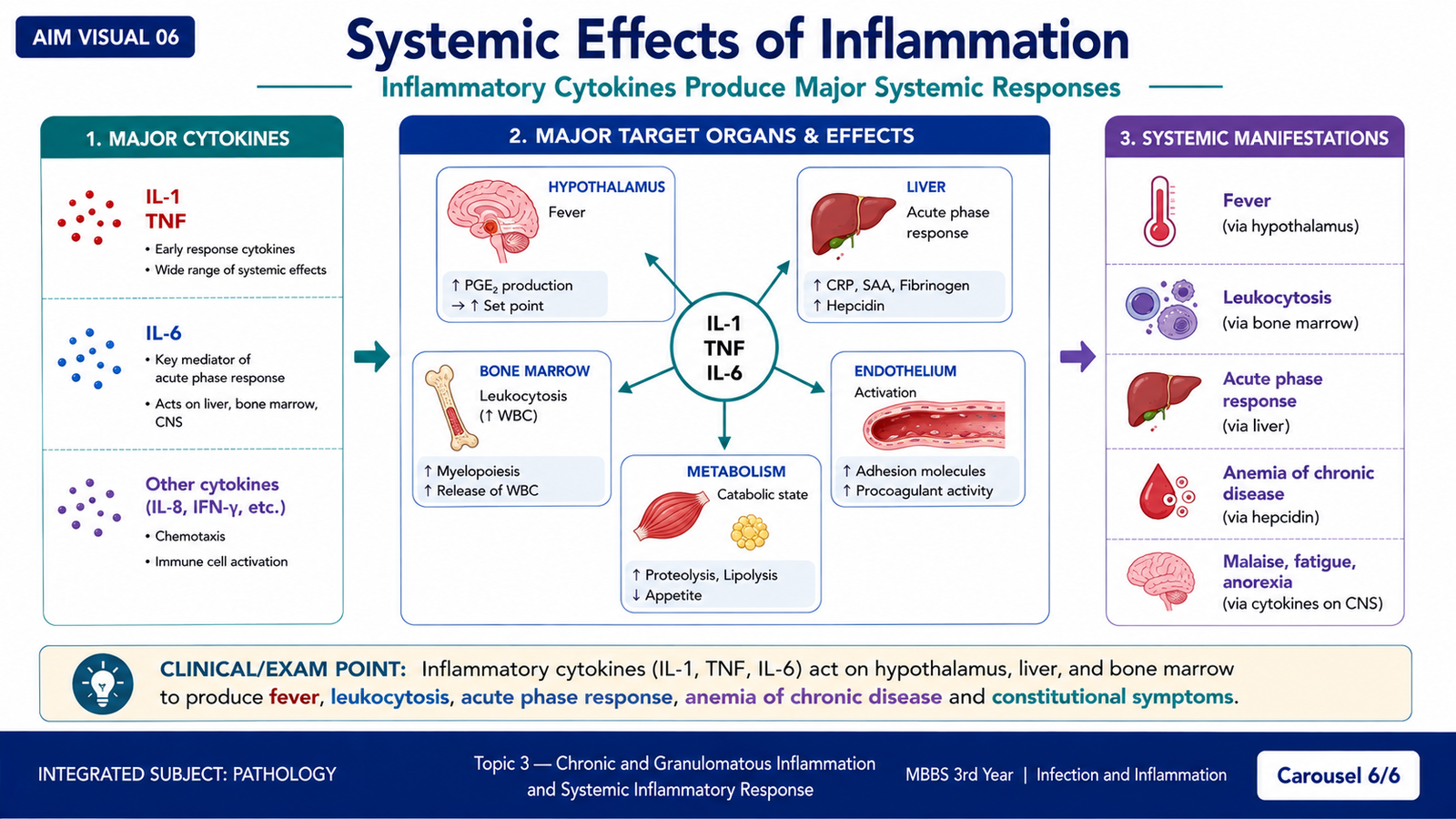
Inflammation begins locally, but inflammatory mediators can enter the circulation and produce effects throughout the body. These changes are collectively called the systemic inflammatory response. Important mediators include cytokines such as IL-1, TNF and IL-6. Their actions on the hypothalamus, liver and bone marrow explain many of the systemic manifestations.
1. Fever
IL-1 and TNF stimulate prostaglandin production in the hypothalamus. Prostaglandins raise the hypothalamic temperature set point, producing fever. Fever is therefore an actively regulated increase in body temperature rather than simple accumulation of body heat.
2. Acute-Phase Protein Production
Cytokines, particularly IL-6, stimulate hepatocytes to increase synthesis of acute-phase proteins. These circulating proteins participate in host defense and provide laboratory evidence of an inflammatory response.
Important acute-phase proteins include:
- C-reactive protein (CRP)
- Serum amyloid A (SAA)
- Fibrinogen
Increased fibrinogen promotes rouleaux formation of red blood cells. Rouleaux sediment more rapidly, contributing to an increased erythrocyte sedimentation rate (ESR).
3. Leukocytosis
Inflammatory cytokines stimulate release of leukocytes from bone-marrow storage pools and increase their production. This causes an increase in the circulating white-cell count known as leukocytosis.
The type of leukocyte increased may vary with the nature of the inflammatory stimulus, but the general principle is that inflammatory mediators act on the marrow to increase the availability of defensive leukocytes.
4. Other Constitutional Effects
Inflammatory cytokines can produce generalized symptoms such as malaise, reduced appetite and increased sleepiness. These manifestations occur because the inflammatory response affects the central nervous system and systemic metabolism rather than remaining confined to the original site of injury.
5. Severe Systemic Inflammatory Response
When inflammatory mediators are produced in very large amounts and circulate widely, their protective effects may become harmful. Excessive systemic cytokine activity can produce widespread vascular and metabolic disturbances and may contribute to severe systemic inflammatory dysfunction.
The important concept is that the same mediators that are useful when acting locally can become damaging when their activation is excessive and widespread.
Integrated Mechanism Flow
↓
2. Persistent activation and recruitment of macrophages and lymphocytes
↓
3. Cytokines, chemokines and macrophage products maintain inflammation
↓
4. Tissue injury occurs while growth factors simultaneously stimulate repair
↓
5. Fibroblast activation, collagen deposition and tissue remodeling may produce fibrosis
↓
6. Difficult-to-eradicate stimuli may produce organized granulomatous inflammation
↓
7. Cytokines entering the circulation produce systemic effects such as fever, acute-phase protein production and leukocytosis.
⭐ AIM High-Yield Review
- Chronic inflammation is prolonged inflammation in which active inflammation, tissue destruction and repair occur together.
- Its characteristic inflammatory cells are macrophages, lymphocytes and plasma cells, rather than the neutrophil predominance typical of acute inflammation.
- Major causes include persistent infections, immune-mediated inflammatory diseases and prolonged exposure to difficult-to-remove harmful agents.
- Macrophages are the central effector cells of chronic inflammation and can promote both tissue injury and repair.
- IFN-γ is an important activator of macrophages during cell-mediated inflammatory responses.
- TNF and IL-1 promote inflammatory reactions and contribute to systemic manifestations.
- The three classic morphological features of chronic inflammation are mononuclear-cell infiltration, tissue destruction and healing by fibrosis.
- A granuloma is an organized collection of activated macrophages, often with epithelioid cells, giant cells and surrounding lymphocytes.
- A giant cell may occur in granulomatous inflammation, but a giant cell alone does not define a granuloma.
- Immune granuloma formation involves persistent antigenic stimulation, T-cell activation, IFN-γ and macrophage activation.
- Fever results when inflammatory cytokines promote hypothalamic prostaglandin synthesis and raise the temperature set point.
- IL-6 is an important stimulus for hepatic production of acute-phase proteins.
- CRP, serum amyloid A and fibrinogen are important acute-phase proteins.
- Increased fibrinogen promotes rouleaux formation and contributes to an increased ESR.
- Leukocytosis occurs because inflammatory mediators stimulate leukocyte release and production in the bone marrow.
🎥 AIM Video Learning
Watch this video after completing the learning material to reinforce chronic inflammation, its causes, cellular features and morphology.