
This chapter follows the KMU learning outcomes and moves from common pyogenic cocci to rickettsial and chlamydial infections in a logical sequence. First understand how each organism produces disease and how the laboratory identifies it; then use the high-yield review for revision.
Topic 17 — Pyogenic, Rickettsial and Chlamydial Infections
A focused introduction to important pyogenic cocci, rickettsial organisms and Chlamydia, emphasizing their defining properties, disease mechanisms and laboratory diagnosis.
Topic Introduction
Pyogenic, rickettsial and chlamydial infections represent different ways in which bacteria cause human disease. Pyogenic bacteria characteristically produce acute inflammation rich in neutrophils and often form pus. Important pyogenic cocci include gram-positive organisms such as Staphylococcus aureus and Streptococcus pyogenes, as well as clinically important gram-negative cocci such as Neisseria species. Rickettsiae are small obligate intracellular bacteria that commonly infect vascular endothelial cells and are transmitted by arthropod vectors. Chlamydiae are also obligate intracellular bacteria but have a distinctive developmental cycle involving elementary and reticulate bodies. The key to understanding these infections is to connect organism properties with tissue injury, clinical features and the specimen or laboratory test used for diagnosis.
A. Pyogenic Infections, Boils and Furuncles
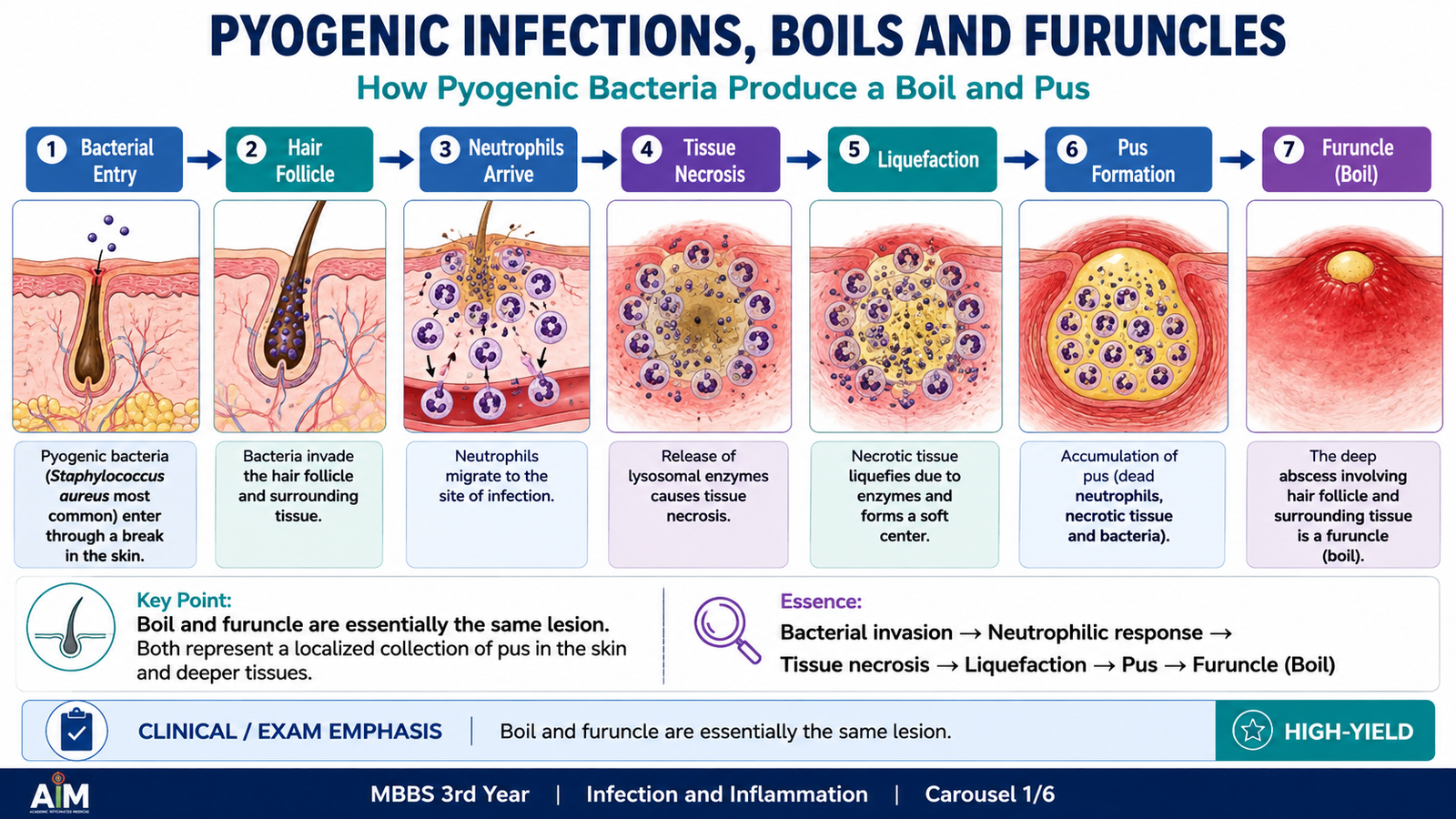
Pyogenic infections are bacterial infections in which the inflammatory response is dominated by neutrophils and commonly results in formation of pus. Pus contains neutrophils, necrotic tissue, microorganisms and protein-rich inflammatory fluid. The ability of pyogenic bacteria to invade tissues, trigger acute inflammation and sometimes produce local tissue destruction explains the characteristic pain, redness, swelling and purulent discharge.
Boil and furuncle
A furuncle, commonly called a boil, is a deep bacterial infection of a hair follicle that extends into the surrounding dermis and subcutaneous tissue. The usual causative organism is Staphylococcus aureus. The infection produces localized tissue necrosis and a collection of pus, creating a painful, tender swelling that may eventually drain.
Bacterial entry into hair follicle → multiplication in local tissue → acute neutrophilic inflammation → tissue necrosis and liquefaction → localized pus-filled lesion → clinically visible boil or furuncle
Important organisms causing pyogenic infections
Several bacteria can produce suppurative inflammation, but a few cocci are particularly important at undergraduate level.
- Staphylococcus aureus: a major cause of skin abscesses, boils, wound infections and other pus-forming lesions.
- Streptococcus pyogenes: commonly causes pyogenic infections of the skin, soft tissues and pharynx.
- Streptococcus pneumoniae: produces acute neutrophilic infections at sites such as the lungs and meninges.
- Neisseria gonorrhoeae: produces a marked neutrophilic inflammatory response in infected mucosal surfaces.
- Neisseria meningitidis: may produce acute suppurative meningitis and invasive bloodstream infection.
B. Important Gram-Positive Cocci
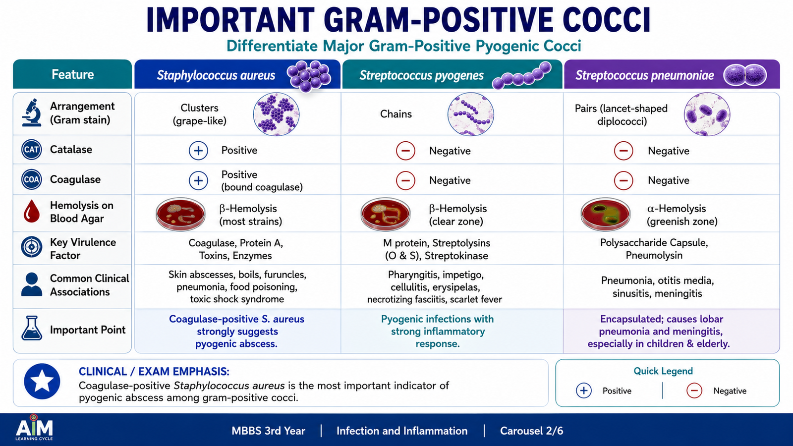
Gram-positive cocci appear purple on Gram staining because their thick peptidoglycan cell wall retains the crystal-violet stain. Their arrangement and biochemical characteristics help separate the major groups. Staphylococci usually appear in clusters, whereas streptococci and enterococci typically appear in chains or pairs. Their different virulence mechanisms produce different patterns of disease.
Staphylococcus aureus
S. aureus is a gram-positive coccus that classically forms grape-like clusters. It is catalase positive and coagulase positive. Its ability to adhere to tissue, escape host defenses and cause local tissue destruction makes it a particularly important cause of pyogenic infection.
Important properties
- Gram-positive cocci in clusters.
- Catalase positive.
- Coagulase positive.
- Frequently colonizes the skin and anterior nares.
- Can produce enzymes and toxins that promote invasion or tissue injury.
Pathophysiology
When S. aureus enters damaged skin or another susceptible site, it multiplies locally and provokes intense neutrophil recruitment. Bacterial factors help the organism resist clearance, while inflammatory cells release enzymes that contribute to tissue liquefaction. The result is the typical localized abscess or pus-filled lesion.
Colonization or tissue entry → bacterial multiplication → virulence-factor activity → intense neutrophil recruitment → tissue destruction → abscess and pus formation
Laboratory diagnosis
The most useful specimen depends on the infected site. Pus from an abscess or infected wound can be examined by Gram stain and cultured. Gram staining may show gram-positive cocci in clusters. Culture commonly produces characteristic colonies, and identification is supported by catalase and coagulase testing. Antimicrobial susceptibility testing may then be performed when clinically required.
Streptococcus pyogenes
S. pyogenes is a gram-positive coccus that typically occurs in chains. It is catalase negative and produces complete, or beta, hemolysis on blood agar. Its surface structures and secreted products help it adhere, spread through tissue and provoke inflammation.
Important properties and pathophysiology
- Gram-positive cocci in chains.
- Catalase negative.
- Beta-hemolytic on blood agar.
- Produces factors that promote tissue spread and interfere with host defense.
- Pyogenic tissue injury is produced largely through bacterial invasion combined with the host inflammatory response.
Laboratory diagnosis
A specimen is taken from the involved site, such as a throat swab or material from an infected lesion. Gram stain may demonstrate gram-positive cocci, while culture and characteristic hemolysis help identification. Appropriate biochemical or antigen-based methods may further support identification when required.
Streptococcus pneumoniae
S. pneumoniae is an encapsulated gram-positive diplococcus. The capsule is an important virulence factor because it reduces effective phagocytosis. The organism can therefore multiply in susceptible tissues and trigger an intense acute inflammatory response.
- Gram-positive, commonly lancet-shaped diplococci.
- Alpha-hemolytic on blood agar.
- Possesses a polysaccharide capsule.
- The capsule promotes survival by limiting phagocytosis.
- Laboratory diagnosis depends on appropriate specimens, microscopy, culture and identification characteristics.
| Feature | S. aureus | S. pyogenes | S. pneumoniae |
|---|---|---|---|
| Arrangement | Clusters | Chains | Diplococci |
| Catalase | Positive | Negative |
C. Important Gram-Negative Cocci
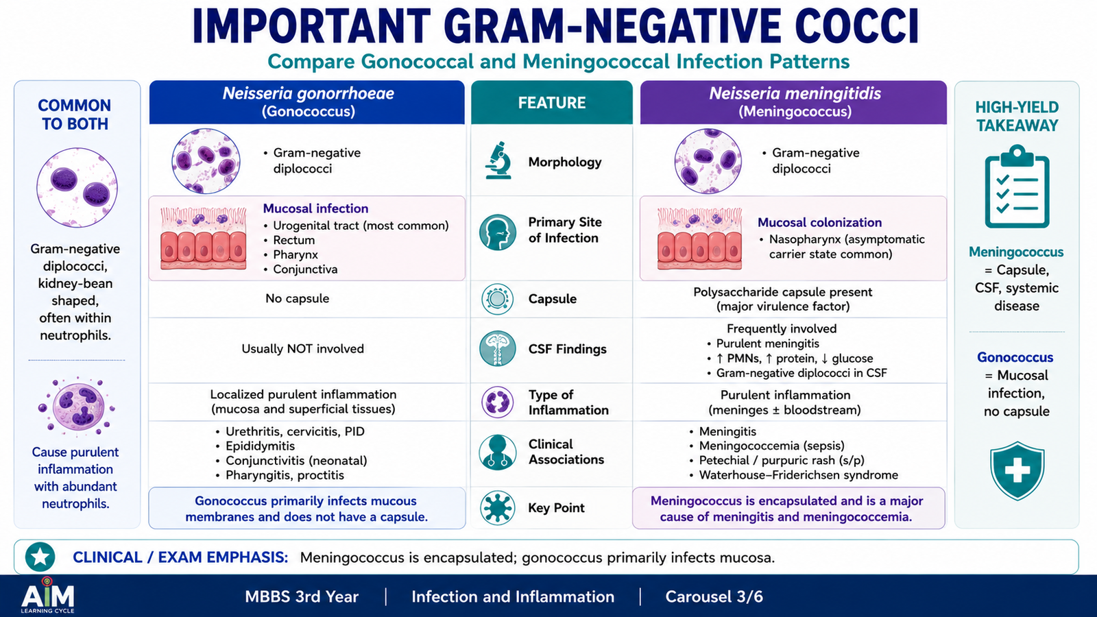
The medically important gram-negative cocci are mainly species of Neisseria. They characteristically appear as gram-negative diplococci. Two major human pathogens are Neisseria gonorrhoeae and Neisseria meningitidis. Although they share several structural features, they infect different sites and produce different disease patterns.
Neisseria gonorrhoeae
N. gonorrhoeae primarily infects mucosal epithelial surfaces. Attachment is followed by local invasion and a strong inflammatory response dominated by neutrophils. This explains why infected secretions can become purulent.
Important properties
- Gram-negative diplococcus.
- Oxidase positive.
- Adapted to human mucosal surfaces.
- Uses surface structures for attachment to epithelial cells.
- Produces marked local neutrophilic inflammation.
Pathophysiology
Laboratory diagnosis
Diagnosis begins with collection of an appropriate specimen from the involved site. Microscopy may show gram-negative diplococci within neutrophils, especially in suitable symptomatic specimens. Culture can be performed on enriched selective media, while nucleic-acid-based testing provides sensitive detection of gonococcal genetic material.
Neisseria meningitidis
N. meningitidis commonly colonizes the nasopharynx but may invade into the bloodstream and meninges. Its polysaccharide capsule helps the organism resist phagocytosis. When large amounts of bacterial endotoxin enter the circulation, severe systemic inflammation and vascular injury can occur.
Important properties
- Gram-negative diplococcus.
- Oxidase positive.
- Possesses a polysaccharide capsule.
- Colonizes the human nasopharynx.
- Can produce meningitis and invasive bloodstream infection.
Laboratory diagnosis
In suspected meningitis, cerebrospinal fluid is an important specimen, while blood may be examined when systemic infection is present. Gram stain can show gram-negative diplococci. Culture and further identification methods confirm the organism, while molecular methods may also demonstrate meningococcal genetic material.
| Feature | N. gonorrhoeae | N. meningitidis |
|---|---|---|
| Morphology | Gram-negative diplococci | Gram-negative diplococci |
| Important site | Mucosal surfaces | Nasopharynx, blood, meninges |
| Capsule | No major polysaccharide capsule | Important virulence factor |
D. Rickettsia — Core Properties and Pathophysiology
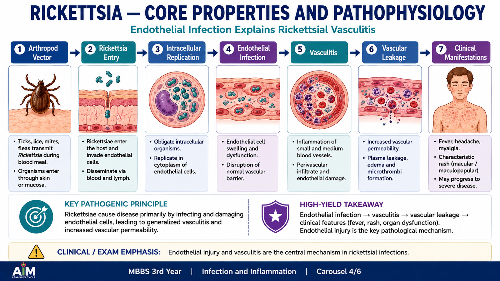
Rickettsiae are small gram-negative-like bacteria that are obligate intracellular organisms. This means they depend on living host cells for replication. Many medically important rickettsial infections are transmitted through arthropods such as ticks, mites, fleas or lice. A major pathological feature of several rickettsial diseases is their preference for vascular endothelial cells.
Important properties
- Small obligate intracellular bacteria.
- Cannot multiply independently in routine cell-free culture media.
- Many infections are associated with arthropod vectors.
- Several important species primarily infect vascular endothelial cells.
- Endothelial injury produces vascular inflammation and increased permeability.
How rickettsial disease develops
After entry into the body, the organism spreads and enters susceptible host cells. In many rickettsial infections, endothelial cells lining small blood vessels are particularly important targets. Intracellular multiplication damages these cells and produces vasculitis, meaning inflammation of the vessel wall. Damaged vessels become more permeable, allowing fluid to escape into surrounding tissue. This vascular injury helps explain systemic manifestations such as fever, headache and rash in several rickettsial illnesses.
Vector exposure → organism enters host → endothelial-cell infection → intracellular multiplication → endothelial injury and vasculitis → increased vascular permeability → systemic illness and characteristic tissue manifestations
E. Important Rickettsial Diseases and Laboratory Diagnosis
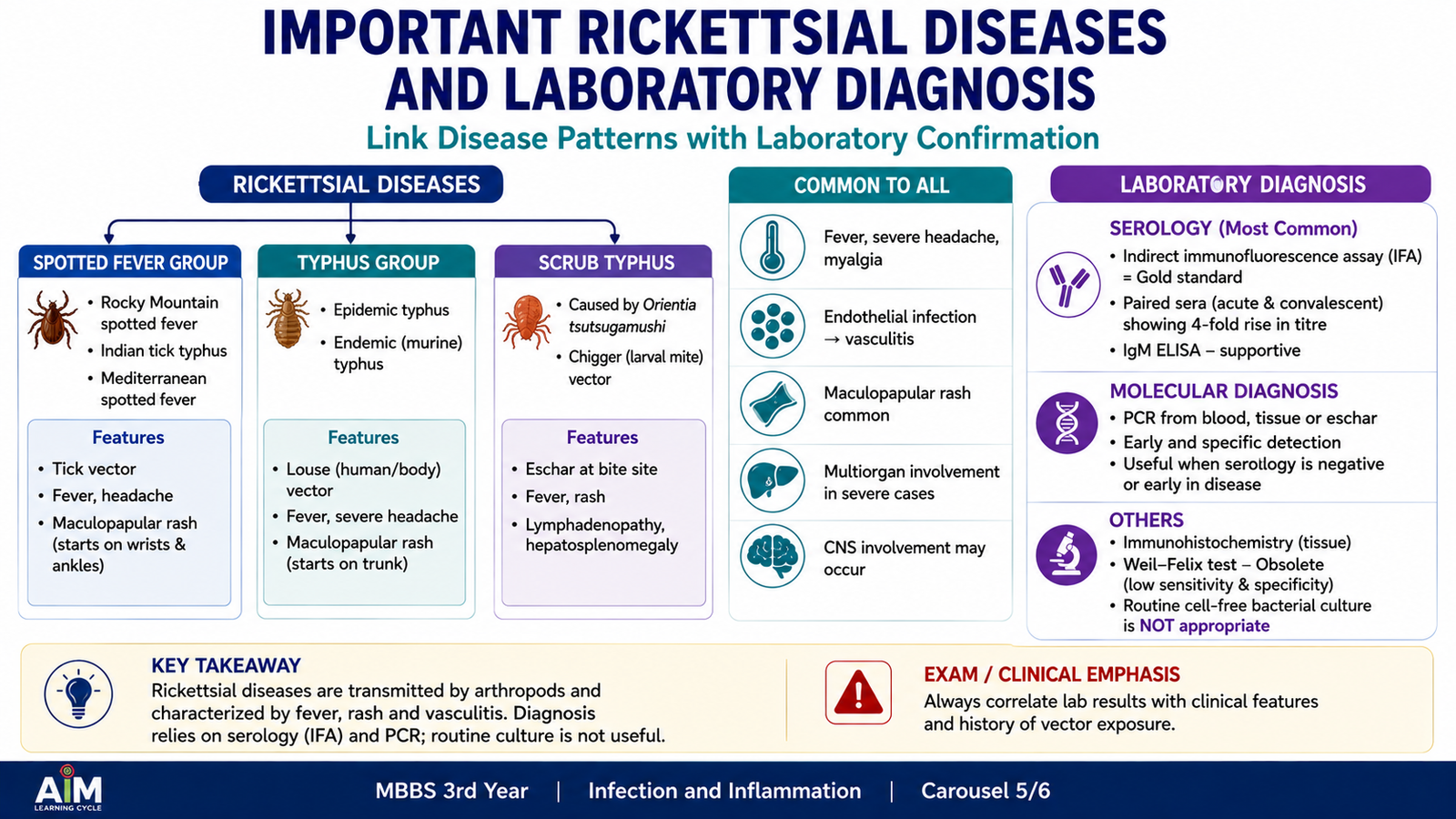
Rickettsial diseases are grouped according to the responsible organisms and their epidemiological pattern. At undergraduate level, the most useful approach is to recognize that many present as an acute febrile illness following vector exposure and that endothelial injury is a central mechanism. The exact clinical pattern varies with the species.
Major disease groups
- Spotted-fever group rickettsioses: commonly associated with tick transmission and characterized by fever with vascular injury that may produce a rash.
- Typhus-group rickettsioses: include infections associated with lice or fleas and may produce fever, headache and rash.
- Scrub typhus: caused by Orientia tsutsugamushi, transmitted by larval mites and characterized by a systemic febrile illness; an eschar may occur at the inoculation site.
The rash seen in several rickettsial illnesses reflects injury to small blood vessels in the skin. In more severe disease, vascular damage in other organs can contribute to systemic complications.
Laboratory diagnosis
Direct routine culture is not usually the first diagnostic approach because rickettsiae require living cells for multiplication. Diagnosis therefore relies mainly on methods that demonstrate either the organism or the immune response to it.
- Serology: detects antibodies directed against the infecting organism and is widely used in diagnosis.
- Molecular detection: PCR can demonstrate rickettsial genetic material in appropriate specimens.
- Direct detection: specialized methods can detect the organism or its antigens in selected settings.
- Clinical correlation: history of possible vector exposure and characteristic clinical findings are important when interpreting laboratory results.
F. Chlamydial Infections
Chlamydiae are obligate intracellular bacteria with a highly characteristic developmental cycle. Unlike ordinary bacteria that simply divide extracellularly, Chlamydia alternates between two forms: the infectious elementary body and the metabolically active reticulate body. This intracellular cycle explains both its biology and the methods used for laboratory diagnosis.
Important properties
- Obligate intracellular bacteria.
- Depend on host cells for replication.
- Possess a distinctive biphasic developmental cycle.
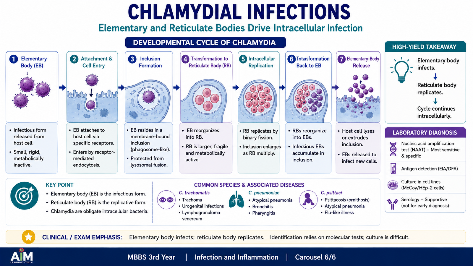
- Elementary body: infectious extracellular form adapted for transmission between cells.
- Reticulate body: intracellular, metabolically active replicative form.
Developmental cycle and pathophysiology
The elementary body attaches to and enters a susceptible epithelial cell. Once inside, it reorganizes into a reticulate body. Reticulate bodies multiply within an intracellular inclusion. They then reorganize into elementary bodies, which are released and infect additional cells. Repeated infection and the accompanying host inflammatory response produce epithelial injury and the clinical manifestations of chlamydial disease.
Elementary body attaches to epithelial cell → enters cell → converts to reticulate body → intracellular replication → reticulate bodies convert back to elementary bodies → release and spread to new cells
Important chlamydial diseases and clinical features
Chlamydia trachomatis infects epithelial cells at several body sites. Different strains are associated with different clinical patterns.
- Genital infection: may produce urethritis or cervicitis. Infection can sometimes be minimally symptomatic while still causing persistent inflammation.
- Ascending genital tract infection: spread from the lower genital tract can produce upper reproductive-tract inflammation.
- Conjunctival infection: certain strains infect ocular epithelium and produce conjunctival inflammation.
- Trachoma: repeated chronic conjunctival infection can lead to scarring and progressive ocular damage.
- Lymphogranuloma venereum: particular strains produce a more invasive infection involving regional lymphatic tissue.
Chlamydia pneumoniae is associated mainly with respiratory tract infection, whereas Chlamydia psittaci can cause respiratory illness following exposure to infected birds.
Laboratory diagnosis
Because Chlamydia is intracellular, the diagnostic method must be appropriate for an organism that cannot be identified reliably by ordinary routine bacterial culture methods. The specimen should contain infected epithelial material from the relevant site.
- Nucleic acid amplification testing: detects chlamydial nucleic acid and is particularly useful for C. trachomatis infection.
- Direct detection methods: chlamydial antigens or organisms may be demonstrated using specialized techniques.
- Serology: may be useful in selected chlamydial infections depending on the clinical context.
- Cell culture: is possible in specialized laboratories because the organism requires living cells, but it is not equivalent to ordinary bacterial culture on standard media.
Integrated Mechanism Flow
Although these bacterial groups differ greatly, each can be understood by connecting microbial properties to tissue injury and laboratory diagnosis.
↓
Characteristic microbial property determines its behavior
Pyogenic cocci → extracellular multiplication and neutrophilic response
Rickettsiae → intracellular endothelial infection
Chlamydiae → intracellular epithelial developmental cycle
↓
Host inflammatory response and tissue injury
↓
Characteristic clinical manifestation
↓
Appropriate specimen is collected
↓
Microscopy, culture, serology or molecular testing supports diagnosis
Important Comparison
| Feature | Pyogenic cocci | Rickettsiae | Chlamydiae |
|---|---|---|---|
| Major biological pattern | Usually extracellular pyogenic infection | Obligate intracellular | Obligate intracellular |
| Important host cell/tissue | Depends on organism and infection site | Vascular endothelium in many diseases | Epithelial cells |
| Typical injury pattern | Neutrophilic inflammation and pus | Vasculitis and vascular leakage | Intracellular infection with epithelial inflammation |
| Routine culture | Useful for many organisms | Not routine; requires living cells | Not routine; requires living cells |
| Important diagnostic approach | Gram stain, culture, identification tests | Serology and molecular methods | Molecular or specialized detection methods |
⭐ AIM High-Yield Review
- A boil and a furuncle describe a deep infection of a hair follicle, most commonly caused by Staphylococcus aureus.
- Pyogenic inflammation is characterized by prominent neutrophils and pus formation.
- S. aureus is a gram-positive coccus in clusters and is both catalase positive and coagulase positive.
- S. pyogenes is a catalase-negative gram-positive coccus associated with beta-hemolysis.
- S. pneumoniae is an encapsulated, typically lancet-shaped gram-positive diplococcus.
- Neisseria gonorrhoeae and N. meningitidis are important gram-negative diplococci.
- Intracellular gram-negative diplococci within neutrophils are an important microscopic clue in suitable specimens from gonococcal infection.
- The meningococcal polysaccharide capsule is an important virulence factor that helps the organism resist phagocytosis.
- Rickettsiae are obligate intracellular bacteria; many are transmitted by arthropod vectors.
- ⭐ Endothelial infection and vasculitis are central to the pathophysiology of many rickettsial diseases.
- Rickettsial diagnosis commonly relies on serology or molecular detection rather than ordinary routine culture.
- Chlamydia has two major developmental forms: the infectious elementary body and replicating reticulate body.
- Chlamydial tissue injury results from intracellular infection together with the host inflammatory response.
- Laboratory diagnosis of C. trachomatis commonly depends on detection of microbial nucleic acid in an appropriate specimen.
- ⭐ The key distinction is: pyogenic cocci commonly produce neutrophil-rich suppuration, rickettsiae characteristically produce endothelial injury, and Chlamydia has a unique intracellular elementary-body/reticulate-body cycle.