
This chapter follows the supplied KMU learning outcomes for tissue healing, regeneration and the cell cycle. First understand how cells regenerate and how damaged tissue is repaired, then use the high-yield section for focused revision.
Topic 7 — Tissue Healing, Regeneration and the Cell Cycle
Module/Theme: Infection and Inflammation
Understand how injured tissues recover through regeneration or repair, how the major phases of healing occur, and how the cell cycle controls the proliferative response of different cell populations.
Topic Introduction
When tissue is injured, the body attempts to restore its structure and function. This recovery can occur by regeneration, in which lost cells are replaced by cells of the same type, or by repair, in which damaged tissue is replaced partly or completely by connective tissue and scar. The final outcome depends mainly on the type of cells involved, the severity of injury and whether the supporting extracellular matrix remains intact. Cell proliferation is therefore central to healing. Understanding the cell cycle and the proliferative capacity of different tissues helps explain why some organs recover almost completely while others heal mainly by fibrosis.
A. Regeneration and Repair: The Two Main Responses to Tissue Injury
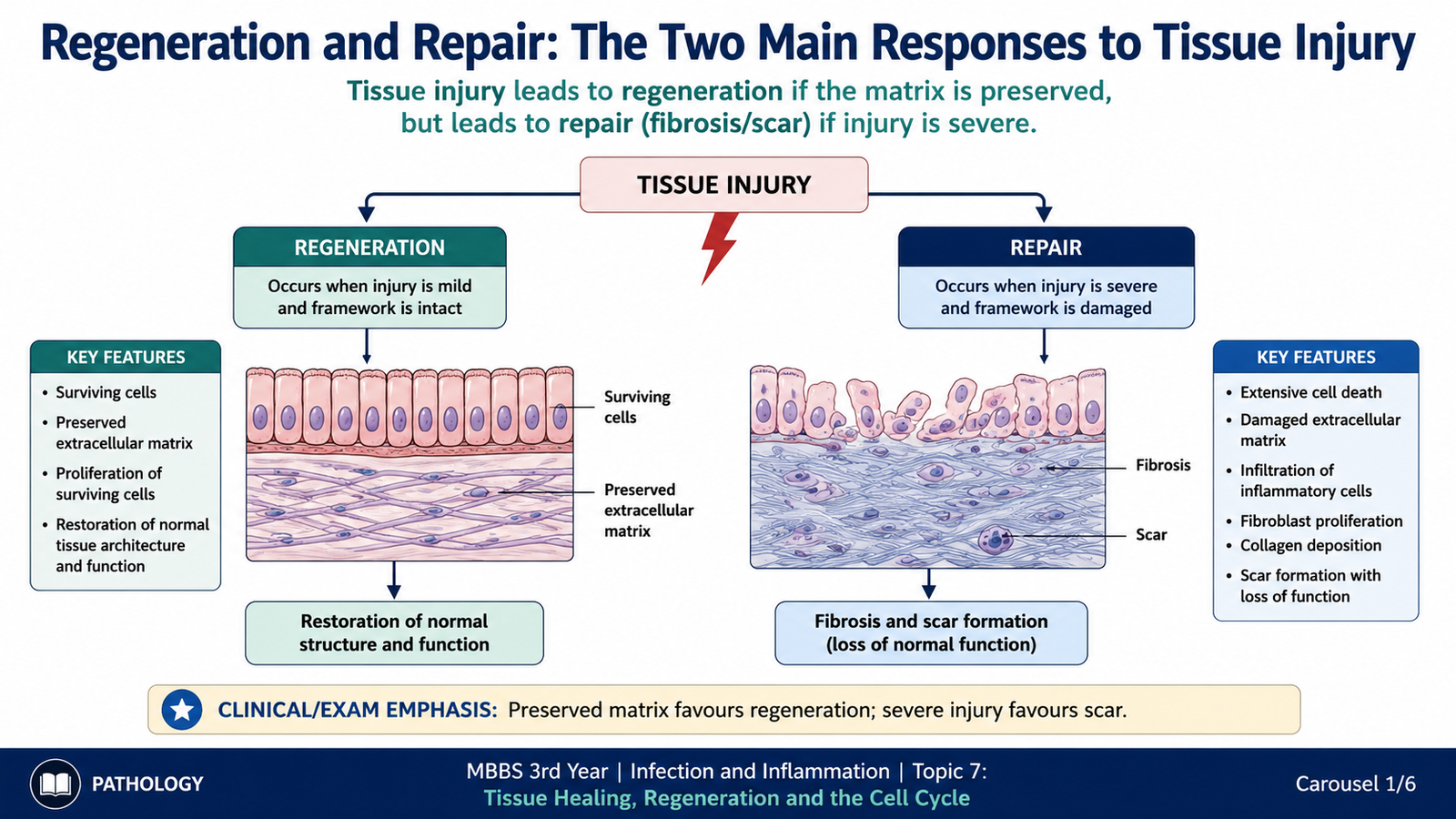
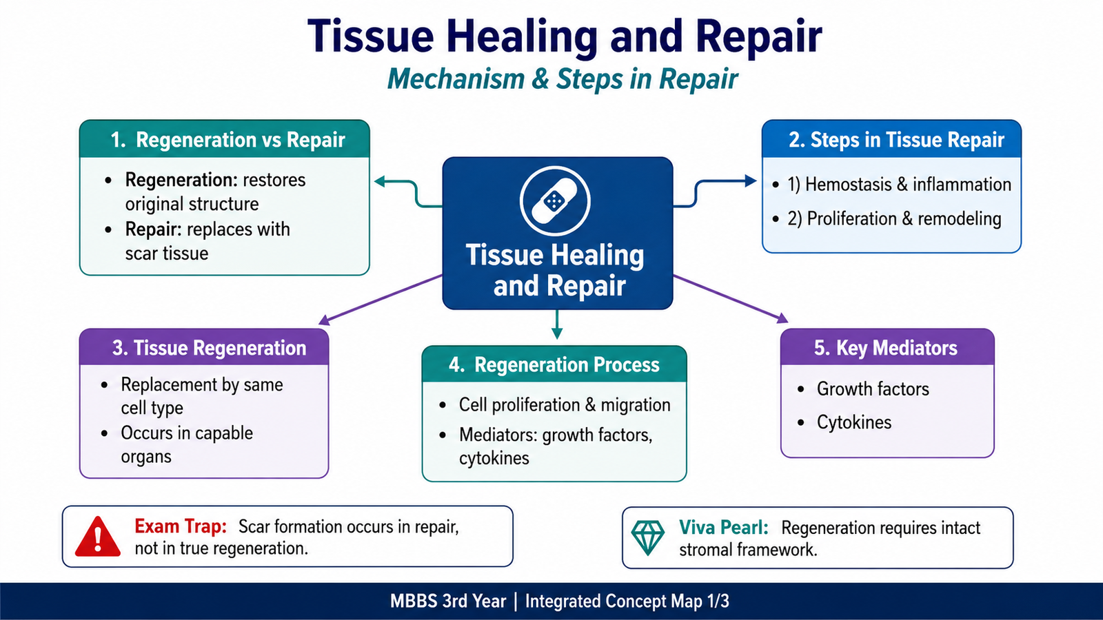
Tissue healing is the coordinated response that follows injury and attempts to restore tissue integrity. The two fundamental mechanisms are regeneration and repair by connective tissue deposition. These processes are related, and both may occur in the same wound.
Regeneration
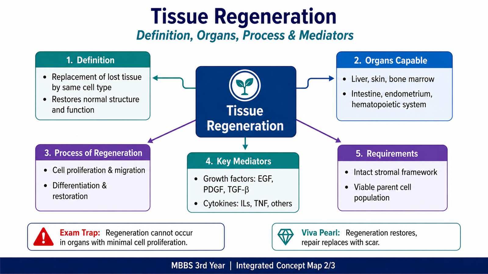
Regeneration means replacement of lost or damaged cells by proliferation of surviving cells and, in some tissues, by activation of tissue stem cells. The regenerated cells are of the same type as those that were lost, so the original tissue structure and function can potentially be restored.
Regeneration is most successful when:
- the injured cells are capable of proliferation;
- the injury is limited or reversible;
- the supporting extracellular matrix framework is preserved;
- appropriate growth signals are available.
Repair
Repair is restoration of tissue integrity mainly by deposition of connective tissue. It becomes particularly important when the injury is severe, the extracellular matrix has been destroyed, or the affected cells have little or no proliferative capacity.
Repair therefore commonly produces a scar. The scar restores structural strength but does not usually reproduce the specialized function of the original tissue.
| Feature | Regeneration | Repair |
|---|---|---|
| Main process | Replacement by the same type of cells | Connective tissue deposition and scar formation |
| Architecture | Can return toward normal | Original architecture may not be completely restored |
| Functional result | Potential restoration of normal function | Structural continuity restored, but specialized function may be reduced |
| Favoured when | Cells can divide and matrix framework is preserved | Damage is extensive or cells cannot regenerate adequately |
B. Major Steps in Tissue Healing and Repair
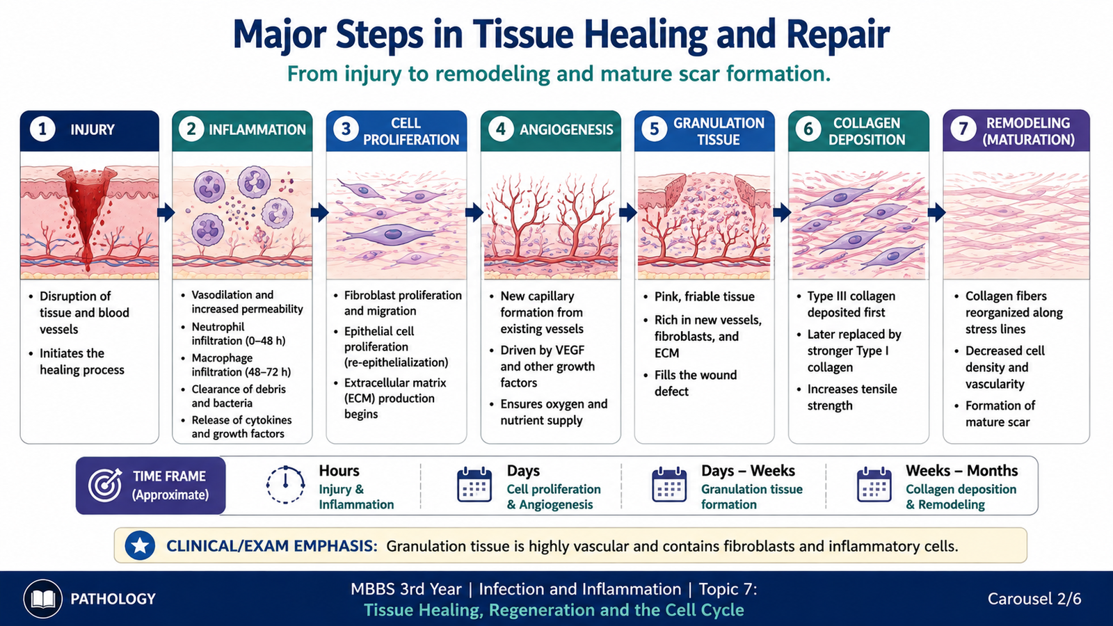
Healing is not a single event. It is a sequence of overlapping responses that remove damaged material, produce new tissue and finally remodel that tissue. The exact balance between regeneration and fibrosis varies with the type and severity of injury.
1. Control of Injury and Inflammation
Tissue injury damages cells and blood vessels. The immediate response limits further damage and creates conditions for healing. Inflammatory cells remove dead tissue and other unwanted material from the injured area. This clearance is important because persistent necrotic tissue interferes with effective repair.
2. Proliferation of Surviving Cells
Cells that remain viable may be stimulated to enter the cell cycle. In tissues capable of regeneration, this proliferation helps replace cells that were lost. Tissue stem or progenitor cells may also contribute when mature cells cannot provide sufficient replacement.
3. Formation of New Blood Vessels
Healing tissue requires oxygen and nutrients. New capillaries therefore develop from existing vessels through angiogenesis. The newly formed vascular tissue contributes to the soft, vascular appearance of early healing tissue.
4. Formation of Granulation Tissue
Granulation tissue is newly formed healing tissue composed mainly of proliferating small blood vessels, fibroblasts and loose extracellular matrix. It fills areas where normal tissue has been lost and provides the framework for further repair.
Its characteristic appearance results from:
- numerous thin-walled newly formed vessels;
- proliferating fibroblasts;
- loose extracellular matrix;
- variable inflammatory cells.
5. Fibroblast Proliferation and Collagen Deposition
When regeneration cannot completely restore the tissue, fibroblasts migrate and proliferate within the damaged area. They synthesize extracellular matrix, particularly collagen. Increasing collagen deposition gradually converts the soft early repair tissue into a stronger fibrous scar.
6. Remodeling
Newly deposited connective tissue is subsequently reorganized. Some extracellular matrix is removed, other components are deposited, and collagen becomes more effectively arranged. As remodeling progresses, the scar becomes less cellular and less vascular and gains tensile strength.
C. Tissue Regeneration and Its Major Mediators
Regeneration requires surviving cells or tissue stem cells to proliferate and replace the lost population. This process is carefully regulated. Cells do not divide simply because tissue has been injured; they respond to signals from growth factors, cell receptors and the extracellular environment.
How Regeneration Occurs
Following tissue loss, local signals stimulate surviving cells to move from a resting state into the cell cycle. These cells replicate their DNA, divide and replace the lost cells. When adequate tissue mass has been restored, proliferative signals decrease and cell growth returns toward its usual level.
Tissue loss → release or activation of growth signals → receptor activation on responsive cells → intracellular signalling → cell-cycle entry → proliferation → restoration of cell population
Growth Factors
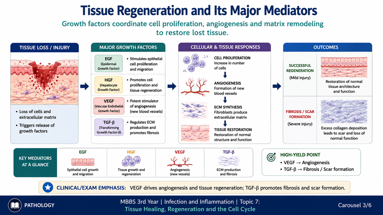
Growth factors are signalling proteins that influence cell proliferation, migration, survival and extracellular matrix production. Their effects depend on the target cell and the receptor expressed by that cell.
Important mediators relevant to regeneration and repair include:
- Epidermal growth factor (EGF) — promotes proliferation of several epithelial cell types.
- Transforming growth factor-alpha (TGF-α) — has growth-promoting effects on epithelial cells.
- Hepatocyte growth factor (HGF) — contributes importantly to hepatocyte proliferation.
- Vascular endothelial growth factor (VEGF) — stimulates formation of new blood vessels.
- Fibroblast growth factors (FGFs) — participate in fibroblast proliferation and angiogenesis.
- Platelet-derived growth factor (PDGF) — promotes migration and proliferation of fibroblasts and other mesenchymal cells.
- Transforming growth factor-beta (TGF-β) — has an important role in connective tissue deposition and fibrosis.
These mediators work together rather than independently. For example, regeneration requires signals that increase cell proliferation, while repair additionally requires signals that promote fibroblast activity, angiogenesis and extracellular matrix deposition.
Role of the Extracellular Matrix
The extracellular matrix is more than structural support. It helps maintain cell organization and provides signals that influence cell growth, movement and differentiation. If its framework remains intact, regenerating cells can repopulate the injured area in an organized manner. If the framework is destroyed, restoration of normal architecture is more difficult and scar formation becomes more prominent.
D. Proliferative Capacity of Different Tissues
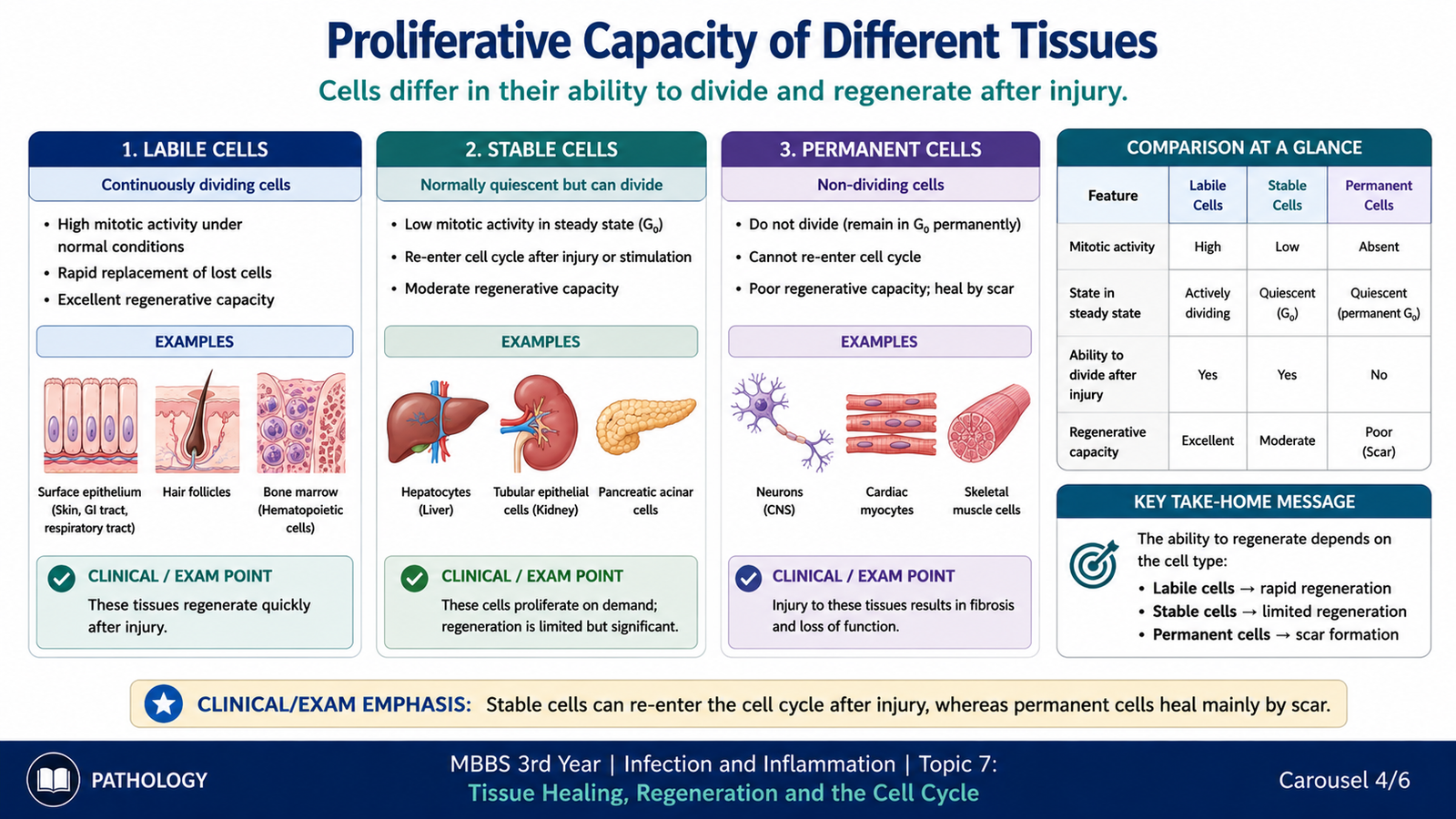
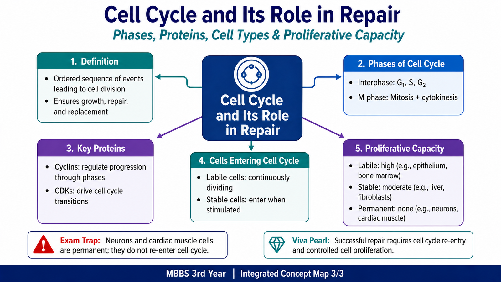
Different tissues have very different capacities for regeneration. This difference is largely determined by whether their cells normally divide continuously, remain resting but can re-enter the cell cycle, or are permanently unable to divide. For this reason, cells are commonly grouped as labile, stable and permanent cells.
Labile Cells
Labile cells divide continuously throughout life because they are repeatedly lost and must be replaced. Their tissues therefore have a strong regenerative capacity as long as the stem-cell compartment and supporting framework remain intact.
Important examples include:
- surface epithelium of the skin;
- epithelial lining of the gastrointestinal tract;
- hematopoietic cells of bone marrow.
Stable Cells
Stable cells normally have a low rate of division and often remain in a resting state. However, they can re-enter the cell cycle when stimulated by injury or loss of tissue. Their regenerative ability is therefore considerable under appropriate conditions.
Examples include:
- hepatocytes;
- renal tubular epithelial cells;
- fibroblasts;
- endothelial cells;
- smooth muscle cells.
The liver is an important example of regeneration by stable cells. Hepatocytes are usually not actively dividing, but after loss of liver tissue they can be stimulated to proliferate and restore tissue mass.
Permanent Cells
Permanent cells are terminally differentiated cells with very limited or absent ability to re-enter the cell cycle. Significant loss of these cells therefore cannot usually be corrected by proliferation of surviving mature cells.
Important examples include:
- neurons;
- cardiac muscle cells.
When permanent cells are destroyed, repair commonly depends on connective tissue deposition. This explains why major myocardial injury, for example, heals predominantly by scar rather than by complete replacement of lost cardiac muscle.
| Cell Group | Usual Proliferative State | Response to Injury | Examples |
|---|---|---|---|
| Labile | Continuously dividing | Strong regenerative response | Skin, intestinal epithelium, hematopoietic cells |
| Stable | Usually resting | Can re-enter cell cycle | Hepatocytes, renal tubular cells, fibroblasts, endothelium |
| Permanent | Terminally differentiated | Little or no mature-cell proliferation | Neurons, cardiac muscle cells |
E. Cell Cycle: Initiation and Major Phases
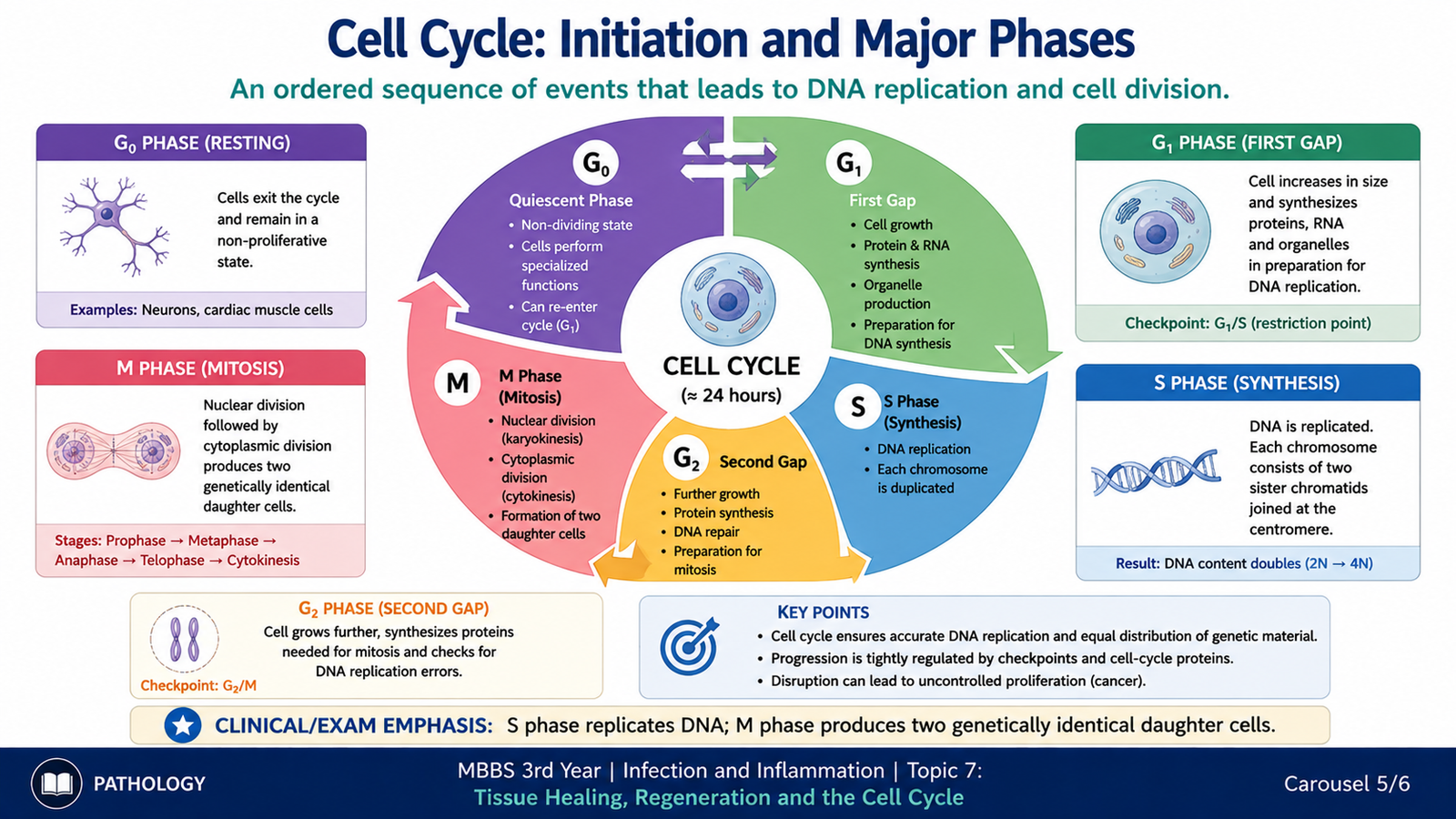
The cell cycle is the organized sequence through which a cell grows, duplicates its DNA and divides into daughter cells. Tissue regeneration depends on cells being able to enter and successfully complete this cycle. Cells that are not actively dividing may remain outside the cycle in a resting state known as G0.
Initiation of the Cell Cycle
Many cells enter the cell cycle in response to extracellular growth signals. Growth factors bind to specific receptors on the cell surface and activate intracellular signalling pathways. These signals increase the activity of proteins that move the cell from a resting state toward DNA synthesis and division.
G1 Phase
During G1, the cell grows and prepares for DNA replication. This phase is particularly important because the cell evaluates whether conditions are suitable for continued proliferation. Regulatory proteins determine whether the cell progresses toward DNA synthesis.
S Phase
The S phase is the period of DNA synthesis. The cell duplicates its genetic material so that each daughter cell can receive a complete set of chromosomes.
G2 Phase
During G2, the cell prepares for mitosis. It continues to grow and checks whether DNA replication has been completed appropriately before division proceeds.
M Phase
The M phase is mitosis, during which duplicated chromosomes are separated and the cell divides to form two daughter cells.
G0 State
G0 is a non-proliferating state outside the active cell cycle. Some cells can remain in G0 temporarily and return to the cycle when stimulated. Stable cells commonly behave in this way. Other highly differentiated cells remain functionally outside the proliferative cycle for prolonged periods.
G0 or resting cell → growth stimulation → G1 → S → G2 → M → daughter cells
F. Proteins That Regulate Cell-Cycle Progression
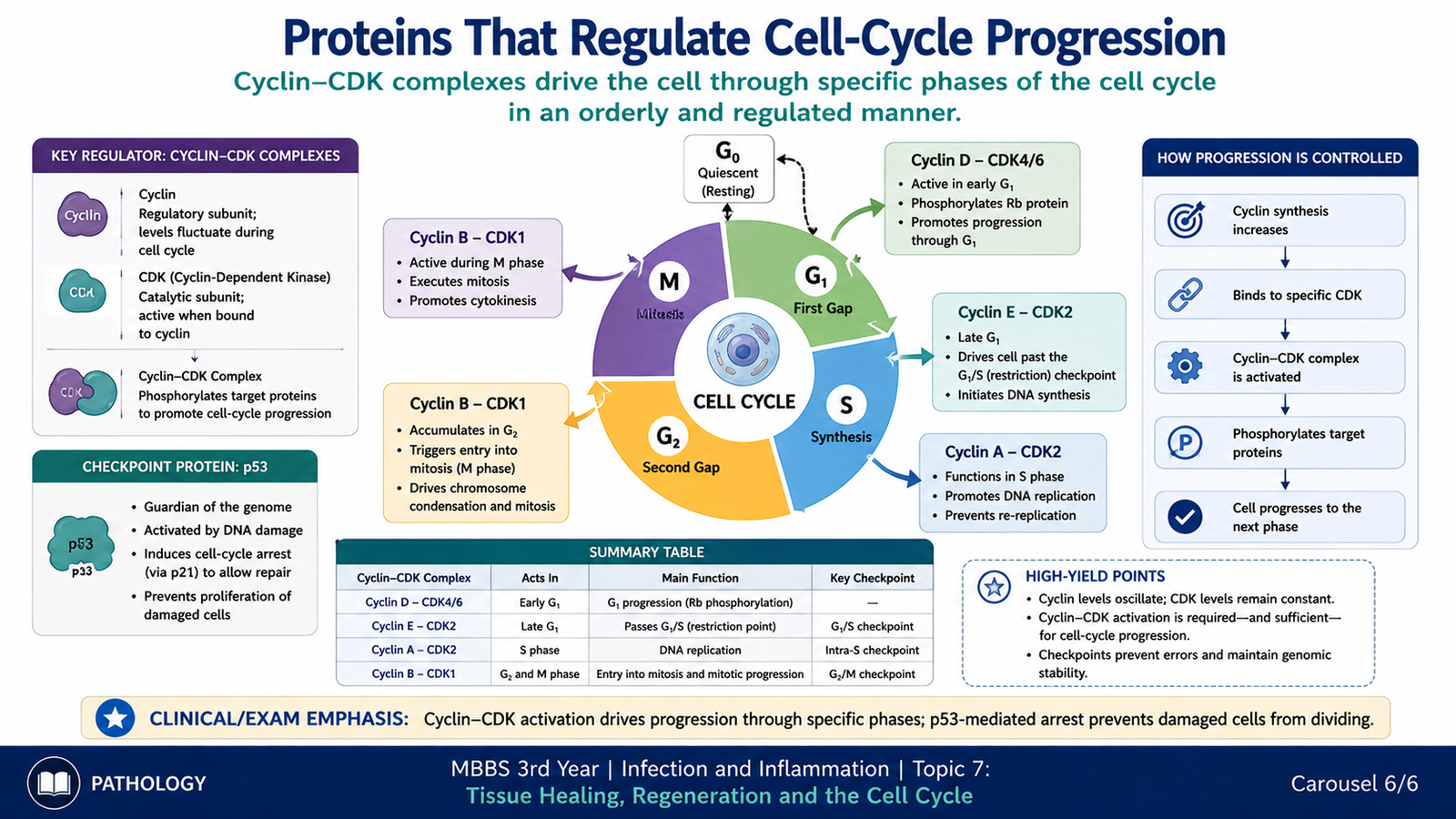
Cell division must be tightly controlled. Progress through the cell cycle is regulated mainly by proteins called cyclins and cyclin-dependent kinases (CDKs). Cyclins change in concentration during different phases of the cycle, while CDKs become active when associated with appropriate cyclins.
Cyclins and Cyclin-Dependent Kinases
Cyclin–CDK complexes phosphorylate target proteins that permit the cell to move through specific stages of the cycle. Their sequential activation provides an ordered system: a cell should not normally enter the next stage before essential events of the previous stage have occurred.
The important principle is:
Important Cyclin–CDK Relationships
At undergraduate level, the major associations help explain how different phases are controlled:
- Cyclin D works mainly with CDK4 and CDK6 during early G1.
- Cyclin E with CDK2 promotes progression from G1 toward S phase.
- Cyclin A with CDK activity contributes to progression through S phase.
- Cyclin B with CDK1 promotes entry into mitosis.
Cell-Cycle Checkpoints
Checkpoints are control points that help prevent inappropriate progression through the cell cycle. They allow the cell to assess whether essential processes such as DNA integrity and DNA replication are satisfactory before division continues.
A particularly important control occurs near the transition from G1 to S phase. Once a cell commits to DNA synthesis, progression toward division becomes much more difficult to reverse. Proteins that inhibit cyclin–CDK activity or respond to DNA damage can delay the cycle when conditions are unsuitable.
Role of p53 in Cell-Cycle Control
The protein p53 is important in the cellular response to DNA damage. When DNA is damaged, p53 can promote expression of proteins that inhibit cyclin–CDK activity, producing temporary cell-cycle arrest. This gives the cell an opportunity to deal with the damage before replication continues. If damage is severe and cannot be adequately managed, p53 can also contribute to pathways leading to cell death.
Integrated Mechanism Flow
The relationship between tissue injury, proliferative capacity and the final healing response can be summarized as follows:
↓
Loss of cells and activation of inflammatory/healing signals
↓
Growth factors stimulate surviving responsive cells
↓
Entry into the cell cycle and cellular proliferation
↓
If cells can proliferate and matrix is preserved → regeneration
↓
If regeneration is inadequate or tissue framework is severely damaged → fibroblast proliferation and extracellular matrix deposition
↓
Scar formation and remodeling
⭐ AIM High-Yield Review
- Regeneration replaces damaged cells with cells of the same type and can restore normal tissue architecture and function.
- Repair restores tissue integrity mainly through connective tissue deposition and scar formation.
- Successful regeneration depends on both the proliferative capacity of cells and preservation of the extracellular matrix framework.
- Major healing events include inflammation and clearance, cell proliferation, angiogenesis, granulation tissue formation, collagen deposition and remodeling.
- Granulation tissue contains newly formed blood vessels, fibroblasts and loose extracellular matrix.
- Growth factors regulate cell proliferation, migration, angiogenesis and extracellular matrix production.
- VEGF is especially important in angiogenesis, while TGF-β is important in extracellular matrix deposition and fibrosis.
- Labile cells continuously divide and include surface epithelia and hematopoietic cells.
- Stable cells are usually resting but can re-enter the cell cycle after stimulation; hepatocytes are an important example.
- Permanent cells, such as neurons and cardiac muscle cells, have very limited proliferative capacity.
- The active cell cycle consists mainly of G1 → S → G2 → M; cells outside the active cycle may be in G0.
- DNA is replicated during the S phase, while cell division occurs during the M phase.
- Cyclins and CDKs regulate orderly progression through different phases of the cell cycle.
- Cell-cycle checkpoints prevent inappropriate progression when important cellular requirements have not been satisfied.
- ⭐ The final balance between regeneration and scar formation is determined by the nature of the injured cells, severity of damage and integrity of the tissue framework.
Tissue Healing, Regeneration & Cell Cycle
Watch this video after completing the learning material to reinforce regeneration versus repair, the stages of tissue healing, growth-factor signalling, proliferative capacity of cells and regulation of the cell cycle.
- Regeneration versus repair by fibrosis
- Inflammation → proliferation → angiogenesis → granulation tissue → scar remodeling
- Growth factors involved in regeneration and repair
- Labile, stable and permanent cells
- G0, G1, S, G2 and M phases of the cell cycle
- Cyclins, CDKs and major cell-cycle checkpoints