
This chapter follows the KMU learning outcomes in a logical sequence. First understand where inflammatory mediators come from and what they do, then connect them with the morphologic patterns, outcomes and defects of inflammation. Revise the high-yield section only after understanding the explanations.
Inflammatory Mediators, Morphologic Patterns and Outcomes of Inflammation
Understand how plasma-derived and cell-derived chemical mediators coordinate inflammation, how different inflammatory responses appear morphologically, and how inflammation may resolve, heal, progress or become defective.
Topic Introduction
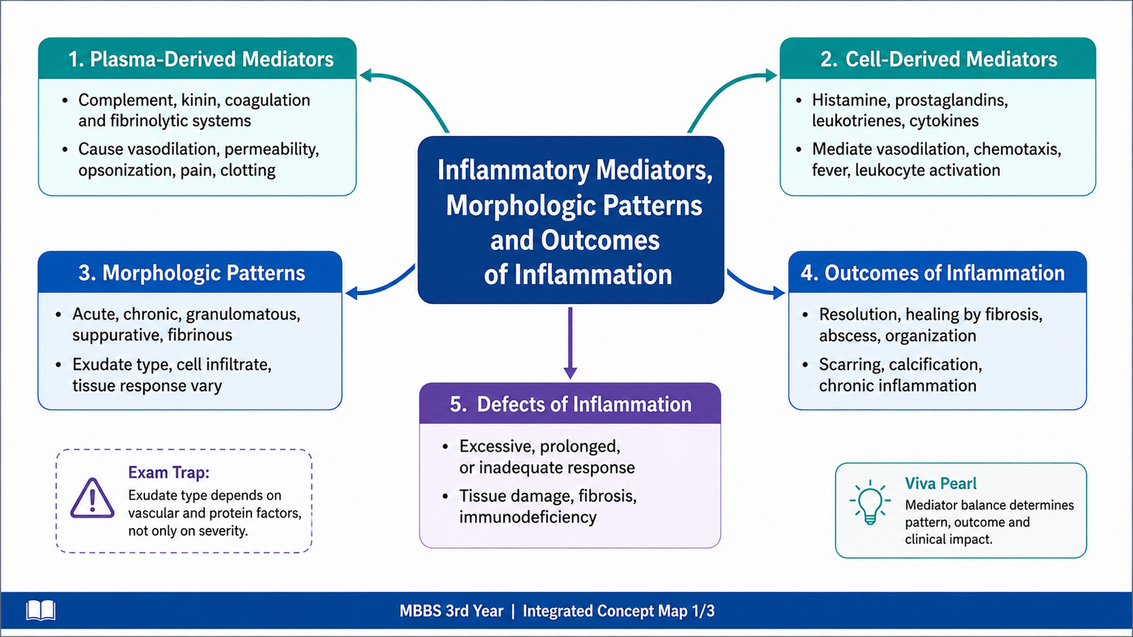
Inflammation is controlled by chemical substances called inflammatory mediators. These mediators are produced either from plasma proteins or from cells and are responsible for vascular dilatation, increased vascular permeability, leukocyte recruitment, pain, fever and tissue injury. The appearance of inflammation in tissues is not always identical; depending on the cause, site and severity, it may form serous fluid, fibrin, pus or an ulcer. The inflammatory response may then completely resolve, heal by fibrosis, form an abscess or progress to chronic inflammation. This chapter explains the major plasma-derived and cell-derived mediators, the cascades that generate them, the important morphologic patterns of inflammation, and the major consequences of defective inflammatory responses.
A. Overview of Inflammatory Mediators
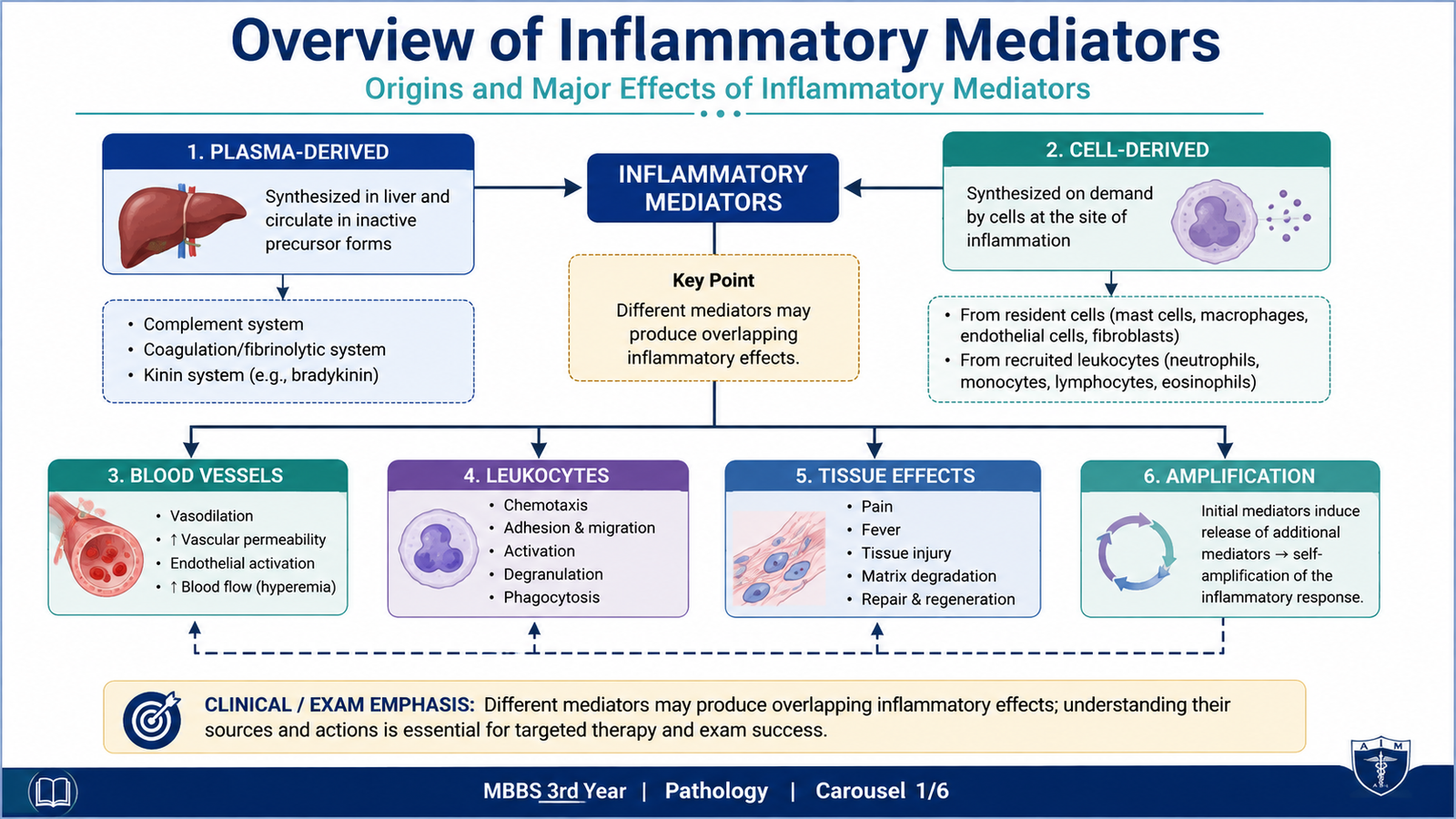
Inflammatory mediators are chemical signals that initiate, amplify and regulate inflammation. They act on blood vessels, leukocytes and surrounding tissues. Some mediators are already present in the circulation as inactive precursors, whereas others are stored inside cells or synthesized rapidly after cellular activation.
Major sources
| Source | Examples | General role |
|---|---|---|
| Plasma-derived | Complement proteins, kinins and products associated with coagulation | Circulate mainly as inactive precursors and become activated in cascades |
| Cell-derived | Histamine, prostaglandins, leukotrienes, cytokines, chemokines, nitric oxide, reactive oxygen species and lysosomal enzymes | Stored in granules or newly synthesized after cellular stimulation |
Important principles
Most mediators act by binding to specific receptors or by modifying enzymatic pathways. Their actions are usually short-lived because uncontrolled mediator activity can damage normal tissue. Different mediators often have overlapping actions. For example, several mediators can increase vascular permeability, while others cooperate in leukocyte recruitment.
- One mediator may have several effects. Histamine, for example, affects vascular tone and permeability.
- Different mediators may produce the same effect. Several mediators contribute to vasodilatation.
- Mediators can stimulate release of other mediators, creating amplification cascades.
- The same mediator may be beneficial or harmful depending on the amount and duration of its action.
- Most mediator activity is tightly controlled to prevent excessive inflammation and tissue injury.
B. Plasma-Derived Mediators and Their Cascades
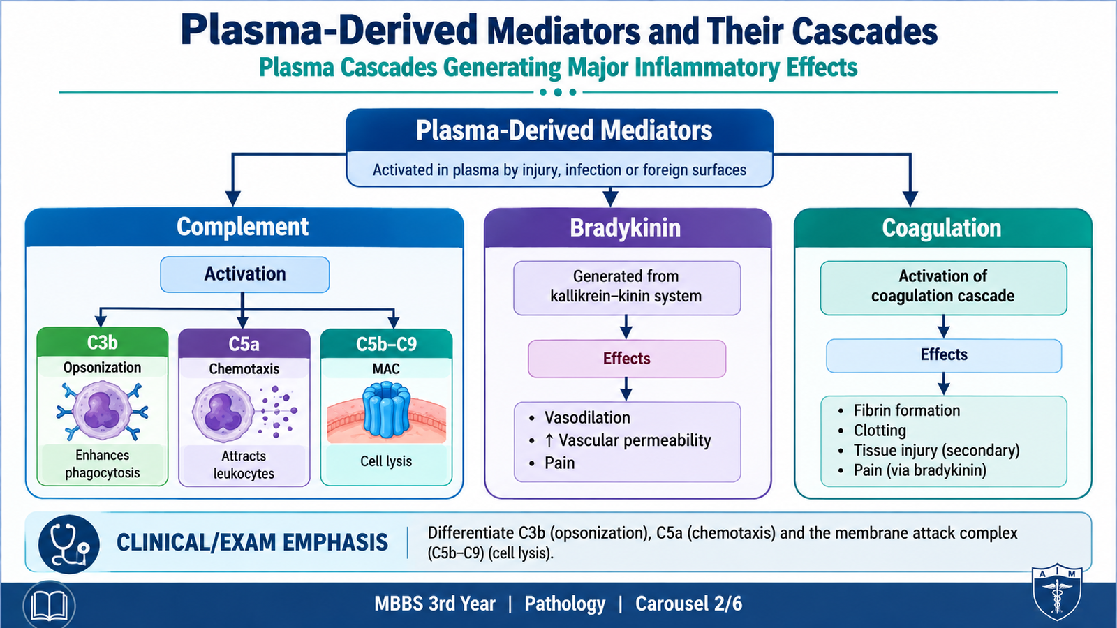
Plasma-derived inflammatory mediators are largely synthesized in the liver and circulate in the blood as inactive precursor proteins. When inflammation begins, these proteins become activated through sequential enzymatic reactions. Because activation of one component can activate several more molecules, these systems behave as cascades and can rapidly amplify the inflammatory response.
1. Complement system
The complement system consists of plasma proteins that become activated in a sequential manner. Complement may be activated by microbial products, antibodies bound to antigens or certain microbial surfaces. Although the initiating pathways differ, they converge on cleavage of C3, which is a central event in complement activation.
Complement activation → C3 cleavage → inflammatory fragments + microbial opsonization → C5 activation → leukocyte recruitment and membrane attack complex formation
Important complement products
- C3a and C5a: increase inflammation partly by stimulating mast-cell histamine release. They are therefore called anaphylatoxins.
- C5a: is also a powerful chemotactic factor and activates leukocytes.
- C3b: coats microbes and promotes their recognition and phagocytosis. This process is called opsonization.
- C5b–C9: form the membrane attack complex, which creates membrane pores and can lyse susceptible microbes.
2. Kinin system
The kinin system produces bradykinin, an important mediator generated from plasma proteins. Bradykinin produces vasodilatation, increases vascular permeability and is particularly important in producing pain. It may also contribute to smooth-muscle contraction.
Plasma precursor activation → kinin cascade → bradykinin → vasodilatation + permeability + pain
3. Coagulation-related pathways
Inflammation and coagulation are closely linked. Tissue injury activates coagulation pathways, and some products generated during coagulation can influence vascular permeability and leukocyte responses. At the same time, inflammation can promote thrombosis by altering endothelial and coagulation activity. Thus, vascular injury, clotting and inflammation can reinforce one another.
C. Cell-Derived Mediators of Inflammation
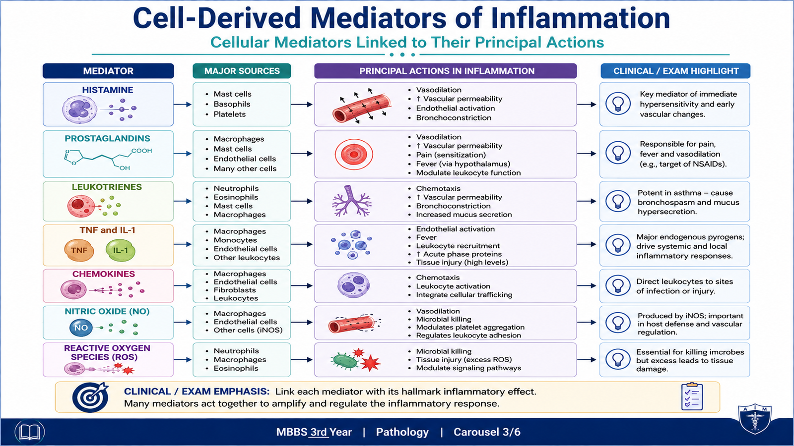
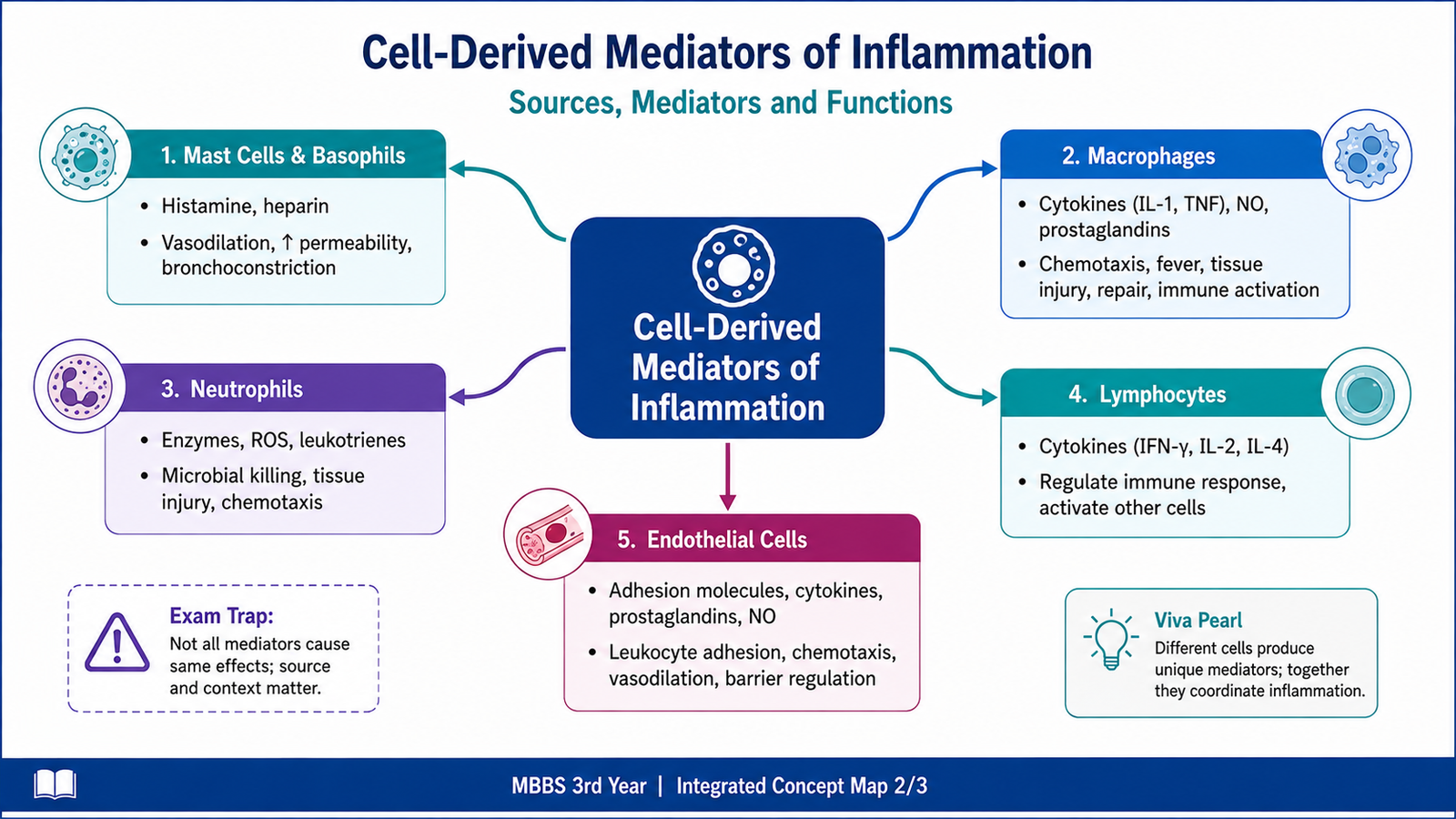
Cell-derived mediators are released mainly by mast cells, macrophages, neutrophils, platelets, endothelial cells and other activated cells. Some are stored preformed in intracellular granules and can be released within seconds. Others are synthesized only after the cell receives an inflammatory stimulus. Together they regulate vascular reactions, leukocyte recruitment, fever, pain and microbial killing.
Histamine
Histamine is one of the earliest mediators released during acute inflammation. Its most important sources are mast cells, although basophils and platelets can also contain histamine. Mast cells release it in response to physical injury, immune reactions and certain complement fragments.
Histamine causes arteriolar vasodilatation and increases the permeability of small venules by producing endothelial contraction. These actions help explain the rapid redness, warmth and edema that develop during the early vascular phase of acute inflammation.
Arachidonic acid metabolites
When inflammatory cells are activated, membrane phospholipids release arachidonic acid. Enzymatic metabolism of arachidonic acid produces two major groups of mediators: prostaglandins through the cyclooxygenase pathway and leukotrienes through the lipoxygenase pathway.
Cell activation → membrane phospholipids → arachidonic acid → cyclooxygenase pathway → prostaglandins
Arachidonic acid → lipoxygenase pathway → leukotrienes
Prostaglandins
- Prostaglandins such as PGE₂ and PGI₂ contribute to vasodilatation.
- PGE₂ contributes importantly to pain sensitization and fever.
- Thromboxane A₂, mainly from platelets, promotes platelet aggregation and vasoconstriction.
- Prostacyclin (PGI₂), mainly from endothelial cells, causes vasodilatation and inhibits platelet aggregation.
Leukotrienes
- LTB₄: promotes leukocyte chemotaxis and activation, especially of neutrophils.
- LTC₄, LTD₄ and LTE₄: increase vascular permeability and cause bronchial smooth-muscle contraction.
Cytokines
Cytokines are proteins produced mainly by activated immune and inflammatory cells. TNF and IL-1 are important pro-inflammatory cytokines. They activate endothelial cells, increase expression of adhesion molecules, promote leukocyte recruitment and contribute to systemic manifestations such as fever.
IL-6 also contributes to systemic inflammatory responses and promotes hepatic synthesis of acute-phase proteins.
Chemokines
Chemokines are a family of small proteins whose major role is to guide leukocyte migration. They form concentration gradients that direct leukocytes toward the site of infection or tissue injury and can also activate leukocyte integrins, helping cells adhere firmly to endothelium before migration.
Nitric oxide
Nitric oxide (NO) is produced by endothelial cells and activated macrophages. Endothelial NO causes vasodilatation by relaxing vascular smooth muscle. Macrophage-derived NO can participate in microbial killing when generated in larger amounts during activation.
Reactive oxygen species and lysosomal enzymes
Activated neutrophils and macrophages generate reactive oxygen species (ROS) and release lysosomal enzymes to destroy microbes. These mechanisms are useful inside phagolysosomes, but if released excessively into surrounding tissues they can injure host cells and extracellular matrix.
| Mediator | Important source | Major inflammatory action |
|---|---|---|
| Histamine | Mast cells | Vasodilatation, increased permeability |
| PGE₂ | Various activated cells | Vasodilatation, pain, fever |
| LTB₄ | Leukocytes | Chemotaxis and leukocyte activation |
| LTC₄/LTD₄/LTE₄ | Leukocytes, mast cells | Permeability, bronchoconstriction |
| TNF and IL-1 | Macrophages and other cells | Endothelial activation, leukocyte recruitment, fever |
| Chemokines | Many inflammatory cells | Directed leukocyte migration |
| Nitric oxide | Endothelium, macrophages | Vasodilatation; microbial killing |
| ROS / lysosomal enzymes | Neutrophils, macrophages | Microbial killing; tissue injury if excessive |
D. Morphologic Patterns of Acute Inflammation
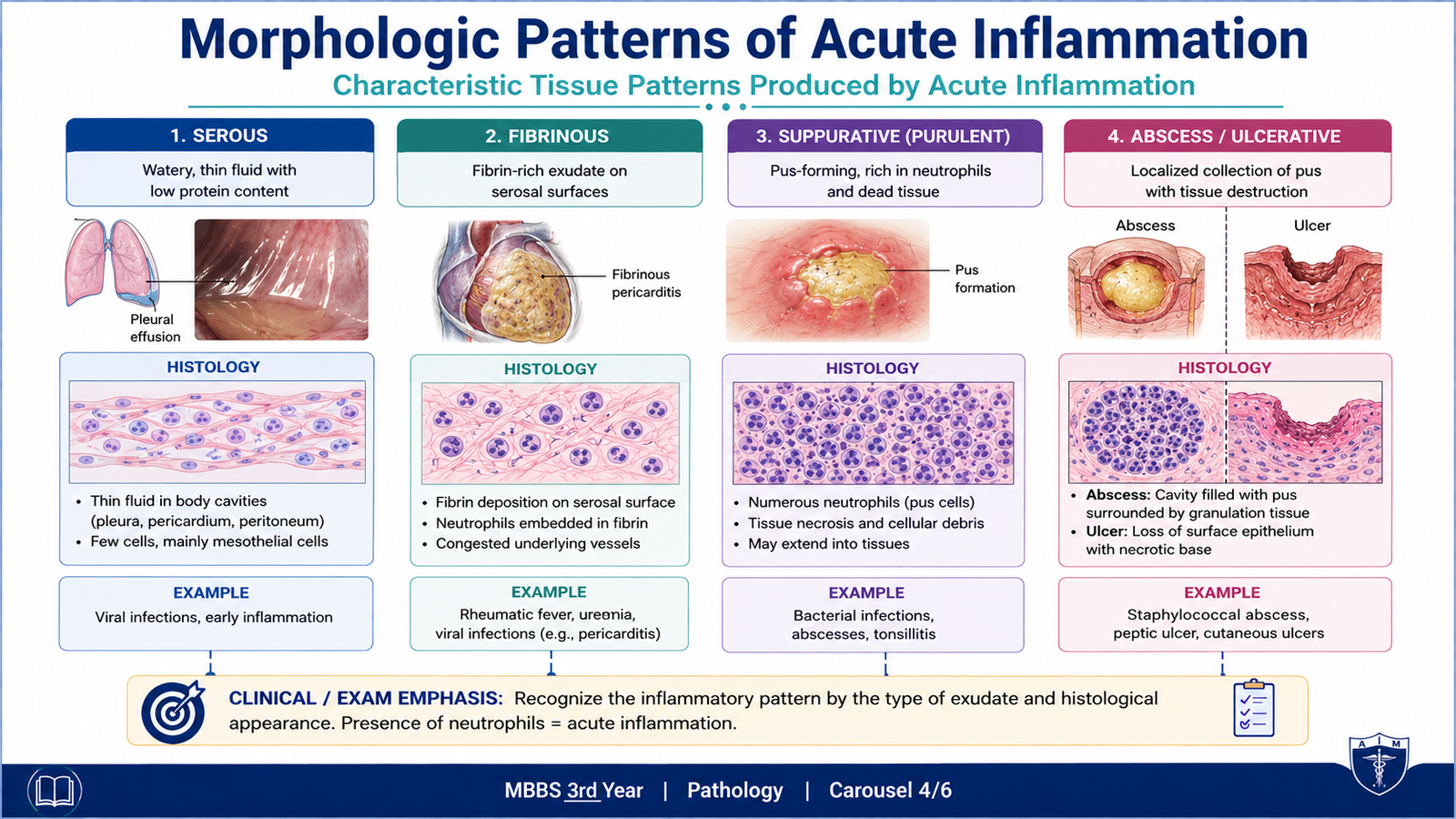
The microscopic and gross appearance of acute inflammation depends on the severity of injury, the cause of inflammation, the tissue involved and the type of exudate produced. Four important patterns are serous inflammation, fibrinous inflammation, suppurative or purulent inflammation, and ulceration.
1. Serous inflammation
Serous inflammation is characterized by the production of a thin, watery, relatively cell-poor fluid. The fluid may originate from plasma that has escaped through more permeable vessels or from secretions of mesothelial cells lining body cavities.
A classic example is a skin blister following a mild burn or viral infection. Microscopically, a fluid-filled space is seen with relatively few inflammatory cells.
2. Fibrinous inflammation
When vascular injury is more severe, larger plasma proteins such as fibrinogen escape from vessels. Fibrinogen is converted to fibrin, producing a fibrin-rich exudate. This is especially important on the lining of body cavities such as the pericardium and pleura.
Microscopically, fibrin appears as an eosinophilic network or amorphous material. If fibrin is removed during recovery, normal structure may be restored. If it persists, fibroblasts and new blood vessels can grow into it, leading to organization and fibrosis.
3. Suppurative or purulent inflammation
Suppurative inflammation is characterized by production of pus, which consists mainly of neutrophils, necrotic cell debris and edema fluid. It commonly occurs with pyogenic bacterial infections.
A localized collection of pus within tissue is called an abscess. An abscess usually contains a central area of necrotic tissue and neutrophils, surrounded by inflammatory cells and, with time, a wall of granulation tissue and fibrosis.
4. Ulceration
An ulcer is a local defect or excavation of a surface caused by shedding of inflamed necrotic tissue. Ulcers develop only where tissue necrosis occurs at or near a surface, such as skin or mucosal surfaces.
During the acute phase, the base and margins contain neutrophils and vascular dilatation. In a chronic ulcer, macrophages, lymphocytes and plasma cells become more prominent, with fibroblast proliferation and fibrosis at the base.
| Pattern | Main material | Key histological feature | Typical concept |
|---|---|---|---|
| Serous | Watery fluid | Cell-poor fluid accumulation | Blister |
| Fibrinous | Fibrin | Eosinophilic fibrin deposits | Inflamed serosal surface |
| Suppurative | Pus | Neutrophils + necrotic debris | Abscess |
| Ulcer | Surface tissue loss | Necrosis with inflammatory infiltrate | Skin or mucosal defect |
E. Outcomes of Acute Inflammation
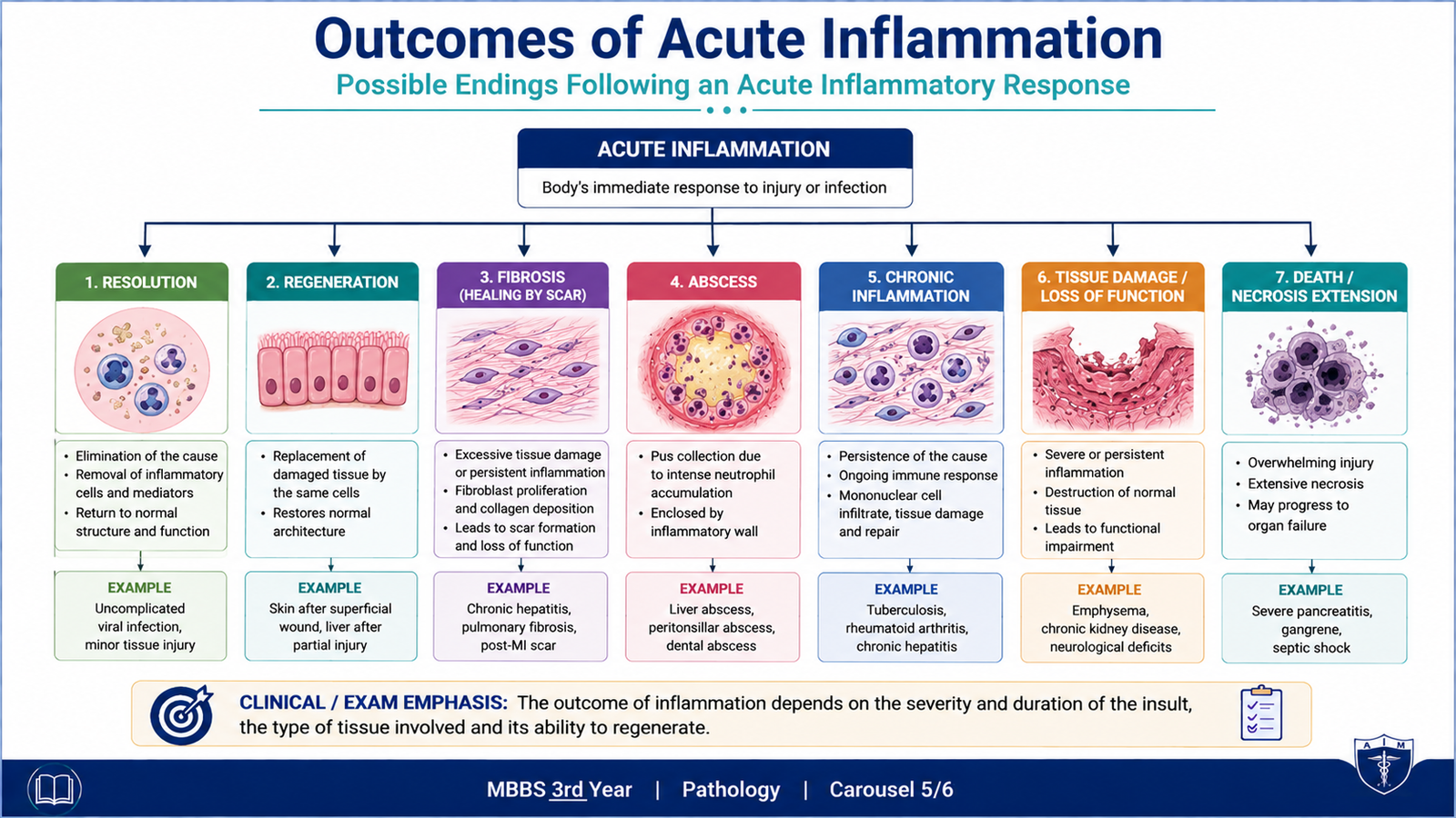
Acute inflammation does not have a single fixed ending. Its outcome depends on the nature of the injury, the amount of tissue damage, the regenerative ability of the affected tissue and whether the harmful stimulus can be removed. The major outcomes are complete resolution, healing by fibrosis, abscess formation and progression to chronic inflammation.
1. Complete resolution
Resolution occurs when the injurious stimulus is eliminated, tissue damage is limited and the affected tissue can regenerate. Vascular permeability returns to normal, edema fluid and inflammatory cells are removed, dead cells are cleared by macrophages, and normal tissue structure is restored.
Elimination of cause → mediator activity stops → neutrophils disappear → macrophages clear debris → tissue regeneration → normal structure restored
2. Healing by fibrosis
Fibrosis occurs when there is substantial tissue destruction, when the affected tissue has limited regenerative capacity, or when a fibrin-rich exudate cannot be completely removed. Fibroblasts produce collagen, replacing the damaged area with scar tissue.
This restores structural continuity but may reduce normal function, particularly if extensive fibrosis develops in an organ.
3. Abscess formation
Abscess formation is particularly associated with pyogenic organisms and extensive neutrophilic inflammation. Tissue destruction produces a localized cavity containing pus. Depending on the site and extent, healing may occur with scarring after the inflammatory material is cleared.
4. Progression to chronic inflammation
Acute inflammation may become chronic when the offending agent persists or when the acute response cannot completely eliminate the cause. Neutrophils become less dominant and are replaced by macrophages, lymphocytes and other mononuclear cells. Continued inflammation is accompanied by tissue destruction and attempts at healing.
F. Defects of Inflammation and Their Consequences
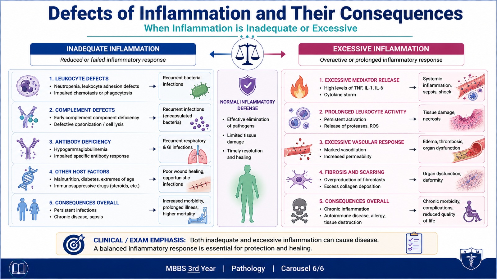
Inflammation is protective only when the required cells, plasma proteins and signaling mechanisms function properly. Defects in these components can impair microbial elimination, delay healing and increase susceptibility to infection. On the other hand, an excessively strong or poorly controlled inflammatory response can itself injure tissues.
Defects affecting leukocytes
Effective inflammation requires leukocytes to reach the site of injury, recognize and ingest microbes, and kill them. A defect at any of these stages weakens host defense.
- Reduced leukocyte numbers: insufficient neutrophils or other leukocytes reduce the cellular response to infection.
- Defective adhesion or migration: leukocytes may fail to leave the circulation and reach infected tissue efficiently.
- Defective phagocytosis: impaired recognition or ingestion allows microorganisms to persist.
- Defective intracellular killing: organisms may survive even after being phagocytosed.
Defects involving plasma mediator systems
Complement abnormalities can interfere with opsonization, chemotaxis or direct microbial killing. Because complement performs several coordinated functions, the clinical consequence depends on which component or pathway is defective.
Consequences of inadequate inflammation
- Repeated or severe infections
- Failure to eliminate microorganisms effectively
- Persistence or spread of infection
- Delayed clearance of damaged tissue
- Impaired or delayed wound healing
Consequences of excessive inflammation
An inflammatory response that is too intense or not properly controlled can damage otherwise normal tissue. Activated leukocytes may release ROS, proteases and other mediators outside their target area. Persistent cytokine and mediator activity can therefore convert a protective response into an important cause of tissue injury.
Defective response → inadequate microbial clearance → persistent infection and poor healing
Excessive response → mediator and leukocyte overactivity → collateral tissue injury
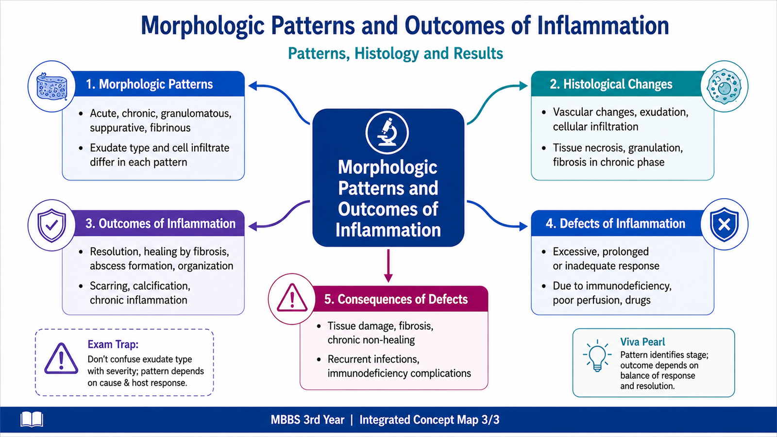
Integrated Mechanism Flow
↓
2. Plasma-derived and cell-derived mediators are generated or released
↓
3. Mediators produce vasodilatation, increased permeability and leukocyte recruitment
↓
4. Fluid, plasma proteins and leukocytes accumulate at the affected site, producing the inflammatory morphology
↓
5. The injurious agent is removed or persists, while inflammatory cells clear damaged tissue
↓
6. The response ends in resolution, fibrosis, abscess formation or chronic inflammation
↓
7. Defective regulation may instead cause recurrent infection, impaired healing or excessive tissue injury
⭐ AIM High-Yield Review
- Inflammatory mediators may be plasma-derived or cell-derived.
- Plasma mediators generally circulate as inactive precursors and are activated through enzymatic cascades.
- ⭐ C3b promotes opsonization; C5a promotes chemotaxis; C5b–C9 forms the membrane attack complex.
- Bradykinin causes vasodilatation, increased permeability and pain.
- Histamine is an important early mediator of vasodilatation and increased vascular permeability.
- ⭐ PGE₂ contributes to pain and fever, while LTB₄ is important in leukocyte chemotaxis.
- TNF and IL-1 activate endothelium and promote leukocyte recruitment and systemic inflammatory effects.
- Chemokines guide leukocytes toward sites of inflammation.
- Serous inflammation produces thin fluid; fibrinous inflammation produces fibrin-rich exudate.
- ⭐ Suppurative inflammation produces pus containing neutrophils and necrotic debris; a localized collection forms an abscess.
- An ulcer is a surface defect produced by shedding of inflamed necrotic tissue.
- The four major outcomes of acute inflammation are resolution, fibrosis, abscess formation and progression to chronic inflammation.
- Resolution is favored when the cause is removed, tissue injury is limited and the tissue can regenerate.
- Defective leukocyte or complement function may cause recurrent infection and impaired healing.
- Excessive mediator and leukocyte activity can cause collateral injury to normal tissues.
🎥 AIM Video Learning — Inflammatory Mediators
Watch this video after completing the learning material to reinforce the major plasma-derived and cell-derived mediators of inflammation and their important actions.