
This chapter follows the KMU learning outcomes in a logical sequence. First understand how a wound becomes a scar, then focus on the growth factors, extracellular matrix and factors that can improve or disturb healing.
Topic 8 — Repair by Scarring, Growth Factors and Extracellular Matrix
Module/Theme: Infection and Inflammation
Understand how damaged tissue is replaced by connective tissue, how growth factors regulate healing, how the extracellular matrix supports repair, and why wounds may heal poorly or produce excessive scars.
Topic Introduction
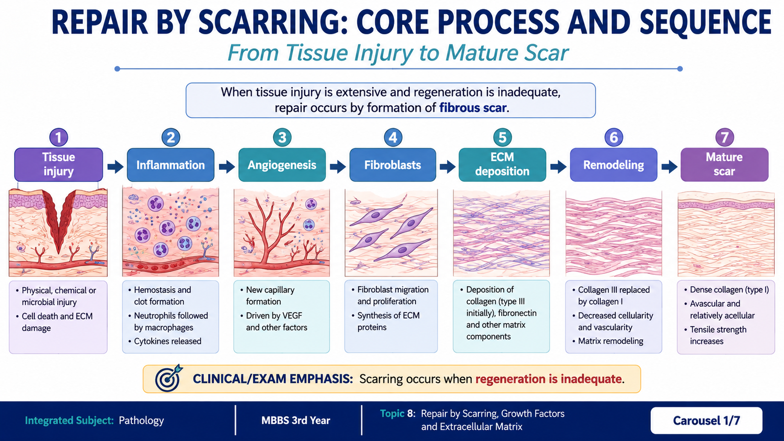
When tissue injury is too severe for complete regeneration, the body restores continuity mainly by depositing connective tissue and forming a scar. This process is called repair by scarring. It requires several coordinated events, including formation of new blood vessels, migration and proliferation of fibroblasts, deposition of extracellular matrix and later remodeling of the scar. These events are controlled by growth factors and by signals from the extracellular matrix. Healing is therefore not simply the production of collagen. It is an organized biological process involving inflammatory cells, vascular cells, fibroblasts, matrix proteins and regulatory mediators. Understanding these steps also explains why infection, poor nutrition, mechanical stress and other factors can delay healing or produce abnormal scars.
A. Repair by Scarring: Core Process and Sequence
Repair by scarring occurs when damaged tissue cannot be completely restored by regeneration. This is especially important when the injury is extensive, when the extracellular matrix framework has been destroyed, or when the affected cells have limited ability to proliferate. In these situations, the defect is progressively filled with vascularized connective tissue and eventually converted into a relatively less cellular collagenous scar.
Main steps of repair by scarring
The steps overlap rather than occurring as completely separate stages. Inflammation prepares the injured site, newly formed vessels provide nutrients, fibroblasts produce extracellular matrix, and the developing scar is subsequently remodeled.
Macrophages are particularly important because they help clear damaged tissue and also release mediators that stimulate angiogenesis, fibroblast migration, fibroblast proliferation and extracellular matrix synthesis.
| Stage | Main event | Purpose |
|---|---|---|
| Inflammation | Removal of damaged tissue and release of mediators | Prepares the wound for repair |
| Angiogenesis | Formation of new vessels | Supplies oxygen and nutrients |
| Fibroblast proliferation | Migration and multiplication of fibroblasts | Provides matrix-producing cells |
| ECM deposition | Collagen and other matrix proteins accumulate | Strengthens and fills the defect |
| Remodeling | Matrix is reorganized and vascularity falls | Produces a mature scar |

B. Granulation Tissue, Angiogenesis and Fibroblast Activity
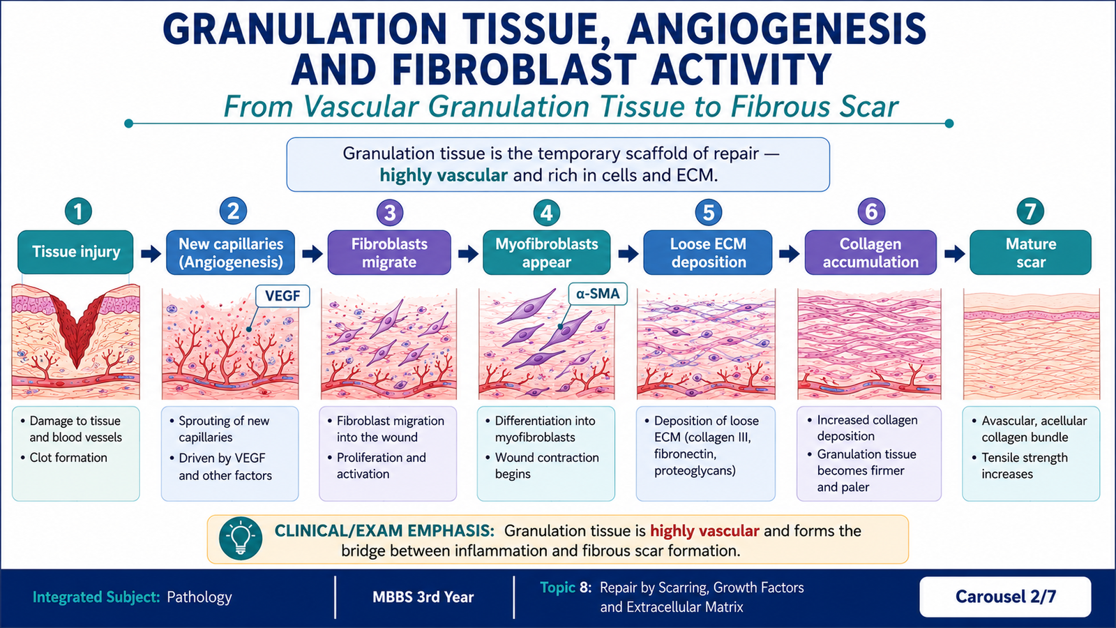
A healing wound initially develops granulation tissue, a soft, vascular connective tissue composed mainly of newly formed capillaries, proliferating fibroblasts and a loose extracellular matrix containing inflammatory cells. Its granular pink-red appearance is produced largely by the numerous small blood vessels.
Angiogenesis
Angiogenesis means formation of new blood vessels from existing vessels. It is essential because repairing tissue has high metabolic requirements and requires oxygen and nutrients. Vascular endothelial growth factor, particularly VEGF, is a major stimulus for this process.
During angiogenesis, vascular dilation and increased permeability are followed by separation of supporting cells from the vessel wall. Endothelial cells then migrate toward the angiogenic stimulus, proliferate, form vascular tubes and recruit supporting cells to stabilize the new vessels.
Fibroblast migration and proliferation
Fibroblasts migrate into the injured area under the influence of growth factors released mainly by macrophages and other cells. They proliferate and begin producing extracellular matrix proteins, especially collagen. Some fibroblasts acquire features of smooth-muscle cells and become myofibroblasts, which help contract the wound.
From granulation tissue to scar
As healing progresses, collagen accumulates and the amount of fluid, inflammatory cells and vascular tissue decreases. The initially highly vascular granulation tissue is therefore gradually converted into a pale, relatively avascular fibrous scar.
C. Growth Factors, Receptors and Signaling in Tissue Repair
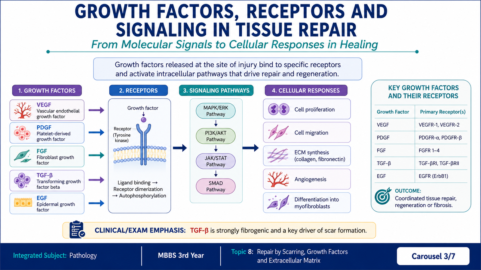
Growth factors are signaling proteins that regulate cell survival, migration, proliferation and production of extracellular matrix. They act by binding to specific receptors on target cells. In wound healing, several growth factors work together rather than acting independently. Their effects depend on which cells express the appropriate receptors and on the surrounding extracellular environment.
Important growth factors in repair
| Growth factor | Important repair actions | Receptor type |
|---|---|---|
| EGF | Stimulates epithelial and other cell proliferation | Receptor tyrosine kinase |
| TGF-α | Promotes epithelial-cell proliferation | EGF receptor, a receptor tyrosine kinase |
| HGF | Stimulates proliferation and migration of several epithelial cells | MET receptor tyrosine kinase |
| VEGF | Promotes angiogenesis and increases vascular permeability | Receptor tyrosine kinase |
| PDGF | Promotes fibroblast migration and proliferation and contributes to ECM production | Receptor tyrosine kinase |
| FGF | Supports fibroblast proliferation and angiogenesis | Receptor tyrosine kinase |
| TGF-β | Stimulates collagen and other ECM production and decreases ECM degradation | Serine/threonine kinase receptor |
How growth-factor receptors produce a cellular response
Many growth factors bind to receptors with intrinsic enzyme activity. Binding activates intracellular signaling pathways that alter gene transcription and cellular behavior. Receptor tyrosine kinases commonly activate pathways such as RAS–MAP kinase and PI3K–AKT. TGF-β acts through serine/threonine kinase receptors and intracellular SMAD proteins.
Growth factor → receptor binding → intracellular signaling proteins → altered gene transcription → cell proliferation, migration, survival or matrix production
The important undergraduate concept is that growth factors convert an extracellular signal into a coordinated cellular response. In repair, this allows inflammatory cells, endothelial cells and fibroblasts to communicate with one another and organize the healing process.
D. Extracellular Matrix: Components and Functions in Repair
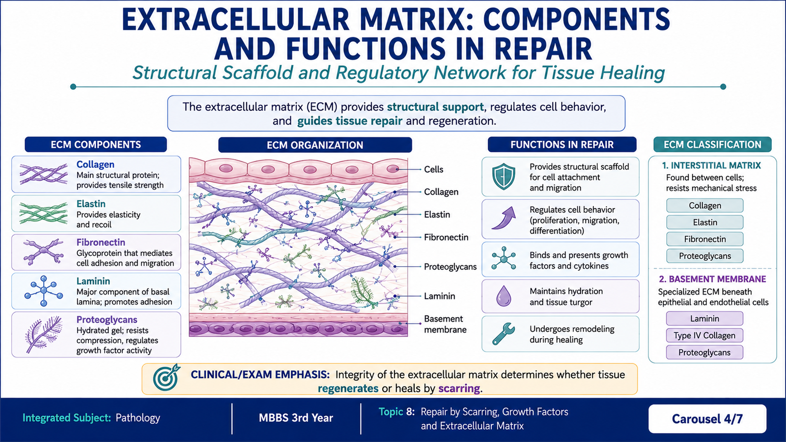
The extracellular matrix (ECM) is the network of macromolecules outside cells that provides structural support and also influences cell growth, movement and differentiation. It is therefore not simply an inert scaffold. During tissue repair, cells interact continuously with ECM proteins, and changes in the matrix influence the behavior of the cells participating in healing.
Main components of the extracellular matrix
The ECM can be considered in terms of three major groups of components: fibrous structural proteins, adhesive glycoproteins, and proteoglycans with glycosaminoglycans.
| ECM component | Examples | Major role |
|---|---|---|
| Fibrous structural proteins | Collagen, elastin | Tensile strength and elasticity |
| Adhesive glycoproteins | Fibronectin, laminin | Cell attachment and interaction with matrix |
| Proteoglycans and glycosaminoglycans | Proteoglycan-rich ground substance | Hydration, resilience and organization of the matrix |
Interstitial matrix and basement membrane
The ECM is organized into two broad patterns. The interstitial matrix is present in connective tissues and between cells. It contains fibrillar collagens, elastin, fibronectin, proteoglycans and other proteins. The basement membrane forms a specialized sheet beneath epithelial and some other cells and contains characteristic matrix proteins, including type IV collagen and laminin.
Role of ECM in tissue repair
The extracellular matrix performs several functions that are essential for successful healing.
- Mechanical support: it provides a framework on which cells can migrate and organize.
- Tensile strength: collagen increases the strength of the developing scar.
- Cell adhesion: adhesive proteins help cells attach to the surrounding matrix.
- Regulation of cell behavior: matrix interactions influence proliferation, migration and differentiation.
- Reservoir for regulatory molecules: the ECM can bind and concentrate growth factors.
- Maintenance of tissue architecture: an intact matrix framework favors restoration of normal structure, whereas extensive matrix destruction favors scar formation.
E. Collagen Deposition, Remodeling and Scar Maturation
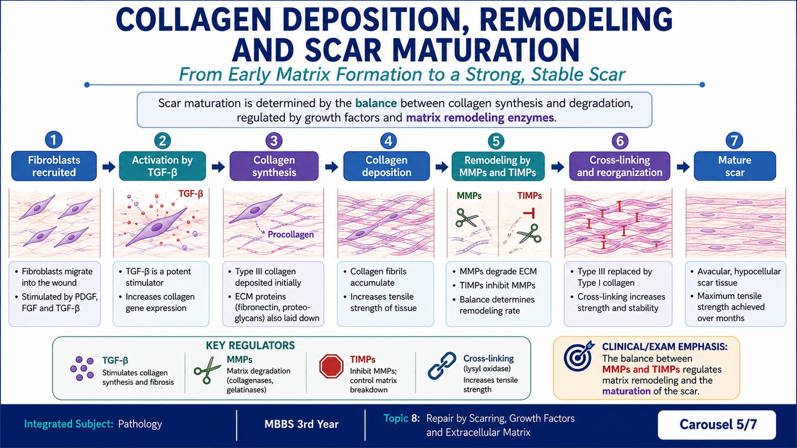
A scar becomes strong not only because collagen is produced, but also because the collagen is progressively organized and remodeled. Fibroblasts synthesize collagen and other matrix components under the influence of mediators such as TGF-β. At the same time, old or excess matrix must be removed. Normal healing therefore depends on a balance between ECM synthesis and ECM degradation.
Collagen deposition
As fibroblasts become active, collagen increasingly replaces the loose provisional matrix of early granulation tissue. The amount of collagen rises, while the number of inflammatory cells and blood vessels gradually falls. The wound therefore changes from soft, red granulation tissue to a firmer and paler scar.
Matrix metalloproteinases
Matrix metalloproteinases (MMPs) are enzymes that degrade components of the extracellular matrix. They participate in remodeling by removing matrix that is no longer needed. Their activity is controlled by endogenous inhibitors called tissue inhibitors of metalloproteinases (TIMPs).
ECM synthesis by fibroblasts ↔ ECM degradation by MMPs
TIMPs restrain excessive MMP activity.
Scar maturation
During maturation, the scar becomes less cellular and less vascular. Collagen fibers are reorganized and cross-linked, increasing tensile strength. However, a healed wound does not usually regain the full mechanical strength of completely uninjured tissue.
Wound contraction also reduces the size of the defect. This is largely mediated by myofibroblasts, which have contractile properties.
F. Factors Affecting Wound Healing
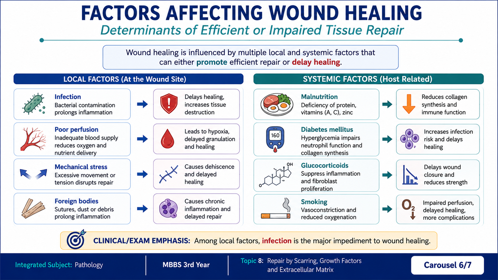
Wound healing can be altered by conditions acting directly at the wound or by systemic factors affecting the patient as a whole. These factors usually delay healing by increasing tissue injury, reducing oxygen or nutrient delivery, interfering with inflammation, or impairing collagen synthesis and remodeling.
Local factors
- Infection: one of the most important causes of delayed healing. Microbial infection prolongs inflammation and causes additional tissue injury.
- Poor blood supply: reduced perfusion limits oxygen and nutrient delivery to cells involved in repair.
- Mechanical stress: excessive movement or tension can separate wound edges and interfere with stable scar formation.
- Foreign bodies: retained material can maintain inflammation and interfere with normal repair.
- Size, site and type of wound: larger tissue defects and wounds with extensive tissue destruction require more granulation tissue and scar formation.
Systemic factors
- Nutrition: protein deficiency impairs synthesis of structural proteins, while vitamin C deficiency interferes with normal collagen formation.
- Metabolic disease: diabetes mellitus is associated with impaired healing through several mechanisms, including vascular abnormalities and increased susceptibility to infection.
- Glucocorticoids: suppress inflammatory responses and reduce collagen synthesis, thereby weakening repair.
G. Abnormalities of Repair and Their Consequences
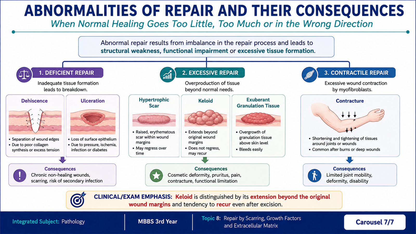
Repair can become abnormal when too little scar tissue is formed, too much extracellular matrix is deposited, or wound contraction becomes excessive. The resulting abnormalities are clinically important because they may cause wound failure, disfigurement or limitation of movement.
Deficient scar formation
If collagen deposition or wound strength is inadequate, the wound may separate. This is called wound dehiscence. Poor healing may also contribute to ulcer formation, particularly when blood supply, sensation or mechanical protection is impaired.
Excessive scar formation
An excessive healing response can produce too much collagen. A hypertrophic scar is raised but remains within the boundaries of the original wound. A keloid grows beyond the boundaries of the original injury because of excessive collagen deposition.
| Feature | Hypertrophic scar | Keloid |
|---|---|---|
| Extent | Confined to original wound | Extends beyond original wound |
| Main abnormality | Excessive collagen deposition | Marked excessive collagen deposition |
Exuberant granulation tissue
Granulation tissue may occasionally grow above the level of the surrounding skin. This excessive tissue can interfere with re-epithelialization because epithelial cells cannot effectively cover the projecting granulation tissue.
Contracture
Wound contraction is normally useful because it reduces the size of the wound. When contraction becomes excessive, however, it produces a contracture. Contractures may restrict movement, particularly when scars cross joints or develop after extensive skin injury.
Integrated Mechanism Flow
↓
Inflammation and macrophage activation
↓
Growth-factor release, especially VEGF, PDGF, FGF and TGF-β
↓
Angiogenesis + fibroblast migration and proliferation
↓
Extracellular matrix and collagen deposition
↓
MMP/TIMP-controlled matrix remodeling and wound contraction
↓
Mature fibrous scar
⭐ AIM High-Yield Review
- Repair by scarring becomes important when complete regeneration cannot restore the injured tissue.
- Granulation tissue contains newly formed capillaries, fibroblasts and loose extracellular matrix.
- Macrophages are major coordinating cells because they remove debris and release mediators of repair.
- ⭐ VEGF is a major mediator of angiogenesis.
- PDGF and FGF promote important fibroblast and repair responses.
- ⭐ TGF-β is a major fibrogenic mediator: it increases ECM production and reduces matrix degradation.
- Most EGF, VEGF, PDGF and FGF receptors are receptor tyrosine kinases; TGF-β signals through serine/threonine kinase receptors.
- ECM consists mainly of structural proteins, adhesive glycoproteins, proteoglycans and glycosaminoglycans.
- Collagen provides tensile strength, while fibronectin and laminin contribute importantly to cell-matrix interactions.
- MMPs degrade extracellular matrix; TIMPs inhibit MMP activity and help regulate remodeling.
- Infection is a major local cause of delayed wound healing because it prolongs inflammation and causes further tissue injury.
- Protein or vitamin C deficiency can impair collagen production and wound repair.
- Glucocorticoids can impair healing by suppressing inflammation and collagen synthesis.
- ⭐ A hypertrophic scar remains within the original wound boundaries; a keloid extends beyond them.
- Excessive wound contraction can produce a contracture and restrict movement.
🎥 AIM Video Learning — Tissue Repair and Wound Healing
Reinforce repair by scarring, granulation tissue, wound-healing events,
factors affecting healing and abnormalities of tissue repair.
Video: Tissue Repair and Wound Healing — Pathology Learning with Dr. Monica