
This chapter follows the KMU learning outcomes in a logical sequence. First understand how each organism produces disease and how it is identified, then revise the characteristic clinical, laboratory and preventive clues in the High-Yield Review.
Topic 18 — Gram-Positive Rods, Zoonotic and Directly Contagious Infections
A structured introduction to medically important gram-positive rods and selected zoonotic and directly transmissible infections, linking organism characteristics with disease mechanisms, clinical recognition, laboratory diagnosis and prevention.
Topic Introduction
Gram-positive rods are a diverse group of bacteria that include both spore-forming and non-spore-forming organisms. Some produce powerful toxins, whereas others invade tissues or survive within host cells. Several important infections in this topic are also zoonoses, meaning that they are naturally transmitted between animals and humans. In addition, rabies, scabies, leprosy and trachoma have important preventive and community-health implications. The easiest way to understand this topic is to connect each organism with its reservoir or source, route of transmission, pathogenic mechanism, characteristic clinical picture, appropriate diagnostic specimen and key preventive measure.
A. Classification and Core Features of Gram-Positive Rods
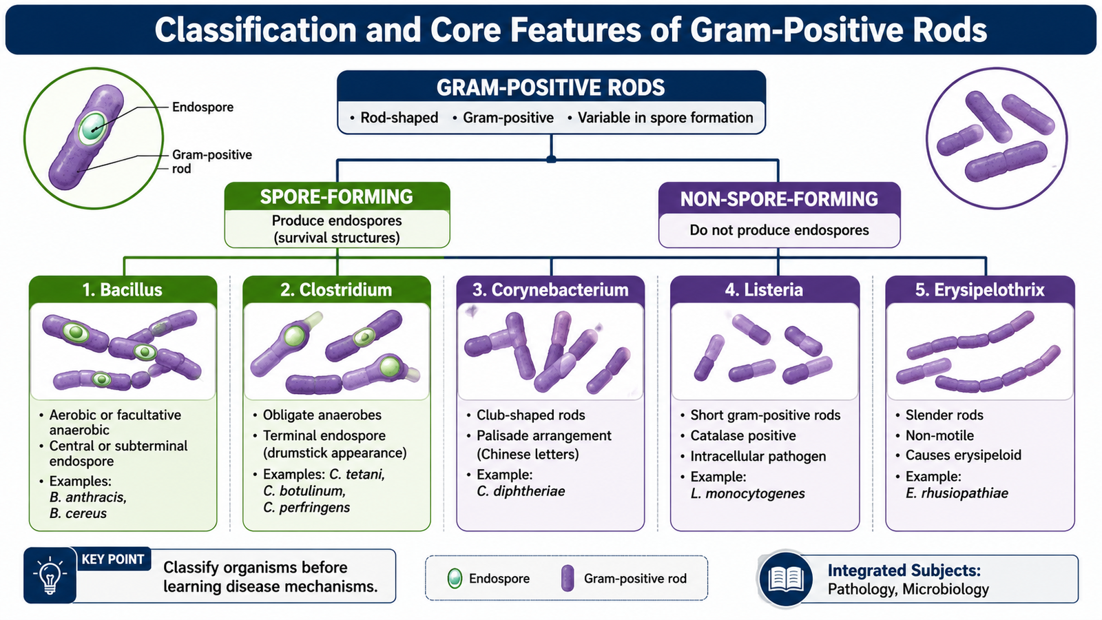
Gram-positive rods are bacteria that usually appear as elongated purple-staining organisms on Gram stain because their thick peptidoglycan cell wall retains the crystal-violet dye. A useful first distinction is whether the organism can form endospores. Endospores are highly resistant dormant structures that allow certain bacteria to survive adverse environmental conditions.
Spore-forming gram-positive rods
The medically important aerobic or facultatively anaerobic spore-forming rods belong mainly to Bacillus, while the important anaerobic spore-formers belong mainly to Clostridium and related clostridial organisms.
- Bacillus anthracis — causes anthrax.
- Bacillus cereus — commonly associated with food poisoning.
- Clostridium tetani — causes tetanus.
- Clostridium botulinum — causes botulism.
- Clostridium perfringens — causes myonecrosis/gas gangrene and some food-borne disease.
- Clostridioides difficile — produces toxin-mediated antibiotic-associated colitis.
Non-spore-forming gram-positive rods
These organisms do not form resistant endospores. Their pathogenic mechanisms therefore depend on features such as toxin production, tissue invasion or intracellular survival rather than spore formation.
- Corynebacterium diphtheriae — toxigenic strains cause diphtheria.
- Listeria monocytogenes — an intracellular pathogen important particularly in pregnancy, neonates, older adults and immunocompromised patients.
- Erysipelothrix rhusiopathiae — an occupationally acquired zoonotic organism associated with animal or animal-product exposure.
B. Bacillus Species and Anthrax
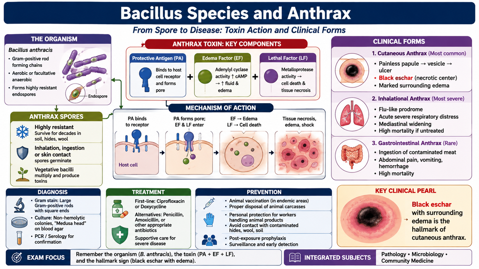
Bacillus species are large gram-positive rods that form spores and generally grow in the presence of oxygen. The major pathogen in this topic is Bacillus anthracis, the cause of anthrax. Humans usually acquire anthrax through exposure to infected animals, contaminated animal products or bacterial spores rather than through ordinary person-to-person contact.
Bacillus anthracis: important properties
- Large gram-positive, spore-forming rod.
- Classically non-motile.
- Possesses a distinctive poly-D-glutamic acid capsule.
- Produces anthrax toxin consisting of protective antigen, edema factor and lethal factor.
How anthrax produces disease
After spores enter through skin, inhalation or the gastrointestinal tract, they germinate within the host. Vegetative bacteria multiply and release toxin components. Protective antigen allows the enzymatic toxin components to enter cells. Edema factor increases intracellular cyclic AMP and promotes marked edema, while lethal factor disrupts cellular signaling and contributes to tissue injury, systemic toxicity and shock.
Exposure to spores → spore germination → bacterial multiplication → toxin production → edema and tissue injury → local or systemic anthrax
Clinical types of anthrax
Cutaneous anthrax follows inoculation through damaged skin. It commonly begins as a papule and progresses to a vesicular lesion and then an ulcer with a characteristic black eschar. Marked surrounding edema may occur. The lesion is classically less painful than its striking appearance might suggest.
Inhalational anthrax follows inhalation of spores. Spores reach the lungs and are transported to regional lymph nodes, where germination can produce severe hemorrhagic mediastinal inflammation and systemic disease.
Gastrointestinal anthrax may follow ingestion of contaminated animal products and can cause gastrointestinal inflammation, ulceration and systemic toxicity.
Laboratory diagnosis
Diagnosis depends on the clinical form. Appropriate specimens may include material from a cutaneous lesion, blood in systemic disease or respiratory specimens when inhalational disease is suspected. Direct microscopy may demonstrate large gram-positive rods. Culture and appropriate confirmatory laboratory methods establish the diagnosis. Because anthrax is potentially hazardous, suspected specimens require appropriate laboratory handling.
Epidemiology and prevention of anthrax
Anthrax is primarily a disease of herbivorous animals. Human risk is therefore greatest in people exposed to infected livestock, animal hides, wool, hair or contaminated animal materials. Prevention focuses on interrupting contact with infected animals and their products and controlling disease in animal reservoirs.
- Recognize and control infection in livestock.
- Avoid unsafe handling of animals that have died from suspected anthrax.
- Use appropriate occupational protection when handling potentially contaminated animal materials.
- Apply appropriate decontamination and public-health control measures when spores are suspected.
- Use vaccination for persons in defined high-risk exposure situations where indicated by public-health practice.
C. Clostridial Infections and Tetanus
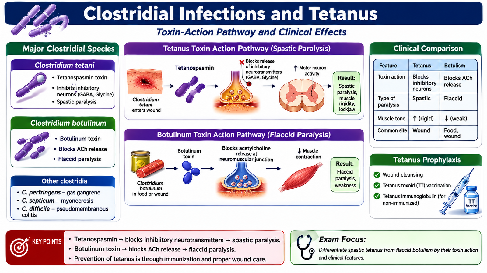
Clostridial organisms are gram-positive, spore-forming rods that characteristically grow under anaerobic conditions. Their spores persist in the environment, while disease usually results after spores or organisms enter a suitable site and produce potent exotoxins. The clinical effect depends on the particular toxin produced.
Major clostridial organisms
| Organism | Main mechanism | Characteristic disease | Diagnostic basis |
|---|---|---|---|
| C. tetani | Neurotoxin blocks inhibitory neurotransmission | Spastic paralysis and tetanus | Primarily clinical diagnosis |
| C. botulinum | Neurotoxin prevents acetylcholine release | Descending flaccid paralysis | Clinical pattern plus toxin-related testing |
| C. perfringens | Toxin-mediated tissue destruction | Myonecrosis with gas formation | Specimen microscopy and anaerobic culture |
| C. difficile | Toxin-mediated colonic injury | Antibiotic-associated colitis | Detection of toxigenic organism or toxin activity |
Tetanus: etiology and pathophysiology
Tetanus is caused by Clostridium tetani. Spores are widely distributed in the environment and may enter through contaminated wounds. Germination is favored when local tissue conditions become anaerobic. The organism remains mainly near the wound, but its toxin tetanospasmin reaches the nervous system.
Tetanospasmin interferes with the release of inhibitory neurotransmitters from inhibitory interneurons. Loss of inhibitory control causes continuous motor-neuron activity, producing muscle rigidity and painful spasms.
Risk factors and clinical features
Risk is increased when a person lacks adequate immunity and develops a wound contaminated with soil or other material containing spores. Deep, devitalized or contaminated wounds provide conditions favorable for germination.
D. Non-Spore-Forming Gram-Positive Rods
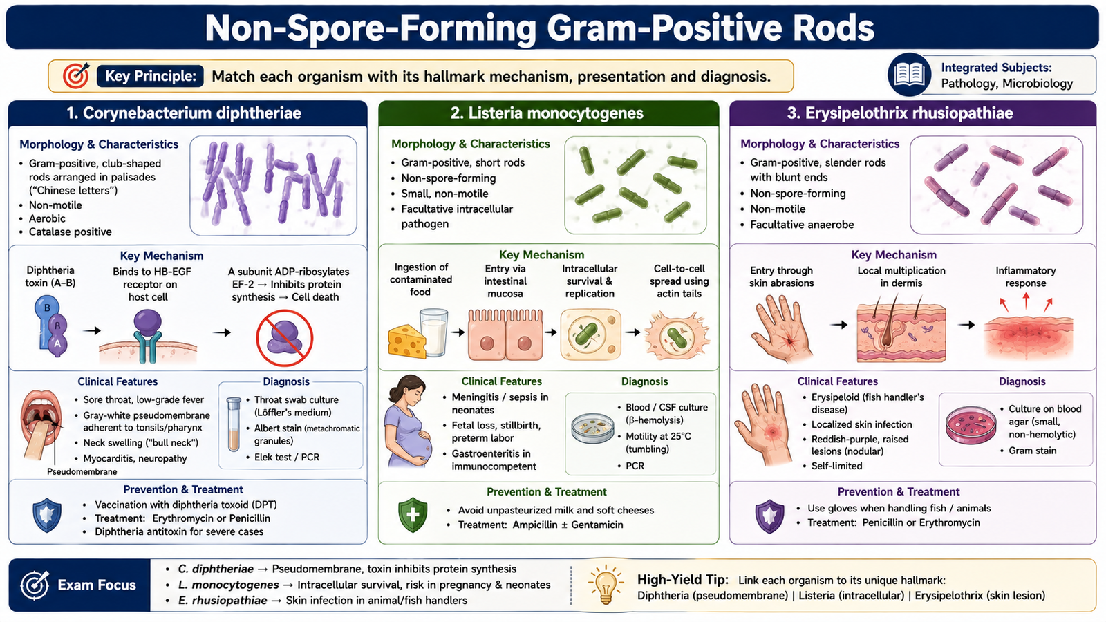
The major non-spore-forming gram-positive rods differ considerably in their mechanisms of disease. Corynebacterium diphtheriae is important because disease is mainly toxin-mediated, whereas Listeria monocytogenes is important because it can survive and multiply within host cells. Recognizing these differences helps explain their clinical and laboratory features.
Corynebacterium diphtheriae
C. diphtheriae is a pleomorphic gram-positive rod. On microscopy, organisms may lie at angles to one another and may show a characteristic arrangement described as resembling Chinese letters. Only strains carrying the gene for diphtheria toxin produce classical toxin-mediated disease.
Diphtheria toxin inhibits host-cell protein synthesis. Local epithelial injury causes necrosis and inflammation, producing a firmly adherent pseudomembrane in respiratory diphtheria. Absorbed toxin can damage distant tissues, particularly the heart and nervous system.
Laboratory investigation begins with an appropriate throat or lesion specimen collected from the affected area. Microscopy and culture help identify the organism, but determining whether the isolate is toxigenic is crucial because toxin production causes the major manifestations of classical diphtheria.
Listeria monocytogenes
L. monocytogenes is a small gram-positive rod capable of intracellular survival. Infection is commonly acquired through contaminated food. After entering host cells, the organism can escape intracellular killing and spread from cell to cell. Cell-mediated immunity is therefore particularly important for host defense.
Important clinical groups include pregnant women, neonates, older adults and people with impaired cellular immunity. Infection may produce febrile illness, bacteremia or central nervous system infection. In pregnancy, maternal disease may be relatively mild while infection can have serious fetal or neonatal consequences.
Diagnosis is made by isolating the organism from an appropriate normally sterile clinical specimen such as blood or cerebrospinal fluid when systemic or central nervous system infection is present.
Erysipelothrix rhusiopathiae
E. rhusiopathiae is a non-spore-forming gram-positive rod associated with animals, fish and animal products. Human infection is therefore often occupational. Local infection typically follows inoculation through injured skin and can produce a painful, well-defined skin lesion known as erysipeloid. Culture of an appropriate lesion specimen can establish the diagnosis.
| Feature | C. diphtheriae | L. monocytogenes | E. rhusiopathiae |
|---|---|---|---|
| Main pathogenic theme | Exotoxin | Intracellular survival | Occupational inoculation |
| Classic clue | Adherent pseudomembrane | Pregnancy/neonatal/CNS disease | Erysipeloid after animal exposure |
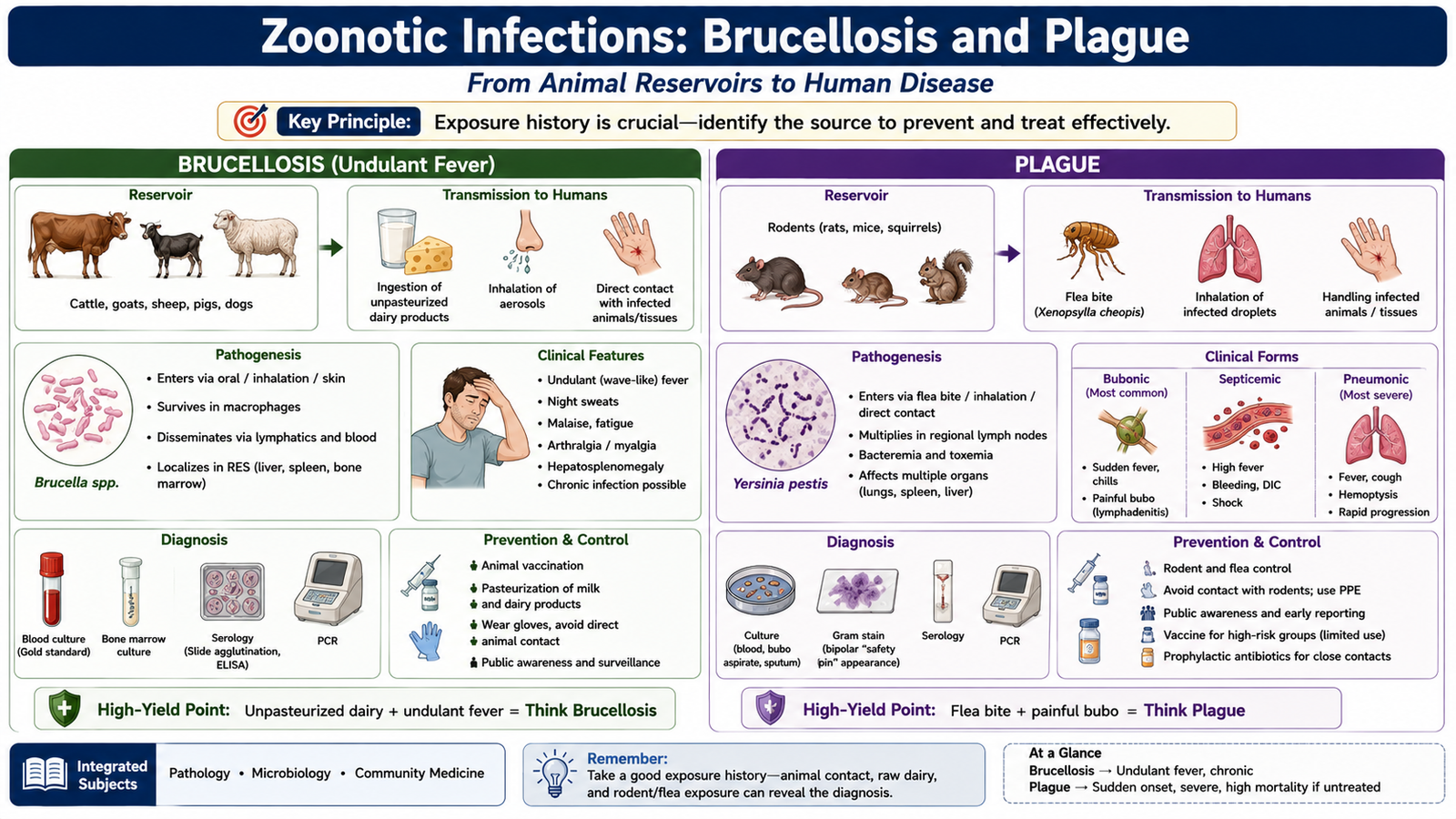
E. Zoonotic Infections: Brucellosis and Plague
A zoonosis is an infection naturally maintained in animals that can be transmitted to humans. Important organisms associated with zoonotic disease include Bacillus anthracis, Brucella species, Yersinia pestis, rabies virus and several other animal-associated pathogens. The route of exposure often provides an important diagnostic clue.
Brucellosis
Brucellosis is caused by Brucella species, which are small gram-negative coccobacilli rather than gram-positive rods. They are included here because brucellosis is a major zoonotic infection in the supplied curriculum. Important animal reservoirs vary by species and include cattle, goats, sheep, pigs and dogs.
Risk factors and transmission
- Consumption of unpasteurized milk or dairy products.
- Direct occupational contact with infected animals or animal tissues.
- Exposure among farmers, veterinarians, slaughterhouse workers and laboratory personnel.
Pathophysiology and clinical features
Brucella organisms enter through mucosal surfaces, the gastrointestinal tract or damaged skin. They can survive within macrophages and spread through the reticuloendothelial system. This intracellular persistence helps explain why disease may become prolonged or relapsing.
Patients may develop fever, sweating, malaise, fatigue, headache, arthralgia or other systemic symptoms. Fever may fluctuate, producing the traditional description undulant fever. Hepatosplenomegaly or focal organ involvement may occur.
Laboratory diagnosis
Blood culture can demonstrate the organism, although growth may be slow. Bone marrow culture may also be useful in appropriate cases. Serological tests detect antibodies and can support diagnosis when interpreted together with the clinical picture and exposure history.
Prevention
- Pasteurization of milk and avoidance of unpasteurized dairy products.
- Control of brucellosis in livestock.
- Protective measures for people handling animals, placentas, aborted material or carcasses.
- Appropriate laboratory precautions.
Plague
Plague is caused by Yersinia pestis, a gram-negative coccobacillus maintained naturally in animal reservoirs, especially rodents, with fleas playing an important role in transmission. Plague is historically important because large epidemics and pandemics caused enormous mortality before the nature of transmission was understood. Its importance today lies in recognizing its zoonotic cycle and preventing transmission.
Main clinical types
- Bubonic plague: follows inoculation, usually through an infected flea bite, and produces painful enlarged lymph nodes called buboes.
- Septicemic plague: occurs when organisms multiply extensively in blood, causing severe systemic illness.
- Pneumonic plague: involves the lungs and is particularly important because respiratory droplets can permit person-to-person transmission.
Laboratory diagnosis
The specimen depends on the clinical form and may include aspirate from a bubo, blood or respiratory material. Microscopy, culture and appropriate confirmatory techniques can identify Y. pestis. Because of the organism’s public-health importance, suspected cases require appropriate infection-control and laboratory precautions.
Prevention of plague
Prevention focuses on breaking the animal–vector–human transmission chain. Rodent and flea control, avoidance of exposure to potentially infected animals, protective measures for persons at occupational risk and prompt recognition of cases are central. Respiratory precautions are important when pneumonic plague is suspected because this form can spread directly between humans.
F. Rabies: Transmission and Pre- and Post-Exposure Prophylaxis
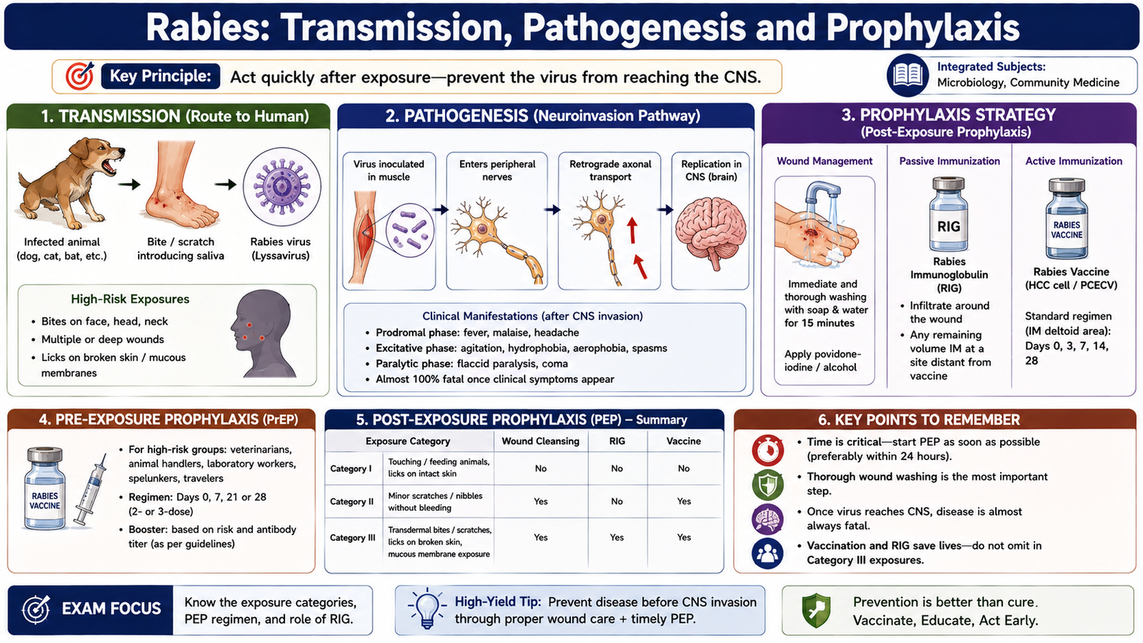
Rabies is an acute viral infection of the central nervous system transmitted mainly through the saliva of an infected mammal, usually by a bite. Once clinical rabies develops, the disease is extremely serious. Prevention therefore depends on acting before symptoms appear, either by vaccinating people at continuing risk or by providing prompt post-exposure prophylaxis after a potentially infectious exposure.
How infection develops
Virus introduced through a bite or contaminated wound initially replicates locally and then enters peripheral nerves. It travels toward the central nervous system, where it produces encephalitis. It subsequently spreads along nerves to tissues including the salivary glands, facilitating further transmission.
Pre-exposure prophylaxis
Pre-exposure prophylaxis is intended for persons who have a continuing or predictable risk of rabies exposure, such as certain laboratory personnel or people with repeated high-risk animal contact. It consists of rabies vaccination before exposure. Pre-exposure vaccination simplifies management after a later exposure but does not mean that a subsequent exposure can be ignored.
Post-exposure prophylaxis
Post-exposure prophylaxis should begin promptly after a significant suspected exposure. Its purpose is to prevent the virus from reaching the nervous system before protective immunity develops.
- Immediate wound care: thoroughly wash and clean the wound to reduce the amount of infectious material.
- Rabies vaccine: provides active immunity according to the appropriate post-exposure schedule.
- Rabies immunoglobulin: provides immediate passive antibodies for indicated exposures in previously unvaccinated persons and is administered so that antibody is available at the exposure site as effectively as possible.
A previously vaccinated person still requires appropriate post-exposure vaccination after a significant exposure, but passive rabies immunoglobulin is generally not required because immune memory is already present.
G. Scabies and Leprosy: Direct Contact and Community Prevention
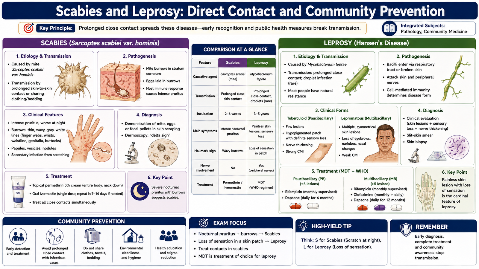
Scabies and leprosy differ greatly in cause and disease mechanism, but both require attention to prolonged or close human contact and community-level prevention. Scabies is an ectoparasitic infestation, whereas leprosy is a chronic bacterial infection caused by Mycobacterium leprae.
Scabies
Scabies is caused by the mite Sarcoptes scabiei. The female mite burrows into the superficial skin and deposits eggs. The host response to mites and their products causes intense itching, classically worse at night.
Transmission occurs mainly through close and sufficiently prolonged skin-to-skin contact. Crowded living conditions and close household contact facilitate spread. Clothing or bedding can contribute in some circumstances, particularly where infestation is heavy.
Preventive measures for scabies
- Identify affected individuals promptly.
- Manage close household or other relevant contacts at the same time when indicated to prevent reinfestation.
- Avoid close skin contact until effective management has been completed.
- Clean or appropriately handle recently used clothing, towels and bedding.
- Reduce transmission in crowded institutions through early recognition and coordinated control.
Leprosy
Leprosy is a chronic infection caused by Mycobacterium leprae, an acid-fast organism with a preference for cooler tissues, particularly the skin and peripheral nerves. The clinical pattern depends strongly on the patient’s cell-mediated immune response.
Risk factors
Risk is associated mainly with prolonged close exposure to an untreated infectious case. Household contact and living in settings where transmission continues increase exposure opportunity. Casual brief contact is much less important than prolonged exposure.
Spectrum or stages of disease
Leprosy lies along an immunological spectrum. At one end, tuberculoid leprosy reflects relatively strong cell-mediated immunity and produces fewer, well-defined lesions with marked sensory loss and relatively few organisms. At the opposite end, lepromatous leprosy reflects weak cell-mediated immunity against the organism and produces numerous, more symmetrical lesions with a high organism burden. Intermediate or borderline forms lie between these poles.
| Feature | Tuberculoid end | Lepromatous end |
|---|---|---|
| Cell-mediated immunity | Relatively strong | Poor |
| Lesions | Few, well defined | Numerous, more diffuse/symmetrical |
| Organism load | Low | High |
Prevention
The central preventive principle is early detection and effective treatment of cases, which reduces continued transmission and prevents progressive nerve damage and disability. Examination and surveillance of appropriate close contacts can support early detection. Community education is also important because stigma and delayed presentation interfere with control.
H. Trachoma: Etiology, Risk Factors, Complications and Prevention
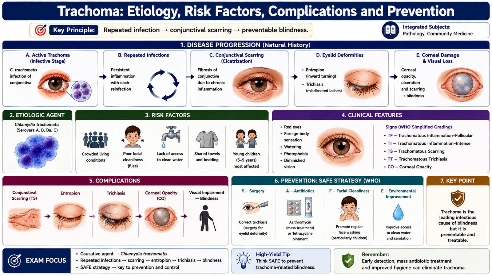
Trachoma is a chronic keratoconjunctival infection caused by particular strains of Chlamydia trachomatis. Repeated infection and inflammation, rather than a single mild episode, are responsible for the progressive scarring that can eventually threaten vision. Transmission is favored where facial hygiene is poor and where environmental conditions facilitate transfer of infected ocular and nasal secretions.
Etiology and transmission
Infectious ocular secretions can be transferred by direct person-to-person contact, contaminated hands or articles, and mechanical carriage by flies. Children often represent an important reservoir of active infection in communities where trachoma is endemic.
Risk factors
- Crowded living conditions.
- Poor facial cleanliness.
- Inadequate access to water for hygiene.
- Poor environmental sanitation.
- Repeated exposure to infected household or community contacts.
How complications develop
Repeated conjunctival infection produces recurrent inflammation. Healing by fibrosis progressively scars the inner eyelid. Scarring can pull the eyelid margin inward, producing entropion. Eyelashes then rub against the cornea, producing trichiasis. Continued corneal trauma and inflammation can lead to corneal opacity and ultimately visual impairment or blindness.
Prevention
Prevention requires both control of active infection and interruption of repeated community transmission. A useful public-health framework is the SAFE strategy:
- S — Surgery for trichiasis to prevent continued corneal injury.
- A — Antibiotics for active infection according to control-program practice.
- F — Facial cleanliness to reduce infected ocular and nasal secretions.
- E — Environmental improvement, including better water access and sanitation to reduce transmission.
Integrated Mechanism Flow
⭐ AIM High-Yield Review
- Bacillus and Clostridium are the major medically important spore-forming gram-positive rod groups.
- B. anthracis is non-motile, possesses a poly-D-glutamic acid capsule and produces protective antigen, edema factor and lethal factor.
- Cutaneous anthrax classically produces a black eschar with prominent surrounding edema.
- C. tetani causes spastic paralysis because tetanospasmin prevents normal inhibitory control of motor neurons.
- Tetanus prevention depends on active immunization; tetanus immune globulin provides passive immunity when indicated after exposure.
- C. botulinum produces descending flaccid paralysis, contrasting with the spastic paralysis of tetanus.
- C. diphtheriae causes toxin-mediated local necrosis and an adherent pseudomembrane; systemic toxin can injure the heart and nerves.
- Listeria monocytogenes is an intracellular gram-positive rod important in pregnancy, neonates, older adults and immunocompromised patients.
- Brucellosis is strongly associated with unpasteurized dairy products and occupational animal exposure.
- Yersinia pestis causes bubonic, septicemic and pneumonic plague; pneumonic plague has important person-to-person respiratory transmission.
- Rabies post-exposure prophylaxis is built around immediate wound cleansing + rabies vaccine ± rabies immunoglobulin according to previous vaccination and exposure status.
- Scabies commonly produces intense nocturnal itching and spreads mainly through close skin contact.
- Leprosy primarily affects the skin and peripheral nerves; tuberculoid disease has stronger cell-mediated immunity than lepromatous disease.
- Repeated trachoma can progress through scarring → entropion → trichiasis → corneal damage → visual loss.
- The SAFE approach to trachoma prevention emphasizes Surgery, Antibiotics, Facial cleanliness and Environmental improvement.
🎥 AIM Recommended Video
Gram-Positive Bacilli of Medical Importance
Review Bacillus, Clostridium, Listeria and Corynebacterium with their important diseases, pathogenesis and laboratory features.
📌 Focus while watching: spore-forming vs non-spore-forming rods, anthrax toxins, tetanus pathogenesis, diphtheria toxin and Listeria.