
📖 2 Learning Material
📖 Step 2 — Learning Material
This topic uses the AIM Learning Cycle to help MBBS students understand the structural and developmental organization of the stomach by integrating Anatomy, Histology and Embryology.
🔹 1️⃣ Introduction
The stomach is a vital organ of the gastrointestinal tract that acts as a reservoir and digestive chamber between the esophagus and small intestine. It is located mainly in the left upper quadrant of the abdomen and plays an essential role in mechanical and chemical digestion. Structurally, the stomach is highly specialized to mix food, secrete digestive enzymes, and regulate gastric emptying. Its rich blood supply and lymphatic drainage are clinically important in conditions such as peptic ulcer disease and gastric carcinoma. Understanding the development of the stomach also explains congenital abnormalities such as pyloric stenosis. This topic integrates Anatomy, Histology, and Embryology to provide a complete structural and functional understanding of the stomach.
🔹 2️⃣ Foundation Concepts
Key Definitions
- Stomach: A muscular, J-shaped organ that stores food and begins protein digestion.
- Gastric glands: Specialized glands in the stomach mucosa responsible for secretion of acid, enzymes, and mucus.
- Fundus: Dome-shaped superior part of stomach.
- Pylorus: Terminal region of stomach connecting to duodenum.
- Stomach bed: Structures lying posterior to stomach that support it.
- Lesser sac (omental bursa): Peritoneal space located behind the stomach.
Essential Terminology
- Cardia: Region where esophagus enters stomach
- Fundus: Superior dome
- Body: Main central region
- Pyloric antrum: Distal stomach portion
- Pyloric canal: Narrow terminal part
- Gastric pits: Openings of gastric glands
- Fundic glands: Glands found in body and fundus
- Pyloric glands: Mucus-secreting glands in pyloric region
Basic Overview
- Stomach lies between esophagus and duodenum
- Located mainly in epigastric and left hypochondriac regions
- Performs storage, mixing, digestion, and controlled emptying
- Contains specialized histological layers
- Receives rich arterial blood supply
- Develops from foregut during embryonic life
🔹 3️⃣ Core Learning — Curriculum Coverage
ANATOMY
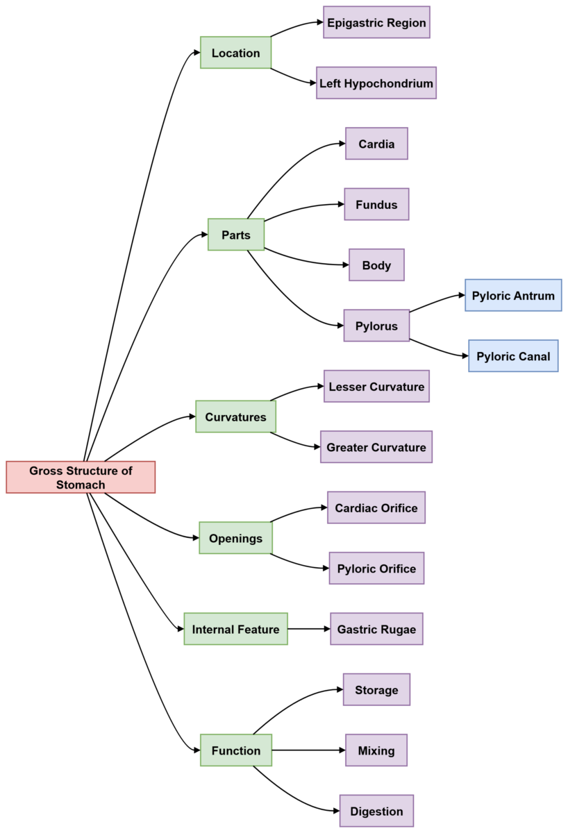
Gross Structure of Stomach
🧠 CORE
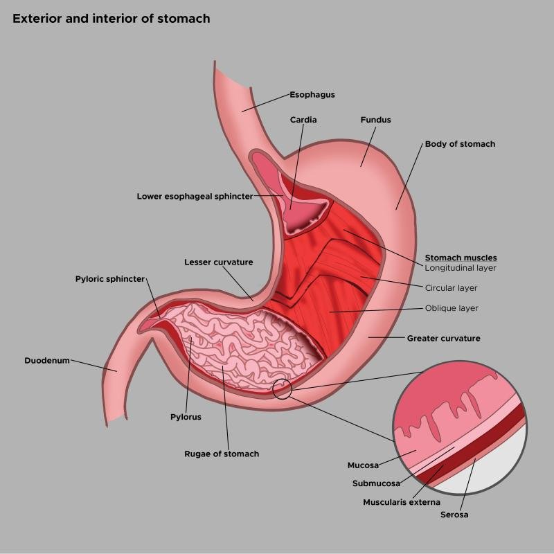
- The stomach is a J-shaped muscular organ located between esophagus and duodenum.
- Lies mainly in left upper abdomen (epigastric and left hypochondriac regions).
- Has two surfaces:
- Anterior surface
- Posterior surface
- Has two curvatures:
- Lesser curvature (right border)
- Greater curvature (left border)
- Divided into four parts:
- Cardia
- Fundus
- Body
- Pylorus
- Has two openings:
- Cardiac orifice
- Pyloric orifice
- Internally contains gastric rugae (folds).
- Main function: storage, mixing, and digestion of food.
🔬 CONCEPT EXPLAINED
Structure
The stomach is positioned between the esophagus superiorly and the duodenum inferiorly. It has a curved shape, allowing expansion during food intake. The lesser curvature faces right, while the greater curvature faces left.
Mechanism
When food enters, the stomach expands due to muscular layers. The rugae flatten, allowing storage of large volumes of food without increasing pressure.
Structure → Function
The curved shape and expandable walls allow the stomach to act as a temporary food reservoir and mixing chamber.
⚠️ IF DAMAGED
Cause: Loss of stomach muscle tone
Effect: Reduced mixing of food → impaired digestion → delayed gastric emptying
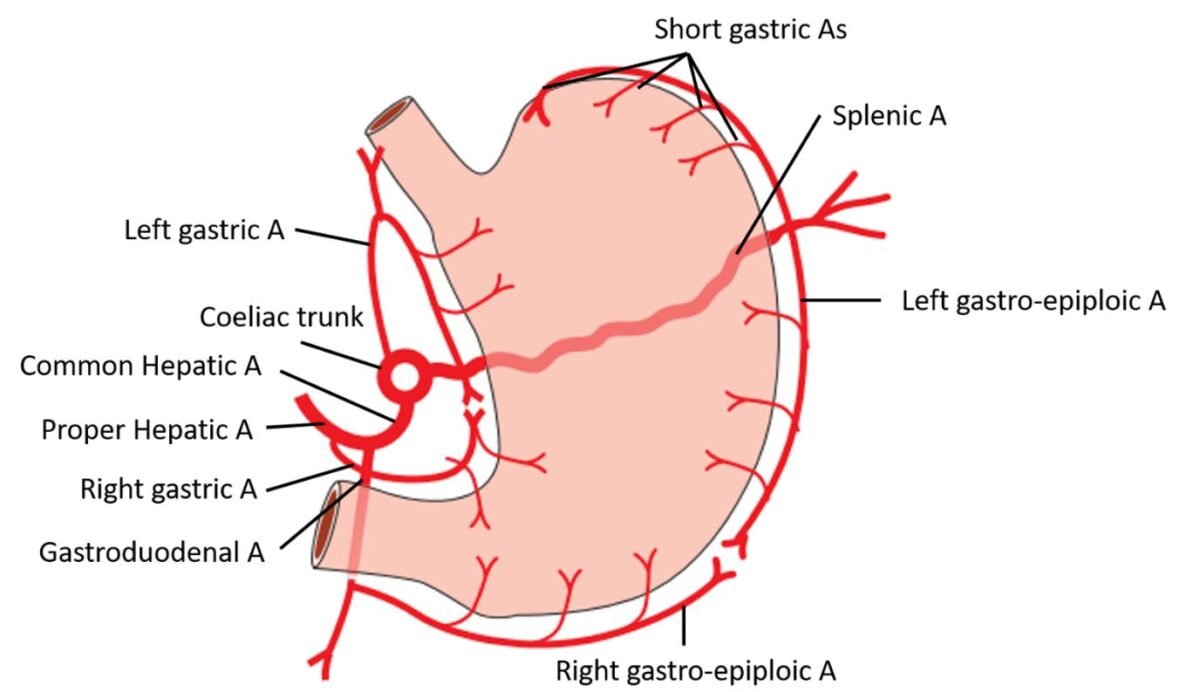
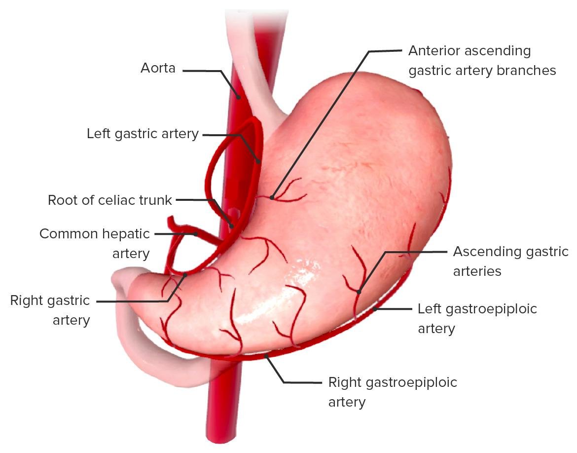
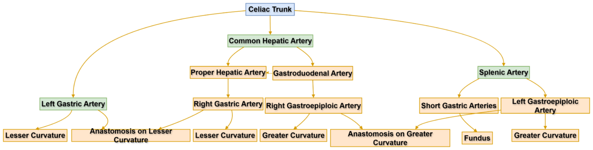
Blood Supply of Stomach
🧠 CORE
- Stomach receives rich arterial blood supply.
- Arteries arise mainly from celiac trunk.
- Lesser curvature supplied by:
- Left gastric artery
- Right gastric artery
- Greater curvature supplied by:
- Right gastroepiploic artery
- Left gastroepiploic artery
- Fundus supplied by:
- Short gastric arteries
- Venous drainage parallels arteries.
- Veins drain into portal vein.
- Rich supply supports secretion and healing.
🔬 CONCEPT EXPLAINED
Structure
Multiple arteries supply different parts of the stomach, forming arterial anastomoses along curvatures.
Mechanism
Blood carries oxygen and nutrients to gastric glands for acid and enzyme production.
Structure → Function
Rich blood supply ensures continuous secretion and tissue repair.
⚠️ IF DAMAGED
Cause: Arterial rupture or ulcer erosion
Effect: Severe bleeding → hematemesis → shock
Lymphatic Drainage of Stomach
🧠 CORE
- Lymph drains along arteries.
- Major lymph nodes:
- Left gastric nodes
- Right gastric nodes
- Gastroepiploic nodes
- Pyloric nodes
- Final drainage into:
- Celiac lymph nodes
- Important pathway for:
- Cancer spread
- Lymph follows vascular pattern.
🔬 CONCEPT EXPLAINED
Structure
Lymph vessels accompany arteries along stomach curvatures.
Mechanism
They remove tissue fluid and immune cells.
Structure → Function
Arrangement allows immune surveillance and drainage.
⚠️ IF DAMAGED
Cause: Malignant spread
Effect: Metastasis to regional lymph nodes
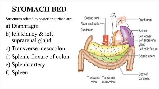
Anatomy of Stomach Bed
🧠 CORE
Stomach bed refers to structures posterior to stomach.
Posterior relations include:
- Pancreas
- Left kidney
- Left suprarenal gland
- Spleen
- Splenic artery
- Transverse mesocolon
- Left colic flexure
- Diaphragm
Supports stomach posteriorly.
🔬 CONCEPT EXPLAINED
Structure
The stomach rests on multiple organs collectively called the stomach bed.
Mechanism
These structures stabilize the stomach during digestion.
Structure → Function
Posterior support maintains proper orientation of stomach.
⚠️ IF DAMAGED
Cause: Posterior gastric ulcer
Effect: Pancreatic involvement → severe back pain
HISTOLOGY
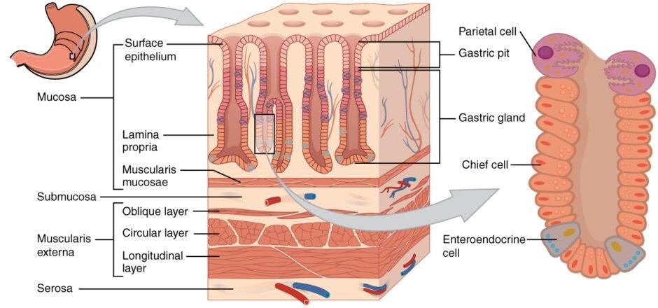
Layers of Stomach Wall
🧠 CORE
Stomach wall has four layers:
- Mucosa
- Submucosa
- Muscularis externa
- Serosa
Mucosa contains:
- Epithelium
- Lamina propria
- Muscularis mucosa
Muscularis externa has:
- Inner oblique layer
- Middle circular layer
- Outer longitudinal layer
Special feature: three muscle layers
🔬 CONCEPT EXPLAINED
Structure
The stomach wall has extra muscle layer (oblique), unlike most GIT organs.
Mechanism
Multiple muscle layers allow strong mixing movements.
Structure → Function
Three muscle layers enable churning of food into chyme.
⚠️ IF DAMAGED
Cause: Muscle damage
Effect: Poor mixing → indigestion
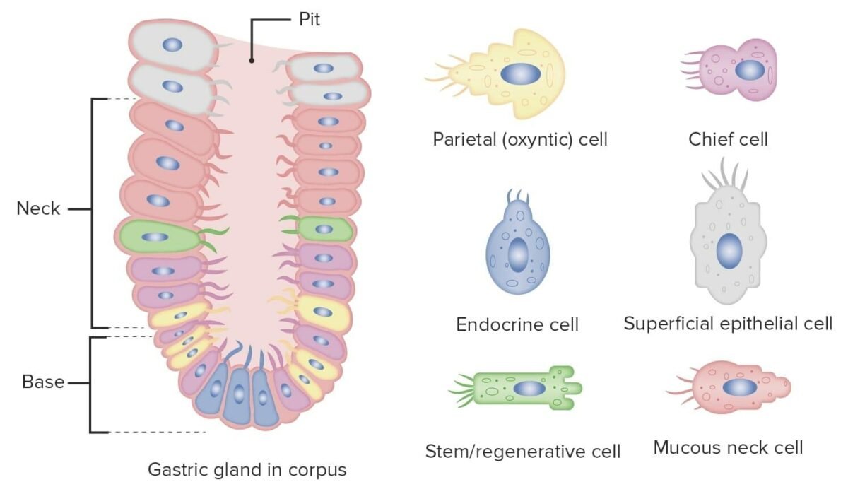
Gastric Glands and Types
🧠 CORE
Three major gastric gland types:
- Fundic glands
- Cardiac glands
- Pyloric glands
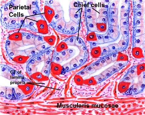
Fundic glands contain:
- Parietal cells
- Chief cells
- Mucous cells
Parietal cells produce:
- Hydrochloric acid
- Intrinsic factor
Chief cells produce:
- Pepsinogen
🔬 CONCEPT EXPLAINED
Structure
Gastric glands are tubular structures opening into gastric pits.
Mechanism
Cells secrete substances needed for digestion.
Structure → Function
Different cells perform specialized digestive roles.
⚠️ IF DAMAGED
Cause: Parietal cell loss
Effect: Vitamin B12 deficiency → pernicious anemia
Fundic Mucosa
🧠 CORE
- Found in fundus and body
- Contains fundic glands
- Rich in:
- Parietal cells
- Chief cells
- Produces:
- Acid
- Enzymes
Main digestive region.
🔬 CONCEPT EXPLAINED
Fundic mucosa is specialized for acid and enzyme production needed for digestion.
⚠️ IF DAMAGED
Acid secretion decreases → digestion impaired.
Pyloric Mucosa
🧠 CORE
- Found in pylorus
- Contains pyloric glands
- Mainly mucus secreting
- Produces gastrin hormone
Protective function.
🔬 CONCEPT EXPLAINED
Mucus protects mucosa from acid damage.
⚠️ IF DAMAGED
Loss of mucus → ulcer formation.
EMBRYOLOGY
Development of Stomach
🧠 CORE
- Develops from foregut
- Appears during 4th week
- Rotates 90° clockwise
- Greater curvature forms from dorsal border
- Lesser curvature forms from ventral border
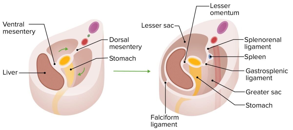
- Dorsal mesogastrium forms:
- Greater omentum
- Ventral mesogastrium forms:
- Lesser omentum
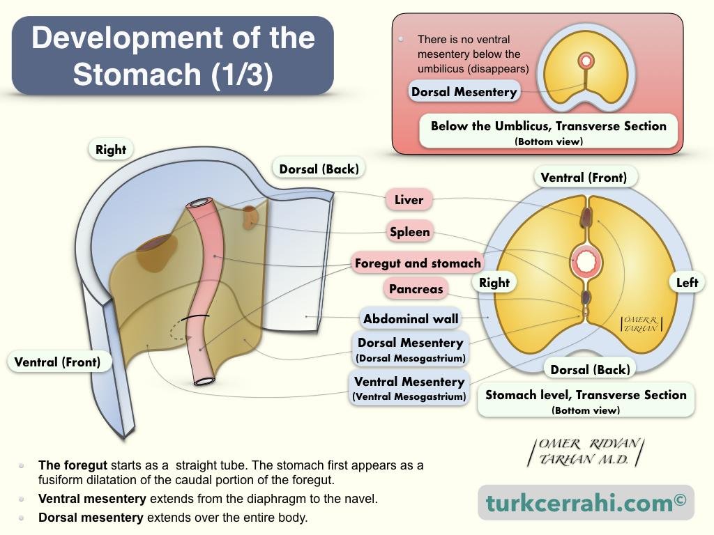
🔬 CONCEPT EXPLAINED
Structure
Initially tube-shaped stomach enlarges and rotates.
Mechanism
Rotation shifts stomach to left side.
Structure → Function
Correct positioning supports digestive efficiency.
⚠️ IF DAMAGED
Rotation failure → abnormal stomach position.
Developmental Anomalies of Stomach
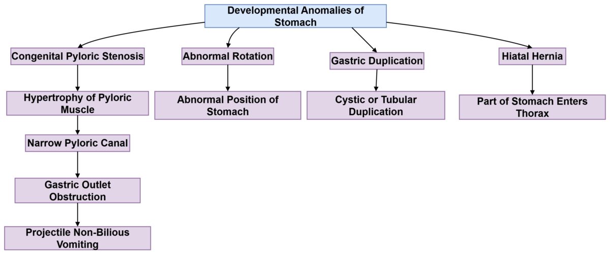
🧠 CORE
Common anomalies:
- Congenital pyloric stenosis
- Gastric duplication
- Hiatal hernia
- Abnormal rotation
🔬 CONCEPT EXPLAINED
Hypertrophy of pyloric muscle leads to obstruction.
⚠️ IF DAMAGED
Cause: Narrow pylorus
Effect: Projectile vomiting in infants
⚙️ 4️⃣ Functional Flow
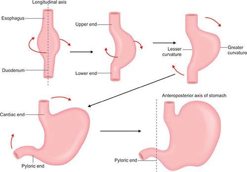
Development of Stomach Rotation
- Foregut dilates to form stomach.
- Dorsal border grows faster.
- Stomach rotates 90° clockwise.
- Greater curvature shifts left.
- Lesser curvature shifts right.
- Final anatomical position established.
🩺 5️⃣ Clinical Correlation
Common exam-relevant conditions:
Peptic Ulcer
- Due to mucosal damage
- Common along lesser curvature
- May bleed from gastric arteries
Gastric Carcinoma
- Spreads through lymphatics
- Early metastasis to celiac nodes
Congenital Pyloric Stenosis
- Hypertrophy of pyloric muscle
- Projectile vomiting in infants
Pernicious Anemia
- Loss of intrinsic factor
- Vitamin B12 deficiency
📌 6️⃣ Summary Points
- Stomach is a J-shaped organ.
- Has four parts: cardia, fundus, body, pylorus.
- Supplied mainly by celiac trunk branches.
- Fundic glands contain parietal and chief cells.
- Stomach wall has three muscle layers.
- Develops from foregut.
- Rotates 90° clockwise.
- Greater omentum develops from dorsal mesogastrium.
- Pyloric stenosis causes projectile vomiting.
- Lymph drains into celiac nodes.