
📖 Step 2 — Learning Material
1️⃣ Introduction
The duodenum and pancreas form a highly coordinated functional unit responsible for digestion and metabolic regulation. The duodenum is the first part of the small intestine, receiving chyme from the stomach and secretions from the liver and pancreas. The pancreas lies posterior to the stomach and functions as both an exocrine gland (digestive enzymes) and an endocrine gland (hormone secretion).
Understanding their structure and development is essential because many clinical conditions — such as duodenal obstruction, annular pancreas, pancreatitis, and diabetes mellitus — arise from structural or developmental abnormalities. The spatial relationships of these organs are also critical for interpreting abdominal imaging and understanding disease spread.
2️⃣ Foundation Basics
Key Definitions
- Duodenum — First part of small intestine connecting stomach to jejunum.
- Pancreas — Mixed gland performing digestive (exocrine) and hormonal (endocrine) functions.
- Ampulla of Vater — Opening where bile duct and pancreatic duct empty into duodenum.
- Brunner’s glands — Specialized mucus-secreting glands of the duodenum.
- Islets of Langerhans — Endocrine cell clusters within pancreas.
- Foregut derivative — Structure formed from embryonic foregut.
- Midgut derivative — Structure formed from embryonic midgut.
- Ventral pancreatic bud — Embryonic structure forming part of pancreas.
- Dorsal pancreatic bud — Larger embryonic pancreatic structure.
Essential Terminology
- C-shaped loop — Shape of duodenum surrounding pancreatic head.
- Retroperitoneal organ — Organ lying posterior to peritoneum.
- Papilla — Small mucosal projection where ducts open.
- Pancreatic acini — Enzyme-secreting units of pancreas.
- Annular pancreas — Pancreatic tissue surrounding duodenum.
Basic Overview
- Duodenum has four parts.
- Pancreas has head, neck, body, tail.
- Duodenum receives:
- Gastric chyme
- Bile
- Pancreatic enzymes
- Pancreas performs:
- Digestive enzyme secretion
- Hormone secretion
- Development involves rotation and fusion of pancreatic buds.
3️⃣ Core Learning — Curriculum Coverage
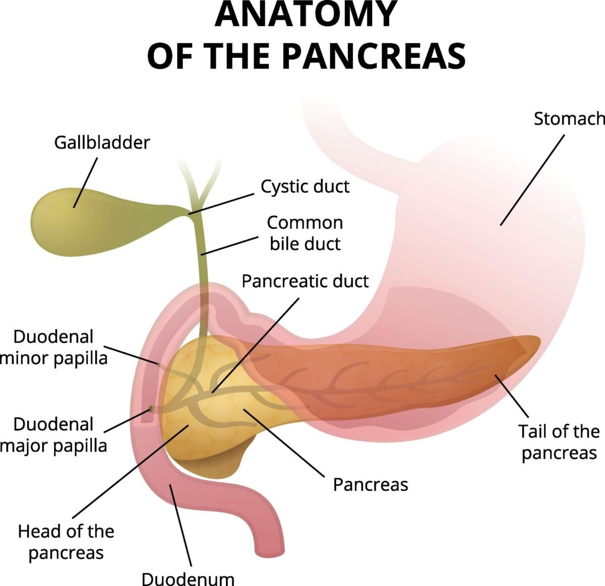
Gross Structure of Duodenum
🧠 CORE
Definition
Duodenum is the first part of the small intestine, extending from pylorus to jejunum.
Major Components
- Length ≈ 25 cm
- Shape — C-shaped loop
- Divided into:
- 1st part — Superior
- 2nd part — Descending
- 3rd part — Horizontal
- 4th part — Ascending
- Mostly retroperitoneal
- Contains:
- Major duodenal papilla
- Minor duodenal papilla
- Suspended at end by ligament of Treitz
Primary Function
- Neutralizes gastric acid
- Receives bile and pancreatic secretions
- Initiates intestinal digestion
🔬 CONCEPT EXPLAINED
Structure
The duodenum begins at the pylorus of stomach and curves around the head of pancreas, forming a characteristic C-shape.
The descending part contains openings of bile and pancreatic ducts. The ascending part joins the jejunum at the duodenojejunal flexure, supported by ligament of Treitz.
Mechanism
- Chyme enters duodenum from stomach.
- Duodenal glands secrete mucus.
- Pancreatic enzymes enter via pancreatic duct.
- Bile enters via bile duct.
- Acid is neutralized.
- Digestion continues.
Structure → Function
- C-shaped structure allows close relation with pancreas.
- Retroperitoneal position stabilizes the organ.
- Papillae location allows mixing of bile and enzymes efficiently.
⚠️ IF DAMAGED
Cause → Effect:
- Duodenal ulcer → wall erosion → bleeding
- Obstruction → vomiting → electrolyte imbalance
- Papilla damage → bile flow obstruction → jaundice
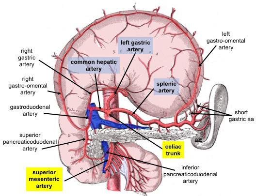
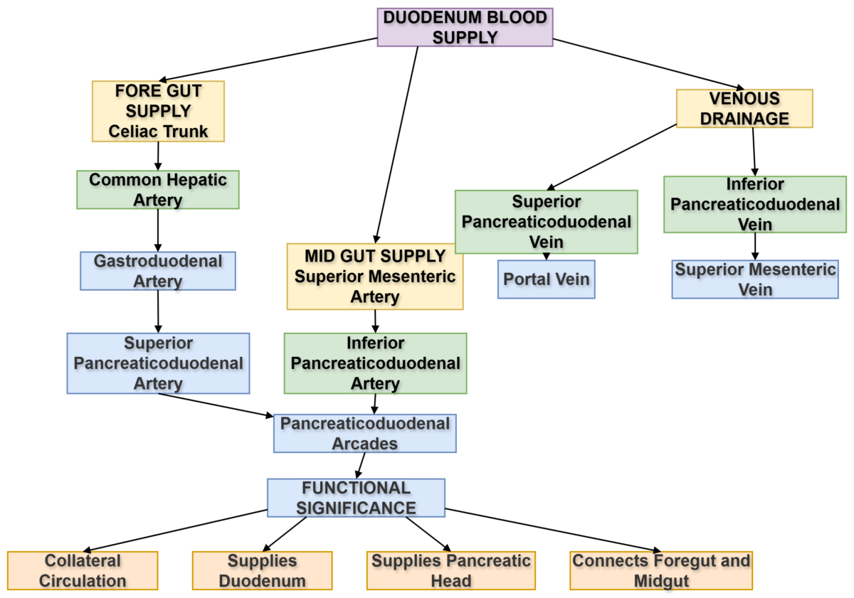
Blood Supply of Duodenum
🧠 CORE
Primary Arterial Supply
- Superior pancreaticoduodenal artery
- From gastroduodenal artery
- Inferior pancreaticoduodenal artery
- From superior mesenteric artery
Parent Arteries
- Celiac trunk
- Superior mesenteric artery
Primary Function
- Provides oxygen and nutrients
- Supports digestion
🔬 CONCEPT EXPLAINED
Structure
Duodenum lies at junction of foregut and midgut, so receives dual blood supply from both arterial systems.
Mechanism
- Blood enters via pancreaticoduodenal arcades.
- Supplies mucosa and muscular wall.
- Supports glandular secretion.
- Maintains tissue health.
Structure → Function
- Dual supply ensures continuous perfusion.
- Anastomoses provide collateral circulation.
⚠️ IF DAMAGED
- Arterial blockage → ischemia
- Ischemia → ulcer formation
- Severe damage → perforation
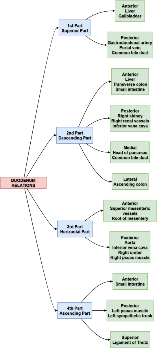
Relations of Duodenum
🧠 CORE
3D Location
- Lies in upper abdomen
- Surrounds pancreatic head.
Major Relations
Anterior:
- Liver
- Gallbladder
- Transverse colon
Posterior:
- Inferior vena cava
- Aorta
- Right kidney
Medial:
- Pancreas
Lateral:
- Ascending colon
🔬 CONCEPT EXPLAINED
Structure
Each part of the duodenum has specific surrounding organs that define its relations.
Structure → Function
Close relation to pancreas allows direct entry of pancreatic enzymes into intestine.
⚠️ IF DAMAGED
- Tumor → compression of bile duct → obstructive jaundice
- Pancreatic swelling → duodenal obstruction
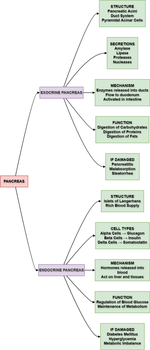
Gross Structure of Pancreas
🧠 CORE
Definition
Pancreas is a mixed gland with exocrine and endocrine components.
Major Parts
- Head
- Neck
- Body
- Tail
- Uncinate process
3D Location
- Posterior to stomach
- Extends from duodenum to spleen
Primary Function
- Produces digestive enzymes
- Releases hormones into blood
🔬 CONCEPT EXPLAINED
Structure
The head lies within duodenal curve, while tail reaches spleen. The pancreas lies retroperitoneally except its tail.
Structure → Function
- Position near duodenum allows efficient enzyme delivery.
- Long shape supports widespread blood supply.
⚠️ IF DAMAGED
- Pancreatitis → enzyme leakage → tissue destruction
- Tumor → bile duct obstruction → jaundice
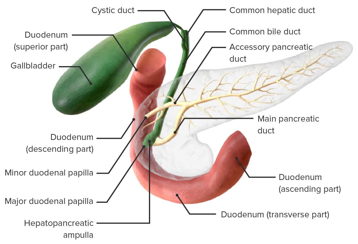
Pancreatic Ductal System
🧠 CORE
Major Ducts
- Main pancreatic duct (Wirsung)
- Accessory pancreatic duct (Santorini)
Opening
- Main duct → Major papilla
- Accessory duct → Minor papilla
Function
- Transfers digestive enzymes to duodenum.
🔬 CONCEPT EXPLAINED
Mechanism
- Acinar cells produce enzymes.
- Enzymes enter duct system.
- Ducts merge.
- Enter duodenum.
⚠️ IF DAMAGED
- Duct blockage → pancreatitis
- Stone obstruction → severe abdominal pain
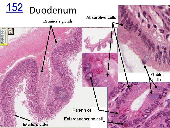
Histology of Duodenum
🧠 CORE
Recognition Features:
- Simple columnar epithelium
- Villi present
- Submucosal Brunner’s glands
- Muscularis externa
Function:
- Secretion of mucus
- Neutralization of acid
🔬 CONCEPT EXPLAINED
Structure → Function:
Brunner’s glands secrete alkaline mucus, protecting mucosa from gastric acid.
⚠️ IF DAMAGED
- Loss of mucus → ulcer formation
Histology of Pancreas
🧠 CORE
Key Structures:
- Acini — exocrine
- Islets — endocrine
- Connective tissue septa
Function:
- Digestive secretion
- Hormone release
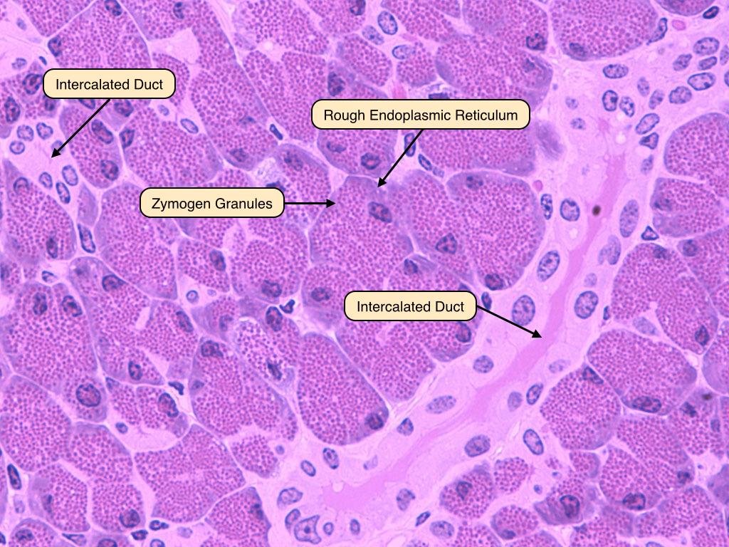
Exocrine Pancreas
🧠 CORE
Cells:
- Acinar cells
- Duct cells
Products:
- Amylase
- Lipase
- Proteases
Function:
- Digestion of nutrients.
Endocrine Pancreas
🧠 CORE
Cells:
- Alpha — Glucagon
- Beta — Insulin
- Delta — Somatostatin
Function:
- Blood glucose regulation.
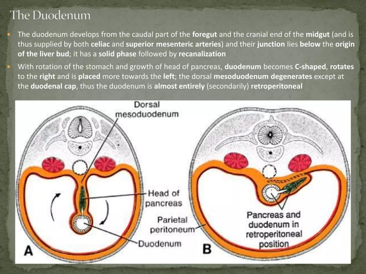
Development of Duodenum
🧠 CORE
Origin:
- Foregut + Midgut
Key Steps:
- Lumen temporarily blocked.
- Recanalization occurs.
- Rotation creates C-shape.
⚠️ IF DAMAGED
- Failure of recanalization → duodenal atresia
Development of Pancreas
🧠 CORE
Origin:
- Dorsal bud
- Ventral bud
Key Steps:
- Ventral bud rotates posteriorly.
- Buds fuse.
- Duct system forms.
⚠️ IF DAMAGED
- Incomplete fusion → annular pancreas
4️⃣ Mechanism Flow
Pancreatic Bud Rotation
- Two buds form.
- Ventral bud rotates.
- Buds fuse.
- Ducts merge.
- Final pancreas forms.
6️⃣ Clinical Correlation
Important Conditions:
- Duodenal atresia
- Annular pancreas
- Pancreatitis
- Pancreatic carcinoma
- Diabetes mellitus
⭐ 7️⃣ Points to Remember
- Duodenum has four parts.
- Mostly retroperitoneal.
- Dual blood supply — foregut + midgut.
- Pancreas is mixed gland.
- Islets regulate blood glucose.
- Brunner’s glands protect mucosa.
- Pancreas develops from two buds.
- Ventral bud rotation is essential.
- Failure leads to annular pancreas.
- Duodenal atresia causes obstruction.