
📖 Step 2 — Learning Material
🔹 1️⃣ Introduction
The liver and spleen are highly vascular organs with important microscopic organization.
The liver lies mainly in the right upper abdomen and performs metabolism, detoxification, bile formation, storage, and plasma protein synthesis.
The spleen lies in the left upper abdomen and filters blood, removes old RBCs, and supports immune defense.
Histology of these organs is important because their function depends directly on their microscopic arrangement.
In exams, students must recognize liver lobules, portal triads, hepatic cords, sinusoids, red pulp, and white pulp.
Clinically, liver damage affects metabolism and bile flow, while splenic damage affects immunity and blood filtration.
🔹 2️⃣ Foundation Concepts
Key Definitions
- Parenchyma: Functional tissue of an organ.
- Stroma: Supporting connective tissue framework.
- Hepatocytes: Main functional cells of liver.
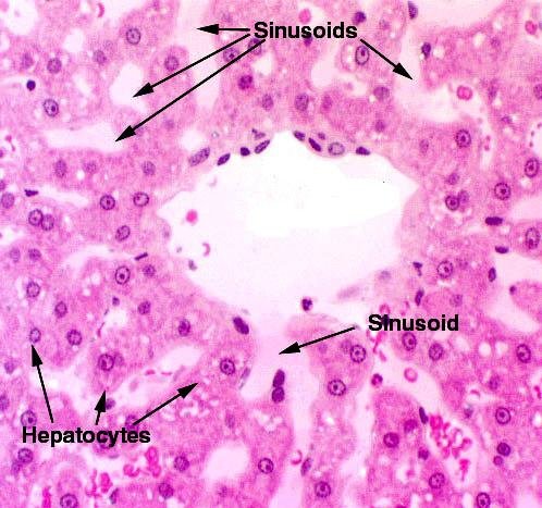
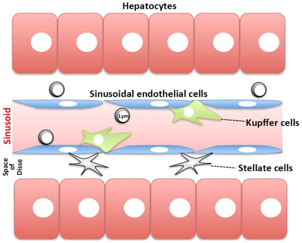
- Sinusoids: Wide blood channels between hepatic cords.
- Portal triad: Group of three structures present at portal areas of liver.
- Liver lobule: Basic microscopic structural unit of liver.
- Red pulp: Splenic tissue involved in blood filtration.
- White pulp: Splenic lymphoid tissue involved in immune response.
- Splenic cords: Cellular cords in red pulp.
- Splenic sinusoids: Vascular channels that filter blood cells.
Basic Overview
- Liver histology is organized around blood flow and bile flow.
- Blood flows from portal triad → sinusoids → central vein.
- Bile flows in the opposite direction from hepatocytes → bile canaliculi → bile ductules.
- Spleen histology is organized around blood filtration and immune surveillance.
- White pulp monitors blood for antigens.
- Red pulp removes old or abnormal RBCs.
🔹 3️⃣ Core Learning — Curriculum Coverage
1: Histological Features of Liver
🧠 CORE
- Liver is covered by a thin connective tissue capsule called Glisson’s capsule.
- The liver is made mainly of hepatocytes.
- Hepatocytes are arranged in plates or cords.
- Blood spaces between hepatocyte cords are called sinusoids.
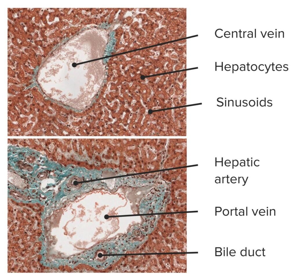
- Each classical liver lobule has a central vein.
- Portal areas are present at the corners of lobules.
- Each portal area contains the portal triad.
- Kupffer cells are macrophages present in sinusoids.
- Bile canaliculi are tiny channels between hepatocytes.
- Liver has dual blood supply: portal vein and hepatic artery.
🔬 CONCEPT EXPLAINED
Microscopically, the liver appears as many small lobules. Each lobule contains plates of hepatocytes radiating toward a central vein.
Between these hepatocyte plates are sinusoids, which carry mixed blood from the portal vein and hepatic artery toward the central vein.
This arrangement allows hepatocytes to directly interact with blood, remove toxins, process nutrients, and produce plasma proteins.
Bile is produced by hepatocytes but flows in the opposite direction to blood. It moves toward bile ductules in the portal areas.
⚠️ IF DAMAGED
- Hepatocyte damage → reduced metabolism and detoxification.
- Sinusoidal damage → impaired blood flow through liver.
- Bile canaliculi obstruction → bile retention → jaundice.
- Kupffer cell dysfunction → reduced removal of bacteria and debris from portal blood.
2: Liver Parenchyma
🧠 CORE
- Liver parenchyma is mainly formed by hepatocytes.
- Hepatocytes are polygonal cells.
- They are arranged in one-cell-thick plates or cords.
- Hepatocytes have central round nuclei.
- Some hepatocytes may be binucleated.
- Their cytoplasm is eosinophilic due to abundant organelles.
- They face blood sinusoids on one side.
- They form bile canaliculi with adjacent hepatocytes.
- They perform metabolic, secretory, and detoxifying functions.
🔬 CONCEPT EXPLAINED
Hepatocytes are arranged so that each cell is close to blood. This is essential because the liver receives nutrient-rich blood from the gastrointestinal tract through the portal vein.
The hepatocyte has two functional surfaces:
- Blood-facing surface: absorbs nutrients, toxins, hormones, and drugs.
- Bile-facing surface: secretes bile into bile canaliculi.
This dual arrangement explains why hepatocytes can process blood contents and also produce bile at the same time.
⚠️ IF DAMAGED
- Hepatocyte necrosis → raised liver enzymes.
- Reduced albumin synthesis → edema.
- Reduced clotting factor synthesis → bleeding tendency.
- Reduced bilirubin handling → jaundice.
- Reduced detoxification → drug toxicity and hepatic encephalopathy.
3: Structural Organization of Liver
🧠 CORE
- Liver is organized into microscopic lobules.
- The classical liver lobule is hexagonal in shape.
- Central vein lies in the center of the lobule.
- Portal triads lie at the corners.
- Hepatocyte cords radiate toward central vein.
- Sinusoids lie between hepatocyte plates.
- Blood flows from portal triad to central vein.
- Bile flows from hepatocytes to portal triad.
- Functional organization explains liver metabolism and bile drainage.
🔬 CONCEPT EXPLAINED
The classical liver lobule is the most commonly described histological unit. It is centered around a central vein.
At the periphery are portal areas containing branches of the portal vein, hepatic artery, and bile duct.
Blood enters the lobule from the portal vein and hepatic artery branches. It passes through sinusoids and reaches the central vein.
Hepatocytes are arranged between these blood channels, allowing maximum contact with blood.
Bile moves in the opposite direction, from hepatocytes toward bile ductules in the portal triad.
⚠️ IF DAMAGED
- Disturbed lobular architecture → impaired liver function.
- Fibrosis around lobules → cirrhosis.
- Sinusoidal blockage → portal hypertension.
- Central vein congestion → hepatic congestion.
- Bile flow obstruction → cholestasis and jaundice.
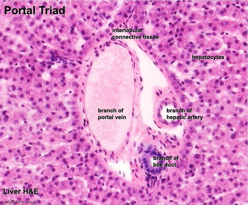
4: Portal Triad Structures and Components
🧠 CORE
The portal triad contains:
- Branch of portal vein
- Branch of hepatic artery
- Branch of bile duct
- Supporting connective tissue
- Lymphatics
- Nerve fibers
Recognition features:
- Portal vein branch is usually largest and thin-walled.
- Hepatic artery branch is smaller and thick-walled.
- Bile duct is lined by cuboidal epithelium.
- Portal triads are located at lobule corners.
- They are surrounded by connective tissue.
🔬 CONCEPT EXPLAINED
The portal triad represents the entry and exit area of the liver lobule.
The portal vein brings nutrient-rich blood from the intestine.
The hepatic artery brings oxygen-rich blood.
The bile duct carries bile away from the liver.
Blood from the portal vein and hepatic artery mixes in the sinusoids and flows toward the central vein.
Bile flows in the opposite direction, from hepatocytes toward the bile duct.
⚠️ IF DAMAGED
- Portal vein obstruction → portal hypertension.
- Hepatic artery compromise → reduced oxygen supply.
- Bile duct obstruction → obstructive jaundice.
- Portal inflammation → hepatitis or cholangitis.
- Fibrosis around portal areas → disturbed liver architecture.
5: Histological Features of Spleen
🧠 CORE
- Spleen is covered by a connective tissue capsule.
- Trabeculae extend inward from the capsule.
- Splenic parenchyma is called pulp.
- It has two main parts: red pulp and white pulp.
- White pulp is lymphoid tissue around central arteries.
- Red pulp contains splenic cords and sinusoids.
- Spleen filters blood, not lymph.
- It removes old RBCs and platelets.
- It produces immune responses against blood-borne antigens.
- It stores blood cells, especially platelets.
🔬 CONCEPT EXPLAINED
The spleen is a blood-filtering lymphoid organ. Unlike lymph nodes, it does not filter lymph; it filters blood.
Its white pulp acts like an immune surveillance area. It detects antigens present in blood.
Its red pulp acts like a mechanical and macrophage-rich filter. Old, rigid, or abnormal RBCs are trapped and removed.
This microscopic arrangement allows the spleen to combine immune function with blood quality control.
⚠️ IF DAMAGED
- Loss of splenic function → increased risk of infection.
- Poor removal of old RBCs → abnormal RBCs remain in blood.
- Splenic enlargement → excessive destruction of blood cells.
- Trauma → severe bleeding because spleen is highly vascular.
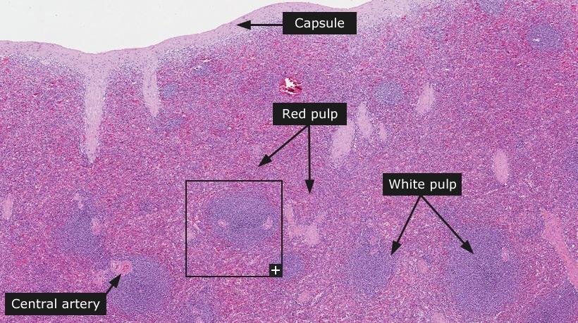
6: Red Pulp of Spleen
🧠 CORE
- Red pulp forms the major part of spleen.
- It is involved in blood filtration.
- It contains splenic cords and venous sinusoids.
- Splenic cords contain macrophages, lymphocytes, plasma cells, and blood cells.
- Sinusoids are lined by elongated endothelial cells.
- Old RBCs are trapped in red pulp.
- Macrophages destroy abnormal RBCs.
- Iron from hemoglobin is recycled.
- Red pulp also stores platelets.
🔬 CONCEPT EXPLAINED
Red pulp acts as a blood filter. Blood passes slowly through splenic cords and sinusoids.
Healthy flexible RBCs pass through narrow sinusoidal spaces and return to circulation.
Old or abnormal RBCs are less flexible. They get trapped in the cords and are phagocytosed by macrophages.
This structure helps maintain healthy circulating blood.
⚠️ IF DAMAGED
- Reduced red pulp function → defective RBC removal.
- Excessive red pulp activity → anemia, leukopenia, or thrombocytopenia.
- Congestion of red pulp → splenomegaly.
- Macrophage dysfunction → poor clearance of damaged blood cells.
7: White Pulp of Spleen
🧠 CORE
- White pulp is lymphoid tissue of spleen.
- It surrounds central arteries.
- T lymphocytes form the periarteriolar lymphoid sheath.
- B lymphocytes form lymphoid follicles.
- Germinal centers may be present in active follicles.
- White pulp responds to blood-borne antigens.
- It produces antibodies through B-cell activation.
- It helps immune defense against encapsulated bacteria.
- It appears basophilic in histological sections.
🔬 CONCEPT EXPLAINED
White pulp is arranged around branches of the splenic artery. The lymphocytes are positioned to monitor blood as it enters splenic tissue.
T cells lie close to central arteries in the periarteriolar lymphoid sheath.
B cells form follicles, which may develop germinal centers during immune activation.
This arrangement allows the spleen to detect blood-borne pathogens and produce an immune response.
⚠️ IF DAMAGED
- Reduced antibody response → increased infection risk.
- Loss of splenic immune function → severe infection by encapsulated organisms.
- Poor antigen detection → weak immune response.
- White pulp atrophy → reduced lymphoid defense.
⚙️ 4️⃣ Functional Flow
A. Blood Flow Through Liver
- Blood enters liver through:
- Portal vein
- Hepatic artery
- Blood reaches portal triads.
- Blood enters sinusoids.
- Blood flows between hepatocyte plates.
- Hepatocytes process nutrients, toxins, and metabolites.
- Blood drains into central vein.
- Central veins join hepatic veins.
- Hepatic veins drain into inferior vena cava.
B. Bile Flow Through Liver
- Hepatocytes produce bile.
- Bile enters bile canaliculi between hepatocytes.
- Bile flows toward portal areas.
- Bile enters bile ductules.
- Bile passes into larger bile ducts.
- Bile reaches gallbladder or duodenum.
Important: Blood and bile flow in opposite directions.
C. Blood Filtration in Spleen
- Blood enters through splenic artery.
- Blood passes near white pulp.
- White pulp detects blood-borne antigens.
- Blood enters red pulp cords and sinusoids.
- Healthy RBCs pass through sinusoids.
- Old or abnormal RBCs are trapped.
- Macrophages remove damaged RBCs.
- Filtered blood leaves through splenic vein.
🩺 5️⃣ Clinical Correlation
Liver
1. Hepatitis
- Cause: inflammation and injury of hepatocytes.
- Effect: impaired metabolism, raised liver enzymes, jaundice.
2. Cirrhosis
- Cause: chronic liver injury with fibrosis.
- Effect: distorted lobular architecture and portal hypertension.
3. Obstructive Jaundice
- Cause: obstruction of bile ducts.
- Effect: bile cannot drain properly, causing jaundice.
4. Portal Hypertension
- Cause: increased resistance to portal blood flow.
- Effect: splenomegaly, ascites, varices.
Spleen
1. Splenomegaly
- Cause: increased work of spleen in infections, hemolysis, or portal hypertension.
- Effect: enlarged spleen with increased blood cell destruction.
2. Hypersplenism
- Cause: excessive splenic activity.
- Effect: anemia, leukopenia, thrombocytopenia.
3. Asplenia / Splenectomy
- Cause: absent or removed spleen.
- Effect: increased risk of severe infection, especially by encapsulated bacteria.
4. Splenic Rupture
- Cause: trauma.
- Effect: life-threatening internal bleeding due to high vascularity.
📌 6️⃣ Summary Points
- Liver parenchyma is mainly formed by hepatocytes.
- Classical liver lobule has a central vein in the center and portal triads at the corners.
- Portal triad contains portal vein, hepatic artery, and bile duct.
- Blood flows from portal triad to central vein.
- Bile flows from hepatocytes to portal triad, opposite to blood flow.
- Sinusoids allow close contact between blood and hepatocytes.
- Kupffer cells are macrophages present in liver sinusoids.
- Spleen has red pulp and white pulp.
- Red pulp filters blood and removes old RBCs.
- White pulp is lymphoid tissue for immune response.
- Spleen filters blood, not lymph.
- Loss of splenic function increases risk of infection by encapsulated bacteria.