
📖 Step 2 — Learning Material
This section provides structured learning content for the topic. Follow each part in sequence to build clear conceptual understanding and maintain logical learning progression.
🔹 Introduction
Salivary glands are essential accessory organs of the digestive system responsible for producing saliva, which maintains oral health and initiates digestion. These glands are located around the oral cavity and include the parotid, submandibular, and sublingual glands. Their secretions help lubricate food, protect teeth, regulate oral hygiene, and facilitate digestion through enzymatic action. Development of salivary glands begins early in embryonic life through epithelial proliferation. Clinically, disorders such as dry mouth (xerostomia), salivary stones, and nerve damage significantly affect digestion and oral health, making this topic highly relevant for clinical understanding.
🔹 Foundation Concepts
Key Definitions
- Saliva — A watery secretion produced by salivary glands that lubricates food and begins digestion.
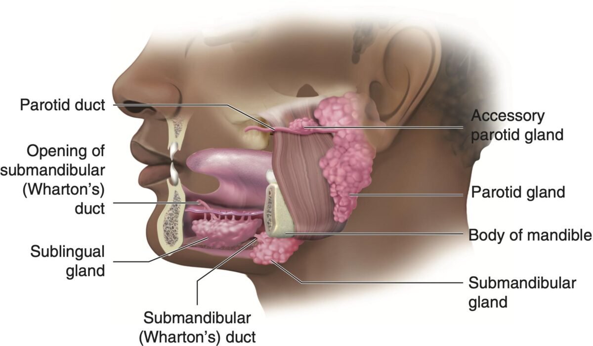
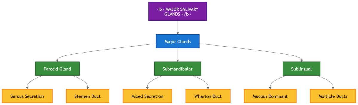
- Major salivary glands — Parotid, submandibular, and sublingual glands.
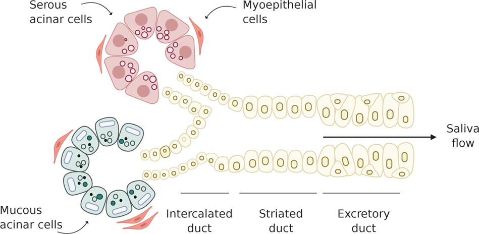
- Serous secretion — Watery secretion rich in enzymes.
- Mucous secretion — Thick secretion rich in mucin.
- Mixed glands — Glands containing both serous and mucous cells.
- Salivary amylase — Enzyme that initiates carbohydrate digestion.
- Acinus — Secretory unit of salivary gland.
- Duct system — Channels that modify saliva composition.
🔹 3️⃣ Core Learning — Curriculum Coverage
A. Major Salivary Glands — Gross Anatomy
1️⃣ Definition of Major Salivary Glands
- Major salivary glands are large paired exocrine glands that produce saliva.
- They release saliva into the oral cavity through ducts.
- They support digestion, lubrication and oral protection.
2️⃣ Names of Major Salivary Glands
- Parotid gland
- Submandibular gland
- Sublingual gland
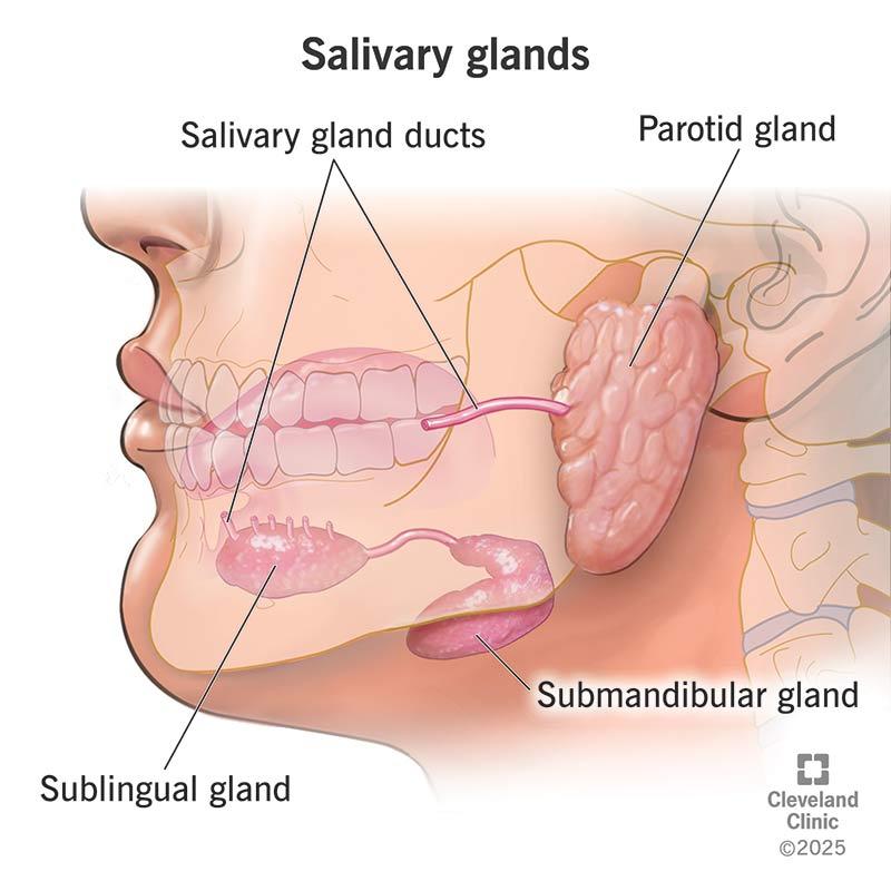
3️⃣ General Location Around Oral Cavity
- Parotid: near the ear
- Submandibular: below the mandible
- Sublingual: floor of the mouth
4️⃣ Relative Size of Glands
- Parotid: largest
- Submandibular: medium
- Sublingual: smallest
5️⃣ Type of Secretion
| Gland | Type of Secretion |
|---|---|
| Parotid | Serous |
| Submandibular | Mixed |
| Sublingual | Mucous dominant |
🧩 Master Concept Map
1. Parotid Gland — Gross Anatomy
Structure
- Largest salivary gland.
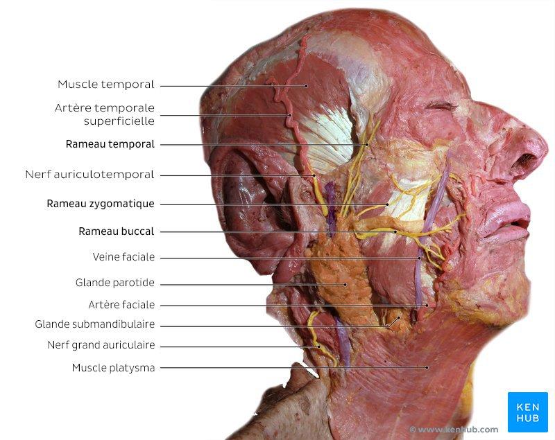
- Located anteroinferior to the ear.
- Lies between ramus of mandible and sternocleidomastoid muscle.
- Enclosed in parotid fascia.
- Traversed by facial nerve.
Parotid Duct / Stensen Duct
- Emerges from anterior border.
- Runs over masseter muscle.
- Turns medially and pierces buccinator muscle.
- Opens into oral cavity opposite the upper second molar tooth.
Blood Supply
Superficial temporal artery, a branch of external carotid artery.
Nerve Supply
- Glossopharyngeal nerve (CN IX) — parasympathetic secretion.
- Auriculotemporal nerve — carries secretomotor fibers.
Structure → Function
Large serous acini produce watery saliva and enable rapid secretion during chewing.
Important Relations
- Lateral: skin, superficial fascia, parotid fascia.
- Medial: ramus of mandible, masseter muscle.
- Anterior: mandible, masseter.
- Posterior: sternocleidomastoid muscle, mastoid process.
- Contents: facial nerve, external carotid artery, retromandibular vein.
Surfaces
- Superficial surface: skin and superficial fascia.
- Anteromedial surface: ramus of mandible and masseter muscle.
- Posteromedial surface: mastoid process, sternocleidomastoid, styloid apparatus.
- Superior surface / base: external acoustic meatus.
⚠️ If Damaged
Parotid inflammation: viral infection such as mumps may cause painful swelling and reduced salivation.
Facial nerve injury: parotid surgery may cause loss of facial expression, facial asymmetry and difficulty closing the eye.
2. Submandibular Gland — Gross Anatomy
Structure
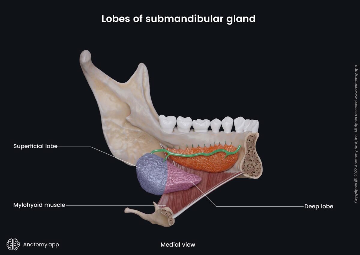
- Lies in the submandibular triangle.
- Has a superficial part and a deep part.
- Superficial part: large portion below mandible, occupying submandibular triangle.
- Deep part: small portion extending forward in the floor of mouth and curving around posterior border of mylohyoid muscle.
- Lies medial to mandible.
- Mixed gland with serous dominance.
- Wharton duct opens beside the lingual frenulum.
Blood Supply
Facial artery.
Nerve Supply
Facial nerve via chorda tympani.
Structure → Function
It produces the majority of resting saliva. Mixed secretion supports lubrication and digestion.
Important Relations
- Superficial part superior: body of mandible.
- Superficial part inferior: skin, platysma, deep fascia.
- Superficial part medial: mylohyoid and hyoglossus muscles.
- Superficial part lateral: mandible.
- Deep part: related to hyoglossus muscle, lingual nerve and submandibular duct.
- The lingual nerve loops around the duct, which is clinically important.
Submandibular Duct / Wharton Duct
- Originates from the deep part of gland.
- Runs forward in the floor of mouth.
- Lies between hyoglossus and mylohyoid muscles.
- Crossed by the lingual nerve.
- Opens near the lingual frenulum.
⚠️ If Damaged
Submandibular stone formation / sialolithiasis: duct obstruction causes pain during meals, swelling of gland and reduced salivation. This gland is most commonly affected by stones.
3. Sublingual Gland — Gross Anatomy
Structure
- Smallest salivary gland.
- Located beneath mucosa of floor of mouth.
- Produces mucous-rich saliva and helps lubrication of oral cavity.
Blood Supply
Lingual artery.
Nerve Supply
Facial nerve via chorda tympani.
Duct System
- Has multiple ducts called ducts of Rivinus.
- Ducts open directly into the floor of the mouth along the sublingual fold.
- Sometimes one duct may join the submandibular duct.
Relations
- Superior: mucous membrane of floor of mouth.
- Inferior: mylohyoid muscle.
- Medial: genioglossus muscle.
- Lateral: mandible.
- This shallow position allows direct opening into the oral cavity.
⚠️ If Damaged
Sublingual duct blockage: duct obstruction may cause swelling in the floor of mouth, difficulty swallowing and reduced lubrication.
B. Embryology — Development of Salivary Glands
🧠 Core
- Salivary glands develop from oral ectoderm.
- Development begins as epithelial buds from oral cavity lining.
- Buds grow into underlying mesenchyme.
- The proximal part forms ducts.
- Terminal parts form secretory acini.
- Development occurs at different weeks for each gland.
- Branching produces the lobular structure of glands.
- Functional maturation occurs before birth.
🔬 Concept Explained
Salivary glands develop from epithelial cells lining the primitive oral cavity. These cells proliferate and form solid epithelial buds that grow into surrounding mesenchyme. With continued growth and branching, these buds form ducts and secretory units that later produce saliva.
📍 Developmental Origin
- Germ layer origin: ectoderm of oral cavity.
- Supporting tissue: surrounding mesenchyme.
- Adult link: oral epithelium forms duct system; terminal buds form secretory acini.
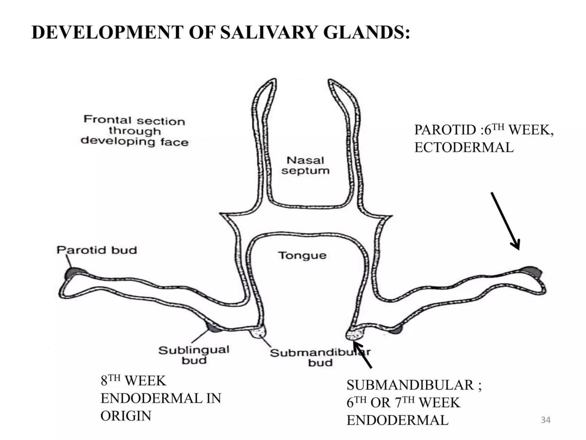
📍 Timeline of Development
- Parotid gland: begins around 6th week; first major salivary gland to develop.
- Submandibular gland: begins around 7th week.
- Sublingual gland: begins around 8th week; last major gland to develop.
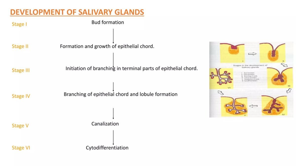
📍 Developmental Steps
Oral epithelium thickens → bud formation → bud elongates into mesenchyme → branching begins → central cells break down and lumen forms → duct system develops → terminal buds form acini → gland matures before birth.
🔗 Structure → Function Link
Branching development produces a large number of acini and increases secretory surface area. This supports efficient saliva production and functional digestion after birth.
⚠️ If Damaged
Developmental failure: defective epithelial growth may cause absence of gland, reduced saliva production and difficulty in lubrication. Duct blockage may cause swelling and reduced salivary flow.
C. Secretion and Regulation of Saliva — Physiology
A. Secretion of Saliva
- Saliva is secreted by salivary gland acinar cells.
- Initial secretion is called primary saliva.
- Primary saliva is isotonic with plasma.
- As saliva passes through ducts, its composition changes.
- Duct cells modify ion content.
- Final saliva becomes hypotonic.
- Water movement occurs by osmosis.
- Duct permeability to water is low.
🔬 Concept Explained
Saliva formation occurs in two main stages: formation of primary secretion and modification in ducts. These two stages explain how saliva changes from isotonic to hypotonic.
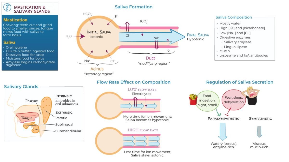
🔄 Mechanism Flow — Formation of Saliva
Acinar cells secrete fluid → Na⁺ enters lumen, Cl⁻ follows and water follows osmotically → primary saliva forms → saliva enters ducts → Na⁺ and Cl⁻ are reabsorbed while K⁺ and HCO₃⁻ are secreted → water movement is limited due to poor duct permeability → final hypotonic saliva forms.
🔗 Structure → Function Link
Acinar cells produce fluid, while duct cells modify its composition. The outcome is efficient saliva production suitable for digestion and lubrication.
B. Nervous Regulation of Salivary Secretion
- Salivary secretion is controlled by the autonomic nervous system.
- Both parasympathetic and sympathetic systems regulate secretion.
- Parasympathetic stimulation produces large-volume watery saliva.
- Sympathetic stimulation produces thick mucous saliva.
- Parasympathetic system plays the dominant role.
- Reflex control is important and sensory input initiates secretion.
Parasympathetic and Sympathetic Effects
- Parasympathetic: increases blood flow, secretion rate and enzyme output, producing watery enzyme-rich saliva.
- Sympathetic: produces a small amount of thick, viscous saliva.
🔄 Salivary Reflex Arc
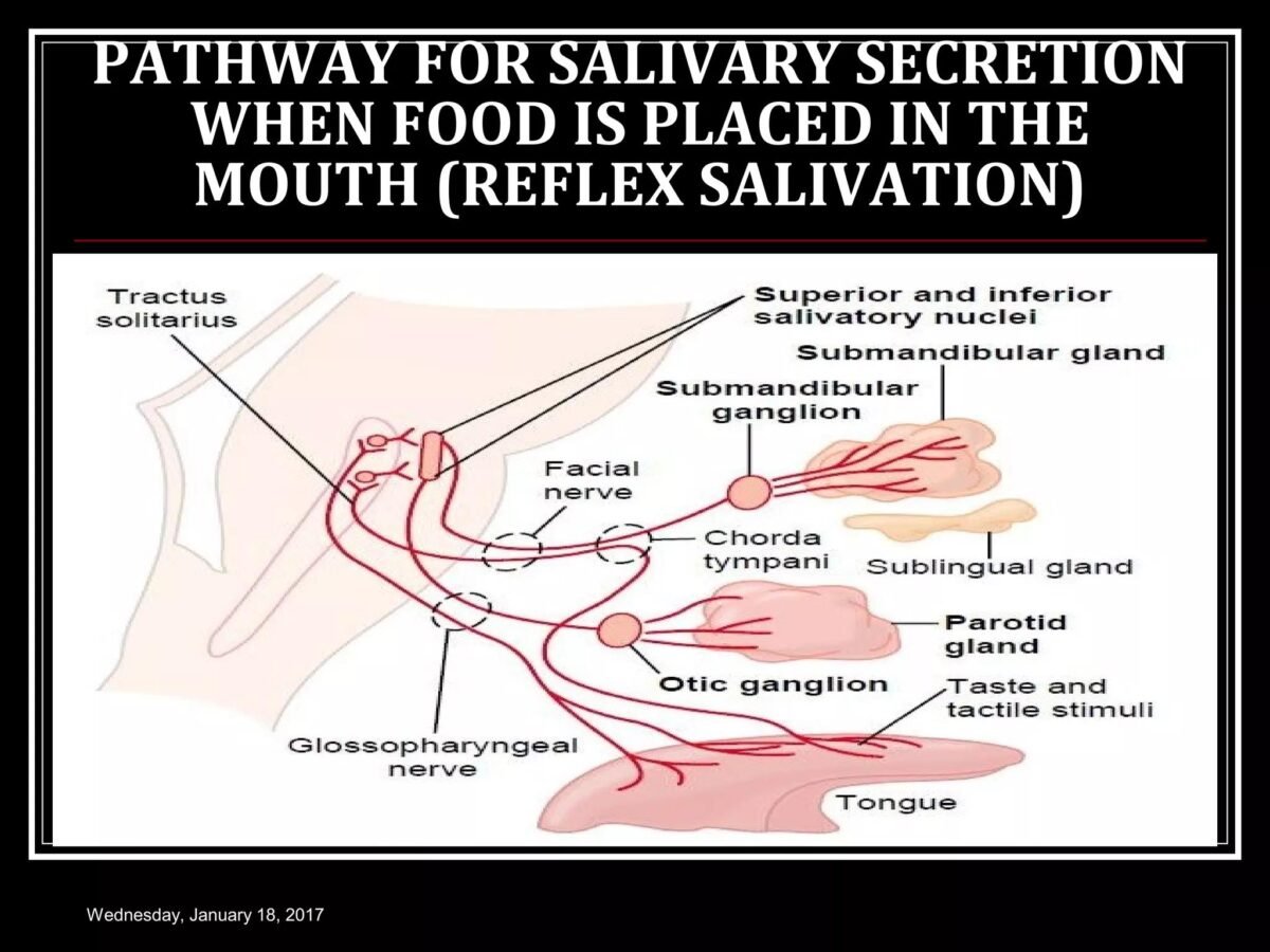
Mechanism Flow — Salivary Reflex Arc
- Stimulus initiation: food enters oral cavity and stimulates taste and tactile receptors.
- Afferent pathway: sensory impulses travel via facial nerve and glossopharyngeal nerve to salivatory nuclei in medulla.
- Central processing: salivatory nuclei are activated and reflex response begins.
- Efferent pathway: parasympathetic fibers arise from superior and inferior salivatory nuclei and reach salivary glands.
- Effector activation: salivary gland cells are stimulated, blood flow increases and secretion increases.
- Response: watery enzyme-rich saliva is produced.
C. Stimuli Increasing Salivary Secretion
- Taste
- Smell
- Chewing
- Nausea
- Conditioned reflex
- Thought of food
These stimuli activate sensory receptors that send signals to the brainstem, which activates parasympathetic pathways and prepares the digestive system before food enters the stomach.
D. Plasma and Saliva Electrolyte Composition
- Electrolyte concentration depends on flow rate.
- Low flow rate: low Na⁺, low Cl⁻, high K⁺.
- High flow rate: higher Na⁺ and Cl⁻.
- Saliva remains hypotonic compared with plasma.
At low flow rate, saliva stays longer in ducts, allowing more Na⁺ reabsorption. At high flow rate, saliva passes quickly and less modification occurs.
E. Function of Salivary Mucus
- Lubrication
- Protection
- Cohesion of food
- Support for swallowing, speech and oral protection
Mucus binds food particles into a bolus, making swallowing easier and preventing injury to oral mucosa.
F. Role of Saliva in Oral Hygiene
- Washes away food particles
- Reduces bacterial growth
- Neutralizes acids
- Protects teeth
Continuous salivary flow prevents accumulation of harmful microorganisms and reduces risk of dental caries.
G. Role in Elimination of Heavy Metals
- Mercury
- Lead
- Certain toxic metals
Toxic substances enter saliva and are removed from the body through swallowing or expectoration.
H. Digestion by Salivary Amylase / Ptyalin
- Begins carbohydrate digestion.
- Acts on starch.
- Produces maltose and dextrins.
- Works best at neutral pH.
- Activity continues briefly in stomach.
Salivary amylase breaks down complex carbohydrates into smaller sugar molecules during chewing, improving efficiency of later digestive processes.
D. Composition, Formation and Functions of Saliva — Biochemistry
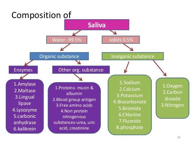
A. Composition of Salivary Secretions
Core: saliva is composed of water, electrolytes, proteins and enzymes, mucins, immunoglobulins, antimicrobial substances and organic molecules.
- Water: about 99%; dissolves food substances, helps form bolus and facilitates swallowing.
- Electrolytes: Na⁺, K⁺, Cl⁻ and HCO₃⁻ maintain ionic balance, pH and enzyme function.
- Enzymes: salivary amylase digests starch; lysozyme provides antibacterial action.
- Mucins: glycoproteins that provide viscosity, lubrication and mucosal protection.
- Immunoglobulins: mainly secretory IgA, which provides immune protection.
- Antimicrobial substances: lysozyme and lactoferrin help maintain oral hygiene.
🔗 Structure → Function Link
| Component | Function |
|---|---|
| Water | Lubrication |
| Electrolytes | pH balance |
| Enzymes | Digestion |
| Mucins | Lubrication |
| IgA | Immunity |
B. Formation of Salivary Components
- Salivary proteins are synthesized in acinar cells.
- Enzymes are produced in rough endoplasmic reticulum.
- Proteins are processed in Golgi apparatus.
- Secretory vesicles store proteins.
- Vesicles release contents into lumen.
- Mucins are synthesized in mucous cells.
- Immunoglobulin A is added to secretion.
Stepwise biochemical process: protein synthesis begins in RER → proteins move to Golgi apparatus → Golgi modifies proteins → proteins are packaged into secretory vesicles → vesicles release contents into lumen → final secretion contains functional molecules.
This process allows saliva to contain digestive enzymes, protective proteins and lubricating mucins. Without this synthesis, saliva would lack functional capability.
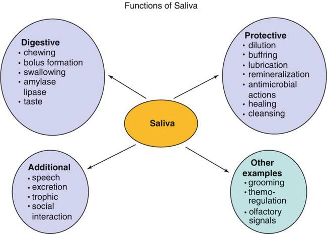
C. Functions of Saliva
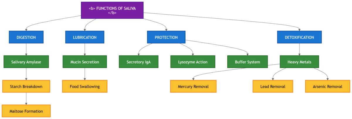
- Lubrication: mucins coat food particles, allowing smooth swallowing and reducing friction.
- Digestion: salivary amylase begins carbohydrate digestion and converts starch into maltose.
- Protection: saliva washes debris, neutralizes acids and prevents infection.
- Taste facilitation: saliva dissolves food molecules so taste receptors are stimulated effectively.
- Maintenance of oral health: continuous flow prevents bacterial accumulation and tooth decay.
- Speech support: lubrication allows smooth tongue movement and clear articulation.
⚙️ Functional Flow
Development → Structure → Secretion → Function → Outcome
Oral ectoderm develops into salivary gland buds (Embryology)
↓
Branching morphogenesis forms ducts and secretory acini (Embryology → Anatomy)
↓
Acinar cells produce primary saliva (Anatomy → Physiology)
↓
Duct system modifies saliva composition (Physiology)
↓
Saliva contains enzymes, mucus and protective proteins (Biochemistry)
↓
Final saliva performs essential functions — lubrication of food, initiation of digestion, protection of oral cavity and maintenance of oral hygiene.
↓
Functional Outcome: efficient chewing, swallowing, digestion and oral protection.
🩺 Clinical Correlation
Xerostomia / Dry Mouth
Cause: dehydration, nerve damage or radiation therapy.
Effect: difficulty swallowing, dental caries and oral infections.
Sialolithiasis / Salivary Stones
Cause: calcium deposition in ducts.
Effect: painful swelling and reduced saliva.
Parotitis / Mumps
Cause: viral infection.
Effect: painful swelling of parotid gland.
📌 Summary Points
- Parotid gland produces serous secretion.
- Submandibular gland produces major resting saliva.
- Sublingual gland produces mucous secretion.
- Parasympathetic stimulation increases saliva.
- Saliva is hypotonic compared to plasma.
- Ducts modify electrolyte composition.
- Salivary amylase starts starch digestion.
- Mucus protects oral mucosa.
- Saliva maintains oral hygiene.
- Reduced saliva leads to dental caries.