Course Content
🔵 THEME 1 — Painful Swallowing
Focuses on anatomy, physiology, and disorders related to swallowing, including oral cavity, salivary glands, esophagus, and neural regulation of deglutition.
🔵 THEME 2 — Pain Epigastrium
Focus: Structural, functional, and clinical basis of epigastric pain. Includes abdominal wall, peritoneum, stomach, pancreas, gastric secretion, and peptic ulcer disease.
🔵 Theme 3 — Jaundice
🔵 Theme 4 — Diarrhoea and Constipation
🔵 Theme 5 — Bleeding Per Rectum
🔵 Theme 6 — Glucose Control (Carbohydrate Metabolism)
🔵 Theme 7 — Obesity (Fat Metabolism)
Gastrointestinal System (GIT) — Year 2 MBBS


💡 Study Tip: For a wider reading view, click the ⬅ arrow beside the course title to hide the course content sidebar.

📖 Step 2 — Learning Material

🔹 1️⃣ Introduction


The appendix and abdominal vessels are important structures involved in gastrointestinal function, blood supply, venous drainage, and immune defense. The appendix is a narrow diverticulum attached to the cecum and is clinically important because appendicitis is one of the most common surgical emergencies. The abdominal aorta and its branches supply oxygenated blood to abdominal organs, while the inferior vena cava returns venous blood to the heart. Lymphatic drainage of abdominal organs plays a major role in immunity, absorption of fats, and spread of infections or malignancies. Understanding the arrangement of abdominal vessels helps students explain ischemia, portal circulation, surgical anatomy, and radiological findings. These structures are located mainly in the posterior abdominal wall and mesenteries. Their design demonstrates strong structure–function relationships essential for maintaining abdominal organ viability.

🔹 2️⃣ Foundation Concepts


Key Definitions

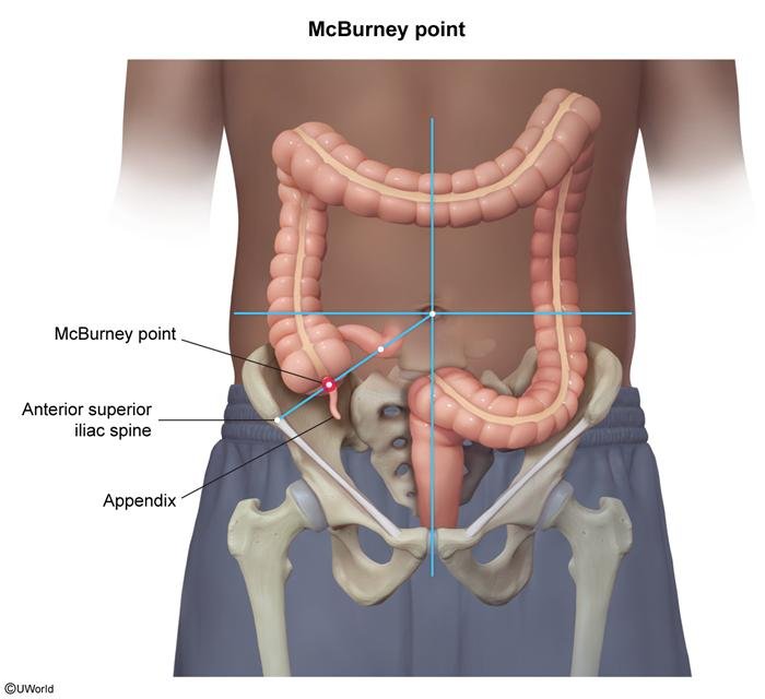
  • Appendix: Narrow worm-like tubular diverticulum attached to cecum.
  • Mesoappendix: Peritoneal fold connecting appendix to ileum and cecum.
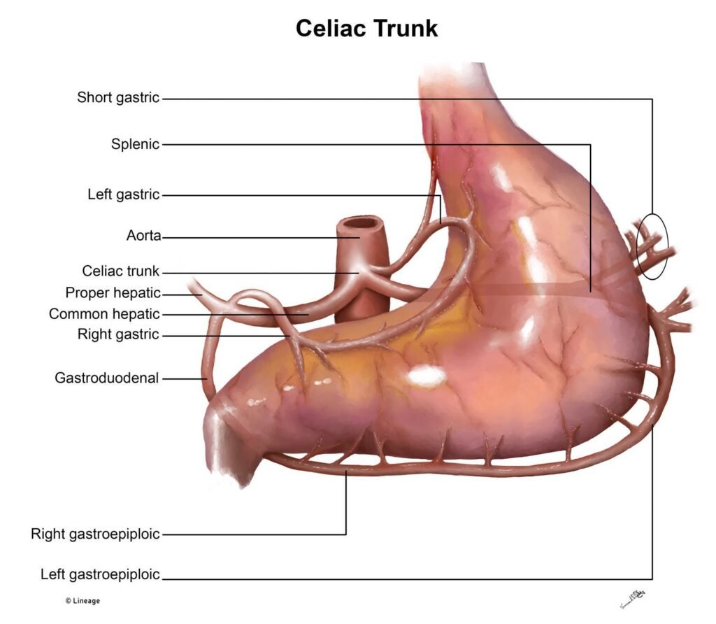
  • Celiac trunk: First major unpaired branch of abdominal aorta supplying foregut.
  • Superior mesenteric artery (SMA): Supplies midgut structures.
  • Inferior mesenteric artery (IMA): Supplies hindgut structures.
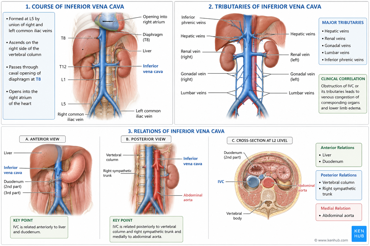
  • Inferior vena cava (IVC): Largest abdominal vein returning blood to right atrium.
  • Cisterna chyli: Dilated lymphatic sac receiving intestinal and lumbar lymph.
  • Lymphatic drainage: Movement of tissue fluid through lymphatic vessels into venous circulation.


Essential Terminology

  • Foregut: Esophagus to proximal duodenum.
  • Midgut: Distal duodenum to proximal 2/3 transverse colon.
  • Hindgut: Distal 1/3 transverse colon to upper anal canal.
  • Tributary: Smaller vessel draining into larger vessel.
  • Anastomosis: Communication between blood vessels.
  • Retroperitoneal: Located posterior to peritoneum.


Basic Overview

  • Appendix arises from posteromedial wall of cecum.
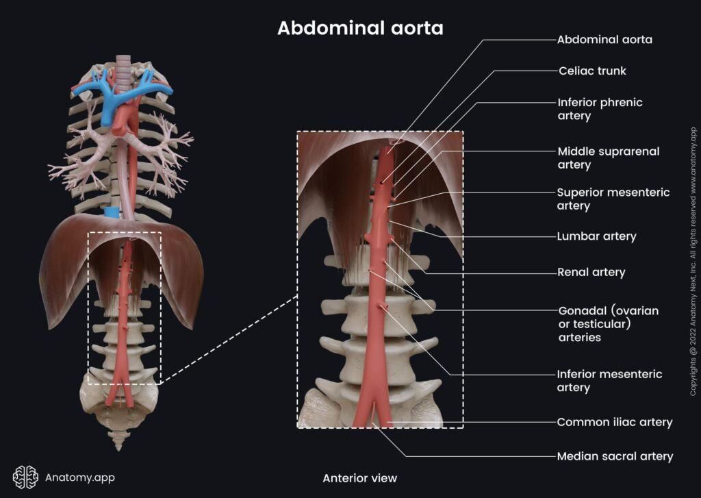
  • Abdominal aorta begins at T12 and ends at L4.
  • Major unpaired branches:
    • Celiac trunk
    • SMA
    • IMA
  • IVC forms at L5.
  • Cisterna chyli is part of lymphatic drainage pathway.
  • Abdominal lymphatics accompany arteries.

🔹 3️⃣ Core Learning — Curriculum Coverage

Appendix — Gross Features, Blood Supply, Mesoappendix and Clinical Correlation


🧠 CORE

  • Appendix is a blind-ended worm-like tubular diverticulum.
  • Arises from posteromedial wall of cecum below ileocecal junction.
  • Average length:
    • 6–10 cm.
  • Located in right iliac fossa.
  • Contains abundant lymphoid tissue.
  • Has narrow lumen.
  • Base remains constant in position.
  • Tip position varies:
    • Retrocecal
    • Pelvic
    • Preileal
    • Postileal.
  • Surface marking:
    • McBurney’s point.
  • Suspended by mesoappendix.
  • Mesoappendix contains:
    • Appendicular artery
    • Veins
    • Nerves
    • Lymphatics.
  • Main arterial supply:
    • Appendicular artery.
  • Parent artery:
    • Ileocolic artery from SMA.
  • Appendicular artery is an end artery.
  • Acute appendicitis is common clinical condition.


🔬 CONCEPT EXPLAINED

The appendix is a narrow blind-ended tube attached to the cecum. It develops as part of the midgut and is suspended by a peritoneal fold called the mesoappendix. The mesoappendix carries vessels, nerves, and lymphatics to the appendix.

The appendix contains abundant lymphoid tissue, especially in children and young adults, which contributes to immune surveillance within the gastrointestinal tract.

Its narrow lumen makes it prone to obstruction by fecoliths or lymphoid hyperplasia. Once obstructed, mucus accumulates, bacterial growth increases, and inflammation develops, leading to appendicitis.

The appendicular artery, a branch of the ileocolic artery from the superior mesenteric artery, runs through the mesoappendix to supply the organ. Because it is an end artery with poor collateral circulation, inflammation can rapidly produce ischemia and gangrene.

The position of the appendix tip varies due to mobility provided by the mesoappendix. Therefore, clinical symptoms of appendicitis may vary depending on appendix position.

Structure → Function

  • Narrow tubular structure → Concentrates lymphoid tissue.
  • Rich lymphoid tissue → Supports immune function.
  • Mesoappendix → Provides mobility and vascular support.
  • End artery supply → Direct blood supply but poor collateral circulation.
  • Variable appendix position → Variable pain patterns in appendicitis.


⚠️ IF DAMAGED

Cause → Effect

  • Luminal obstruction → Bacterial overgrowth → Appendicitis.
  • Inflammation → Venous congestion → Ischemia.
  • End artery compromise → Gangrene of appendix.
  • Rupture → Peritonitis.
  • Retrocecal appendix → Poorly localized pain.
  • Delayed treatment → Sepsis and abdominal infection.

Branches of Abdominal Aorta


🧠 CORE

  • Begins at T12.
  • Ends at L4.
  • Divides into common iliac arteries.
  • Major unpaired branches:
    • Celiac trunk
    • SMA
    • IMA.
  • Paired branches:
    • Renal arteries
    • Gonadal arteries
    • Inferior phrenic arteries.


🔬 CONCEPT EXPLAINED

The abdominal aorta supplies oxygenated blood to abdominal organs and posterior abdominal wall.

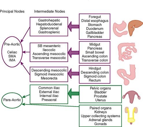
Unpaired branches supply gastrointestinal tract according to embryological divisions:

  • Foregut
  • Midgut
  • Hindgut.

Structure → Function

  • Central large artery → High-volume blood flow.
  • Multiple branches → Organ-specific supply.


⚠️ IF DAMAGED

  • Aortic aneurysm → Risk of rupture.
  • Arterial blockage → Intestinal ischemia.

Celiac Trunk — Course and Distribution


🧠 CORE

  • First unpaired branch of abdominal aorta.
  • Arises at T12 vertebral level.
  • Short thick artery.
  • Emerges just below aortic hiatus of diaphragm.
  • Divides into three main branches:
    • Left gastric artery
    • Splenic artery
    • Common hepatic artery.
  • Supplies foregut-derived organs.
  • Major organs supplied:
    • Lower esophagus
    • Stomach
    • Liver
    • Gallbladder
    • Spleen
    • Pancreas
    • Proximal duodenum.
  • Located in upper abdomen near pancreas.


🔬 CONCEPT EXPLAINED

The celiac trunk is the first major unpaired branch of the abdominal aorta and is responsible for supplying blood to foregut structures. It arises immediately below the diaphragm at the T12 level and rapidly divides into its three terminal branches.

The left gastric artery ascends toward the lesser curvature of stomach and lower esophagus. The splenic artery follows a tortuous course toward the spleen while supplying pancreas and stomach. The common hepatic artery travels toward the liver and gives branches to liver, gallbladder, stomach, and duodenum.

The celiac trunk supplies organs involved in digestion, metabolism, storage, and secretion. These organs require rich vascular supply because of their high metabolic activity.

Structure → Function

  • Short trunk → Rapid distribution of blood to upper abdominal organs.
  • Three large branches → Efficient supply to multiple foregut organs.
  • Rich arterial network → Supports digestion, secretion, and metabolism.
  • Tortuous splenic artery → Accommodates movement of spleen and stomach.


⚠️ IF DAMAGED

Cause → Effect

  • Celiac trunk obstruction → Reduced foregut blood supply.
  • Gastric ischemia → Ulceration and pain.
  • Hepatic ischemia → Liver dysfunction.
  • Splenic artery damage → Internal hemorrhage.
  • Reduced pancreatic blood flow → Pancreatic dysfunction.
  • Severe vascular compromise → Organ ischemia and necrosis.

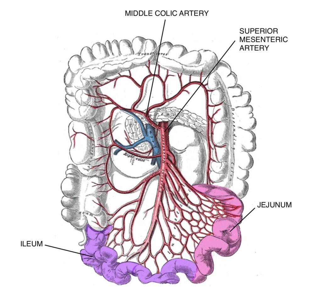
Superior Mesenteric Artery (SMA) — Course and Distribution


🧠 CORE

  • Second unpaired branch of abdominal aorta.
  • Arises at L1 vertebral level.
  • Origin lies inferior to celiac trunk.
  • Passes anterior to:
    • Left renal vein
    • Third part of duodenum.
  • Enters root of mesentery.
  • Descends downward and to the right.
  • Main artery of midgut.
  • Gives intestinal and colic branches.

Major Structures Supplied

  • Distal duodenum
  • Jejunum
  • Ileum
  • Cecum
  • Appendix
  • Ascending colon
  • Proximal 2/3 transverse colon.


🔬 CONCEPT EXPLAINED

The superior mesenteric artery arises from the anterior surface of the abdominal aorta at the L1 level. After originating below the celiac trunk, it descends anterior to the left renal vein and third part of the duodenum before entering the mesentery.

Inside the mesentery, the SMA gives numerous branches to the small intestine and proximal large intestine. These branches form arterial arcades and vasa recta that maintain continuous blood supply during intestinal movement.

The SMA supplies midgut-derived organs which are highly active in digestion and nutrient absorption. Therefore, these organs require rich and continuous arterial supply.

Structure → Function

  • Long mobile course → Reaches extensive midgut structures.
  • Mesenteric position → Allows branches to move with intestines.
  • Multiple arterial branches → Maintains continuous intestinal perfusion.
  • Rich blood supply → Supports digestion and nutrient absorption.


⚠️ IF DAMAGED

Cause → Effect

  • SMA obstruction → Reduced intestinal blood supply.
  • Mesenteric ischemia → Severe abdominal pain.
  • Prolonged ischemia → Bowel necrosis.
  • Reduced intestinal perfusion → Malabsorption.
  • Complete arterial occlusion → Intestinal infarction and gangrene.

Inferior Vena Cava (IVC) — Course, Tributaries and Relations


🧠 CORE

  • Largest vein in the abdomen.
  • Formed at L5 by union of right and left common iliac veins.
  • Ascends on right side of vertebral column.
  • Lies retroperitoneally.
  • Passes through caval opening of diaphragm at T8.
  • Opens into right atrium.

Major Tributaries

  • Hepatic veins
  • Renal veins
  • Gonadal veins
  • Lumbar veins
  • Inferior phrenic veins.

Relations of IVC

Anterior Relations

  • Liver
  • Duodenum.

Posterior Relations

  • Vertebral column
  • Right sympathetic trunk.

Medial Relation

  • Abdominal aorta.


🔬 CONCEPT EXPLAINED

The inferior vena cava is the major venous channel returning deoxygenated blood from the lower limbs, pelvis, abdomen, and abdominal organs to the heart. It forms at the L5 level and ascends vertically on the right side of the abdominal aorta.

The IVC lies posterior to abdominal organs and passes through the diaphragm at the T8 level before entering the right atrium. Because venous blood flows under low pressure, the IVC has thin walls and a large lumen that facilitate efficient venous return.

Multiple tributaries drain into the IVC from abdominal organs and posterior abdominal wall structures. These tributaries ensure continuous drainage of blood from kidneys, liver, diaphragm, gonads, and lumbar region.

Its close anatomical relations with liver, duodenum, and abdominal aorta are clinically important during abdominal surgery, trauma, and radiological procedures.

Structure → Function

  • Large thin-walled vein → Efficient low-pressure venous return.
  • Vertical course → Direct pathway to heart.
  • Multiple tributaries → Efficient drainage of abdominal organs.
  • Retroperitoneal position → Protects major venous channel.


⚠️ IF DAMAGED

Cause → Effect

  • IVC obstruction → Venous congestion.
  • Reduced venous return → Hypotension and shock.
  • Compression of IVC → Lower limb edema.
  • Hepatic vein obstruction → Hepatic congestion.
  • Trauma to IVC → Massive internal hemorrhage.
  • Venous thrombosis → Impaired circulation and organ congestion.

Cisterna Chyli


🧠 CORE

  • Dilated lymphatic sac.
  • Located at L1–L2.
  • Posterior to right crus of diaphragm.
  • Receives:
    • Intestinal trunk
    • Lumbar trunks.
  • Continues as thoracic duct.


🔬 CONCEPT EXPLAINED

The cisterna chyli collects lipid-rich lymph (chyle) from intestines and lower body.

Structure → Function

  • Dilated sac → Temporary lymph reservoir.
  • Continuation into thoracic duct → Returns lymph to venous circulation.


⚠️ IF DAMAGED

  • Lymph leakage.
  • Chylous ascites.


Lymphatic Drainage of Abdominal Organs


🧠 CORE

  • Lymph follows arteries.
  • Drains into pre-aortic lymph nodes.
  • Major groups:
    • Celiac nodes
    • Superior mesenteric nodes
    • Inferior mesenteric nodes.
  • Ultimately drains into cisterna chyli.


🔬 CONCEPT EXPLAINED

Lymphatics remove excess tissue fluid, transport fats, and support immunity.

Organs drain according to arterial supply and embryological origin.

Structure → Function

  • Lymph vessels near arteries → Efficient drainage.
  • Nodes filter pathogens and malignant cells.


⚠️ IF DAMAGED

  • Lymphedema.
  • Spread of abdominal malignancy.
  • Impaired fat absorption.

⚙️ 4️⃣ Functional Flow


Structure Function Outcome
Appendix lymphoid tissue Immune surveillance Gut immune defense
SMA branches Supply small intestine Nutrient absorption
IMA branches Supply distal colon Water absorption
IVC Venous return Maintains circulation
Cisterna chyli Collects lymph Fat transport and immunity

🩺 5️⃣ Clinical Correlation


Acute Appendicitis

  • Most common surgical emergency.
  • Pain shifts from umbilical region to right iliac fossa.
  • May cause perforation.

Mesenteric Ischemia

  • Reduced SMA blood flow.
  • Severe abdominal pain.
  • Bowel necrosis may occur.

Abdominal Aortic Aneurysm

  • Dilatation of abdominal aorta.
  • Risk of fatal rupture.

IVC Obstruction

  • Causes lower limb edema.
  • Venous congestion develops.

Chylous Ascites

  • Leakage of lymph into peritoneal cavity.
  • Occurs after lymphatic injury.

📌 6️⃣ Summary Points


  • Appendix arises from posteromedial wall of cecum.
  • Appendicular artery is an end artery.
  • McBurney’s point is important in appendicitis.
  • Celiac trunk supplies foregut.
  • SMA supplies midgut.
  • IMA supplies hindgut.
  • IVC forms at L5.
  • Cisterna chyli is origin of thoracic duct.
  • Lymphatics generally follow arteries.
  • Mesenteric ischemia is a surgical emergency.
  • Retrocecal appendix may produce atypical symptoms.
  • Lymph nodes filter pathogens and malignant cells.

🎥 7️⃣ Video Explanation

Scroll to Top
Enable Notifications OK No thanks