
📖 Step 2 — Learning Material
🔹 1️⃣ Introduction
- The gall bladder, biliary tree and spleen are important abdominal structures closely related to the liver, stomach, duodenum and pancreas.
- The gall bladder stores and concentrates bile produced by the liver.
- The extrahepatic biliary tree carries bile from the liver and gall bladder to the second part of the duodenum.
- The spleen lies in the left upper abdomen and is related to blood filtration and immune defense.
- These structures are clinically important because gallstones, obstructive jaundice, splenic enlargement and splenic injury are common exam-relevant conditions.
- Understanding their anatomy helps students explain pain patterns, jaundice, surgical landmarks and abdominal trauma findings.
🔹 2️⃣ Foundation Concepts
Key Definitions
- Gall bladder: Pear-shaped sac on the inferior surface of the liver that stores and concentrates bile.
- Bile: Digestive fluid produced by hepatocytes that helps in fat digestion and excretion of bilirubin.
- Biliary tree: Duct system that transports bile from liver and gall bladder to duodenum.
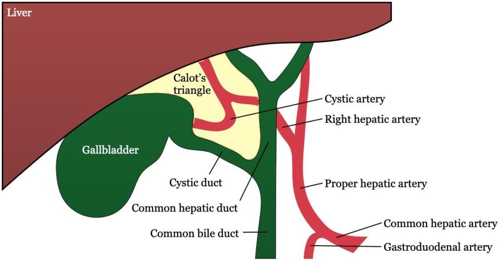
- Calot’s triangle: Anatomical triangle near the neck of gall bladder important in cholecystectomy.
- Cystic duct: Duct connecting gall bladder to common hepatic duct.
- Common bile duct: Duct formed by union of cystic duct and common hepatic duct.
- Spleen: Largest lymphoid organ, located in the left hypochondrium, involved in blood filtration and immunity.
- Hilum of spleen: Medial surface area where splenic vessels enter and leave.
Essential Terminology
- Fundus: Blind rounded end of gall bladder.
- Body: Main part of gall bladder.
- Neck: Narrow part continuous with cystic duct.
- Spiral valve: Mucosal folds in cystic duct.
- Splenic artery: Main arterial supply of spleen, branch of celiac trunk.
- Splenic vein: Venous drainage of spleen, joins superior mesenteric vein to form portal vein.
- Intraperitoneal organ: Organ almost completely covered by peritoneum.
- Peritoneal relation: Relationship of abdominal organs with peritoneum.
🔹 3️⃣ Core Learning — Curriculum Coverage
1: Gross Anatomy of Gall Bladder
🧠 CORE
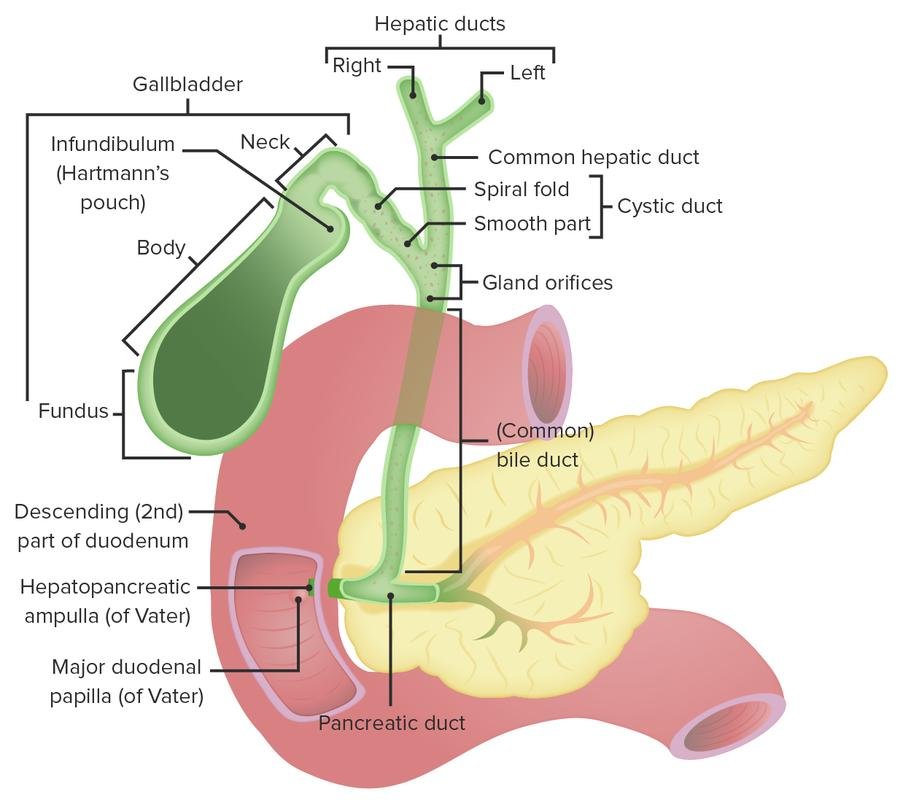
- The gall bladder is a pear-shaped muscular sac.
- It lies on the inferior surface of the right lobe of liver.
- It is situated in the gall bladder fossa.
- It stores and concentrates bile between meals.
- It is about 7–10 cm long.
- Main parts are fundus, body and neck.
- The fundus projects below the inferior border of liver.
- The neck continues as the cystic duct.
- It is related inferiorly to the duodenum and transverse colon.
- Arterial supply is mainly by the cystic artery, usually from the right hepatic artery.
🔬 CONCEPT EXPLAINED
The gall bladder acts as a temporary storage reservoir for bile. Bile is continuously formed by the liver, but bile is mainly needed when fatty food enters the duodenum. Therefore, the gall bladder stores bile when digestion is not active.
Its position on the inferior surface of the liver allows bile from the liver to easily enter the gall bladder through the cystic duct. The muscular wall of the gall bladder contracts when fatty food reaches the duodenum.
Structurally, the gall bladder has three main parts:
- Fundus: Rounded anterior end, projects near the tip of right 9th costal cartilage.
- Body: Main storage part, lies in contact with liver.
- Neck: Narrow part, bends to continue as cystic duct.
The mucosa is folded, allowing expansion when bile accumulates. The wall contains smooth muscle, which contracts to expel bile.
Structure → Function Link
- Pear-shaped sac → allows bile storage.
- Folded mucosa → allows distension.
- Smooth muscle wall → helps bile expulsion.
- Connection with cystic duct → allows bile entry and exit.
- Position under liver → supports direct bile flow from liver.
⚠️ IF DAMAGED
- Gallstone in gall bladder → irritation of wall → right upper quadrant pain.
- Stone blocking cystic duct → gall bladder distension → biliary colic.
- Inflammation of gall bladder → cholecystitis → fever, pain, tenderness.
- Impaired bile release → poor fat digestion → fatty food intolerance.
2: Calot’s Triangle
🧠 CORE
- Calot’s triangle is a small anatomical triangle near the gall bladder neck.
- It is important during removal of gall bladder.
- It helps identify the cystic artery.
- Boundaries include cystic duct, common hepatic duct and inferior surface of liver.
- It is located in the right upper abdomen.
- Main content is usually the cystic artery.
- It may also contain lymph node of Lund.
- It is clinically important because wrong identification may injure bile ducts.
- It is a key exam landmark in biliary anatomy.
Boundaries
- Medial: Common hepatic duct
- Lateral: Cystic duct
- Superior: Inferior surface of liver
Contents
- Cystic artery
- Cystic lymph node
- Small lymphatics and connective tissue
🔬 CONCEPT EXPLAINED
Calot’s triangle is important because the cystic artery usually passes through it to reach the gall bladder. During cholecystectomy, surgeons identify this triangle to safely ligate the cystic artery and cystic duct.
The triangle exists because the cystic duct joins the common hepatic duct at an angle near the liver. This creates a small space where important vessels pass.
Structure → Function Link
- Cystic duct + common hepatic duct + liver surface form a triangular space.
- This space guides identification of the cystic artery.
- Correct identification protects the common bile duct and hepatic ducts.
⚠️ IF DAMAGED
- Cystic artery injury → bleeding.
- Common bile duct injury → bile leakage or obstructive jaundice.
- Wrong duct ligation → impaired bile flow from liver.
- Post-operative bile duct damage → abdominal pain, jaundice, bile peritonitis.
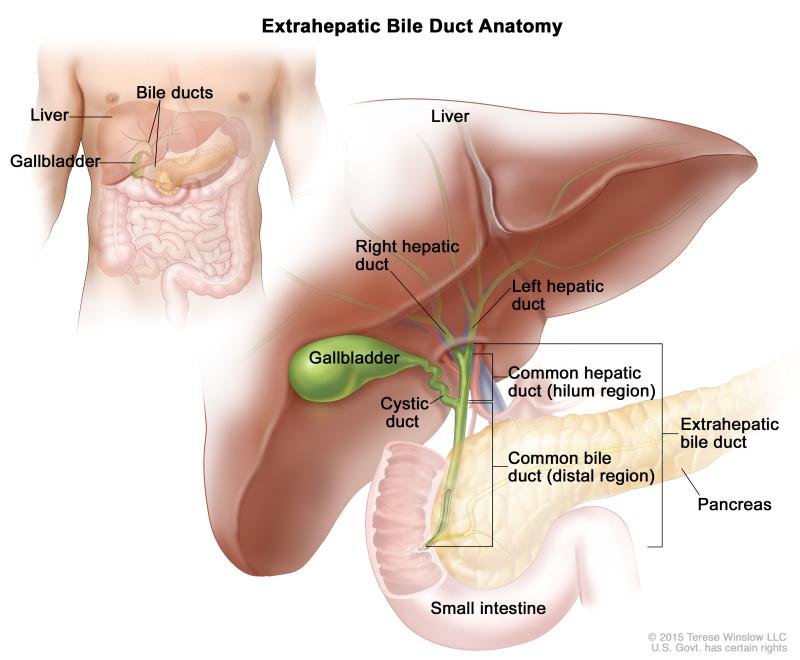
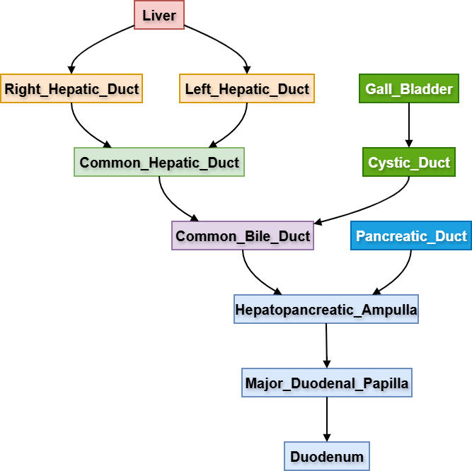
3: Extrahepatic Biliary Tree
🧠 CORE
- The extrahepatic biliary tree carries bile outside the liver.
- It begins with the right and left hepatic ducts.
- Right and left hepatic ducts unite to form the common hepatic duct.
- Cystic duct joins common hepatic duct to form the common bile duct.
- Common bile duct opens into the second part of duodenum.
- It usually joins the main pancreatic duct before opening.
- The opening is at the major duodenal papilla.
- Flow of bile is controlled by sphincters around the terminal duct.
- Main function is bile transport to the intestine.
Major Components
- Right hepatic duct
- Left hepatic duct
- Common hepatic duct
- Cystic duct
- Common bile duct
- Hepatopancreatic ampulla
- Major duodenal papilla
🔬 CONCEPT EXPLAINED
Bile is produced in the liver and must reach the duodenum for fat digestion. The right and left hepatic ducts drain bile from the right and left lobes of the liver. These ducts unite to form the common hepatic duct.
The gall bladder is connected to this system by the cystic duct. When bile is not immediately needed, it flows through the cystic duct into the gall bladder. When digestion starts, gall bladder contraction pushes bile back through the cystic duct into the common bile duct.
The common bile duct descends behind the first part of duodenum and passes near the head of pancreas. It then opens into the second part of duodenum, usually after joining the pancreatic duct.
Structure → Function Link
- Hepatic ducts → collect bile from liver.
- Cystic duct → allows storage and release of bile.
- Common bile duct → delivers bile to duodenum.
- Ampulla near pancreatic duct → coordinates bile and pancreatic juice entry.
- Sphincter control → prevents continuous uncontrolled bile flow.
⚠️ IF DAMAGED
- Stone in common bile duct → obstructive jaundice.
- Blockage near ampulla → bile and pancreatic secretion obstruction.
- Compression by pancreatic head tumor → painless progressive jaundice.
- Bile duct injury → bile leakage and peritonitis.
- Obstruction of bile flow → pale stools, dark urine, itching.
4: Gross Anatomy of Spleen
🧠 CORE
- The spleen is the largest lymphoid organ.
- It lies in the left hypochondrium.
- It is located between the 9th and 11th ribs.
- It is intraperitoneal.
- It has diaphragmatic and visceral surfaces.
- It has anterior, posterior and inferior borders.
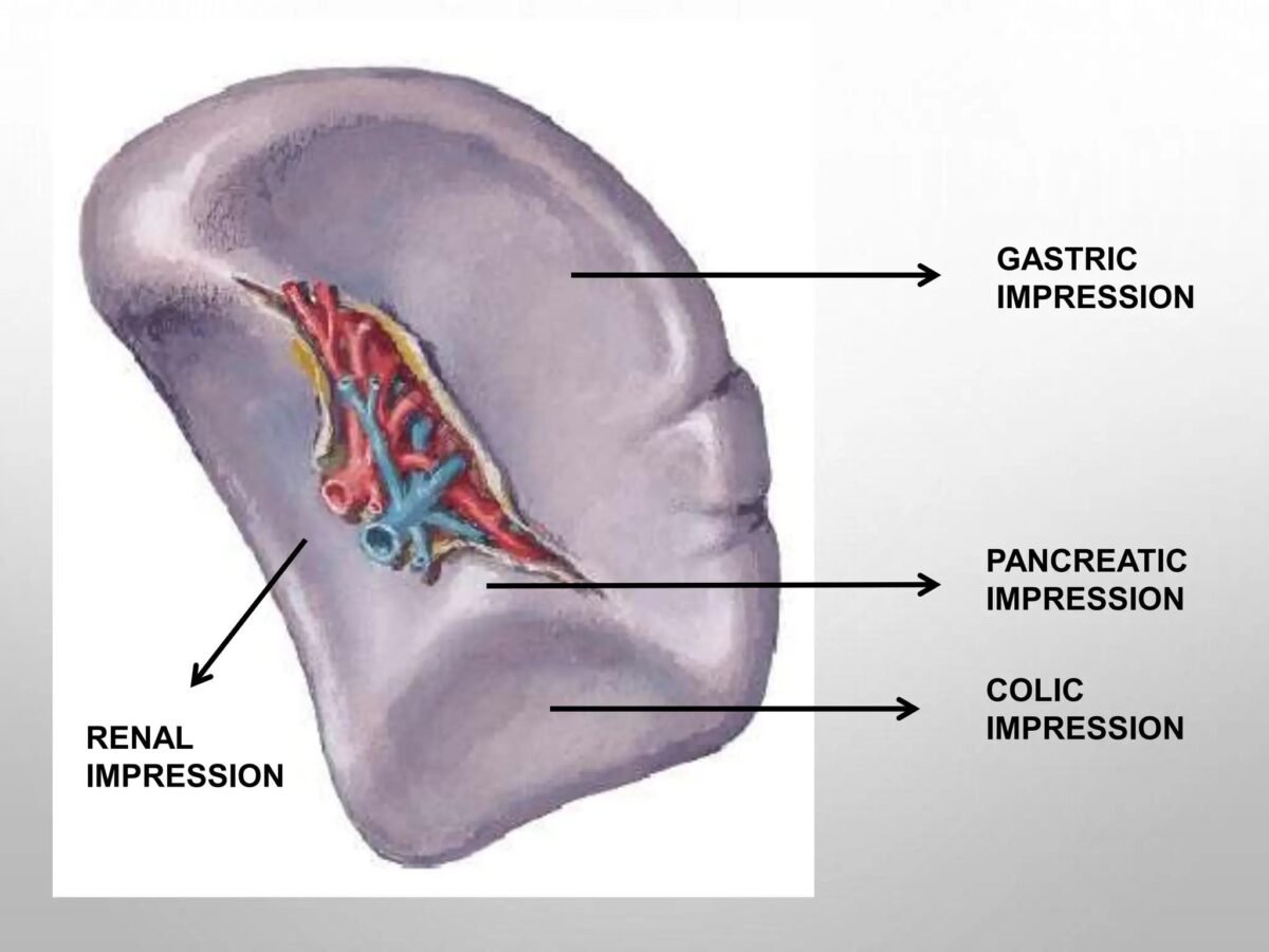
- The visceral surface contains the hilum.
- It is related to stomach, left kidney, colon and pancreas.
- It filters blood and participates in immune response.
- It is commonly enlarged in infections and blood disorders.
3D Location
- Anterior: Stomach
- Posterior: Diaphragm and ribs 9–11
- Medial: Left kidney and tail of pancreas
- Inferior: Left colic flexure
- Lateral: Left lateral abdominal wall
- Superior: Diaphragm
Surfaces
- Diaphragmatic surface: Smooth, convex, related to diaphragm.
- Visceral surface: Irregular, related to abdominal organs.
Visceral Impressions
- Gastric impression: For stomach
- Renal impression: For left kidney
- Colic impression: For left colic flexure
- Pancreatic impression: For tail of pancreas
🔬 CONCEPT EXPLAINED
The spleen is positioned high in the left upper abdomen under the protection of ribs. Its soft and vascular structure allows it to filter blood efficiently. However, because it is fragile and highly vascular, injury can cause severe internal bleeding.
The diaphragmatic surface is smooth because it lies against the diaphragm. The visceral surface is irregular because it touches several abdominal organs.
The hilum lies on the visceral surface and allows entry and exit of splenic vessels. The tail of pancreas reaches close to the hilum, making this relation clinically important.
Structure → Function Link
- Highly vascular organ → filters blood effectively.
- Soft pulp-like tissue → allows blood cell processing.
- Hilum on visceral surface → permits vessel entry and exit.
- Rib protection → protects a fragile organ.
- Relation to immune tissue → supports defense against blood-borne organisms.
⚠️ IF DAMAGED
- Ruptured spleen → severe intraperitoneal bleeding.
- Splenomegaly → enlarged spleen may become palpable.
- Loss of spleen → increased risk of infections.
- Trauma to left lower ribs → possible splenic injury.
- Enlarged spleen → may cause left upper quadrant fullness.
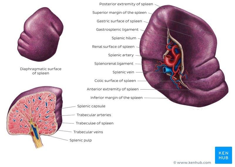
5: Blood Supply of Spleen
🧠 CORE
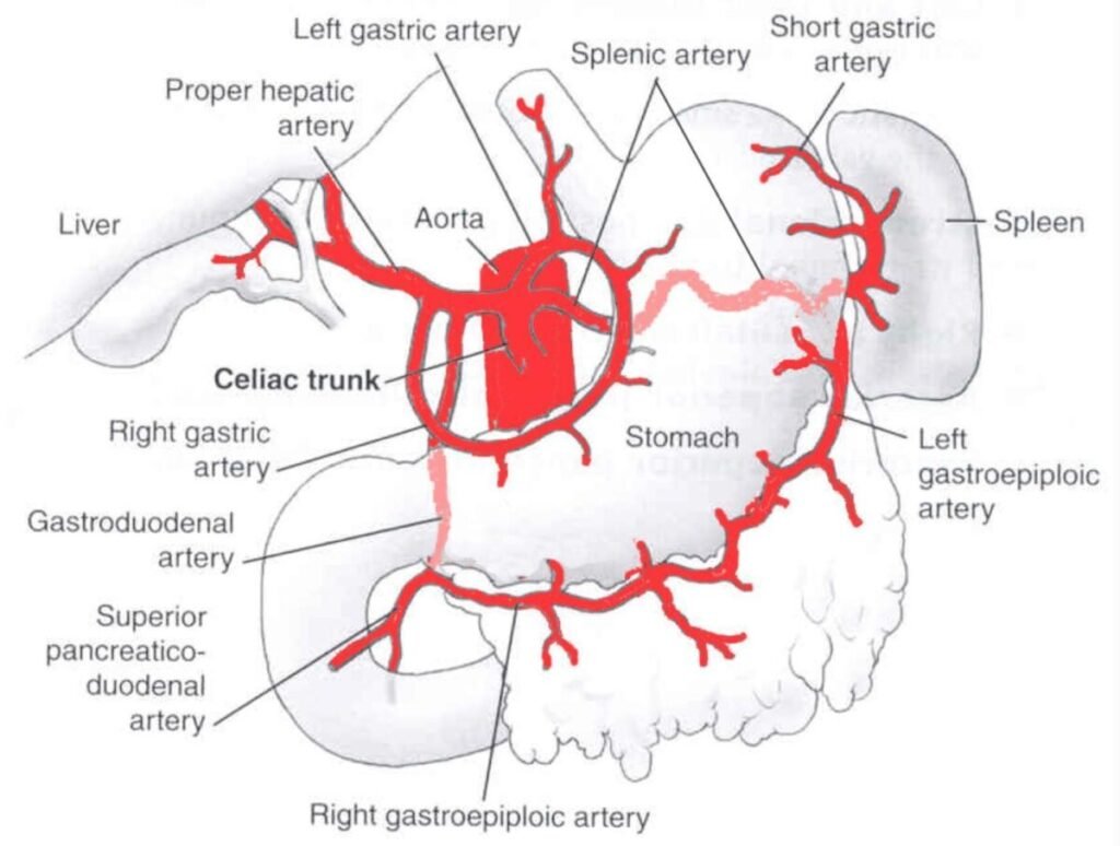
- The spleen is supplied by the splenic artery.
- Splenic artery is a branch of the celiac trunk.
- It runs along the superior border of pancreas.
- It enters the spleen through the hilum.
- It divides into branches before entering splenic tissue.
- Venous drainage is by the splenic vein.
- Splenic vein joins superior mesenteric vein to form the portal vein.
- The spleen has rich blood supply.
- Its vascularity explains severe bleeding in splenic rupture.
Blood Supply Rule
- Primary arterial supply: Splenic artery
- Parent artery: Celiac trunk
- Venous drainage: Splenic vein
- Portal relation: Splenic vein + superior mesenteric vein → portal vein
🔬 CONCEPT EXPLAINED
The splenic artery is a tortuous branch of the celiac trunk. It passes toward the left along the superior border of pancreas and reaches the hilum of spleen. Before entering the spleen, it divides into branches.
The spleen filters large amounts of blood; therefore, it requires a rich arterial supply. This also explains why splenic trauma can cause rapid blood loss.
The venous blood from the spleen drains into the splenic vein. The splenic vein joins the superior mesenteric vein behind the neck of pancreas to form the portal vein.
Structure → Function Link
- Large splenic artery → brings blood for filtration.
- Multiple terminal branches → distribute blood within spleen.
- Splenic vein drainage → connects spleen to portal circulation.
- Close relation to pancreas → explains risk during pancreatic surgery or disease.
⚠️ IF DAMAGED
- Splenic artery injury → rapid hemorrhage.
- Splenic rupture → intraperitoneal bleeding and shock.
- Splenic vein obstruction → portal circulation disturbance.
- Damage near pancreatic tail → risk to splenic vessels.
⚙️ 4️⃣ Functional Flow
A. Flow of Bile
- Hepatocytes produce bile.
- Bile enters small bile canaliculi inside liver.
- Bile passes into right and left hepatic ducts.
- Right and left hepatic ducts form the common hepatic duct.
- During fasting, bile passes through cystic duct into gall bladder.
- Gall bladder stores and concentrates bile.
- After fatty meal, gall bladder contracts.
- Bile passes through cystic duct into common bile duct.
- Common bile duct carries bile to second part of duodenum.
- Bile helps emulsify fats for digestion.
B. Gallstone Obstruction Flow
- Stone forms in gall bladder.
- Stone may enter cystic duct or common bile duct.
- Cystic duct obstruction causes gall bladder distension.
- Distension stimulates pain fibers.
- Patient develops right upper quadrant pain.
- Common bile duct obstruction blocks bile flow to duodenum.
- Bilirubin accumulates in blood.
- Patient develops obstructive jaundice.
C. Splenic Rupture Flow
- Blunt trauma affects left upper abdomen or lower ribs.
- Fragile splenic capsule tears.
- Splenic blood vessels rupture.
- Blood enters peritoneal cavity.
- Patient develops abdominal pain and internal bleeding.
- Severe bleeding may lead to shock.
🩺 5️⃣ Clinical Correlation
1. Gallstones
- Gallstones commonly form in gall bladder.
- They may block the cystic duct or common bile duct.
- Cystic duct blockage causes biliary colic.
- Common bile duct blockage causes obstructive jaundice.
2. Cholecystitis
- Inflammation of gall bladder commonly occurs due to cystic duct obstruction.
- Gall bladder becomes distended and inflamed.
- Patient may develop fever, right upper quadrant pain and tenderness.
3. Obstructive Jaundice
- Occurs when bile cannot flow into the duodenum.
- Causes include common bile duct stone or pancreatic head tumor.
- Features include yellow sclera, dark urine, pale stools and itching.
4. Importance of Calot’s Triangle
- Calot’s triangle helps identify the cystic artery.
- It is important during cholecystectomy.
- Injury to common bile duct may cause bile leakage or jaundice.
5. Splenomegaly
- Spleen may enlarge in infections, hematological diseases and portal hypertension.
- Enlarged spleen may become palpable below the left costal margin.
- It is more vulnerable to rupture.
6. Splenic Rupture
- Usually follows trauma to left upper abdomen or lower ribs.
- Because spleen is highly vascular, rupture can cause severe bleeding.
- It is a surgical emergency.
📌 6️⃣ Summary Points
- Gall bladder lies on the inferior surface of liver.
- Main parts of gall bladder are fundus, body and neck.
- Gall bladder stores and concentrates bile.
- Cystic duct connects gall bladder with common hepatic duct.
- Common hepatic duct + cystic duct = common bile duct.
- Common bile duct opens into the second part of duodenum.
- Calot’s triangle contains the cystic artery.
- Spleen lies in the left hypochondrium between ribs 9–11.
- Spleen has diaphragmatic and visceral surfaces.
- Splenic artery is a branch of the celiac trunk.
- Splenic vein joins superior mesenteric vein to form the portal vein.
- Splenic rupture can cause severe intraperitoneal bleeding.